. 2013 Oct 1;110(40):16259-64.
doi: 10.1073/pnas.1309057110. Epub 2013 Sep 16.

ECHIDNA-mediated post-Golgi trafficking of auxin carriers for differential cell elongation

Affiliations

ECHIDNA-mediated post-Golgi trafficking of auxin carriers for differential cell elongation

Yohann Boutté et al. Proc Natl Acad Sci U S A. .

Abstract

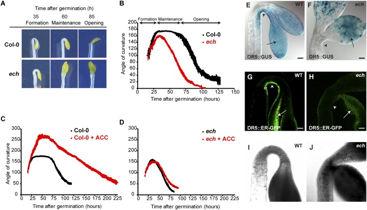
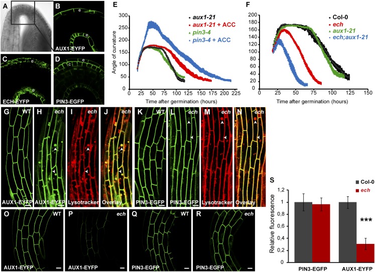
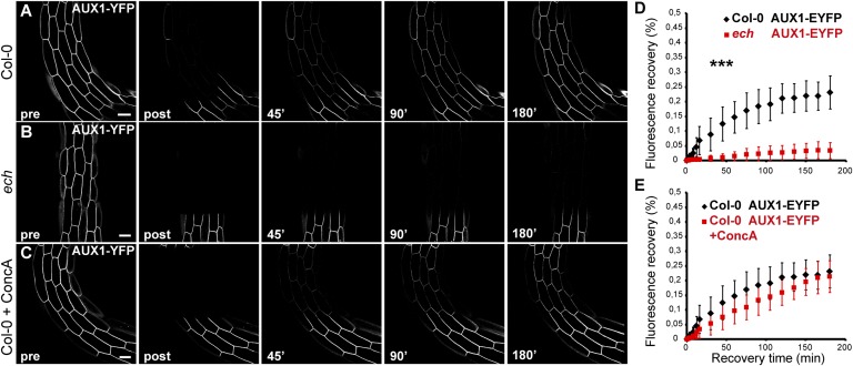
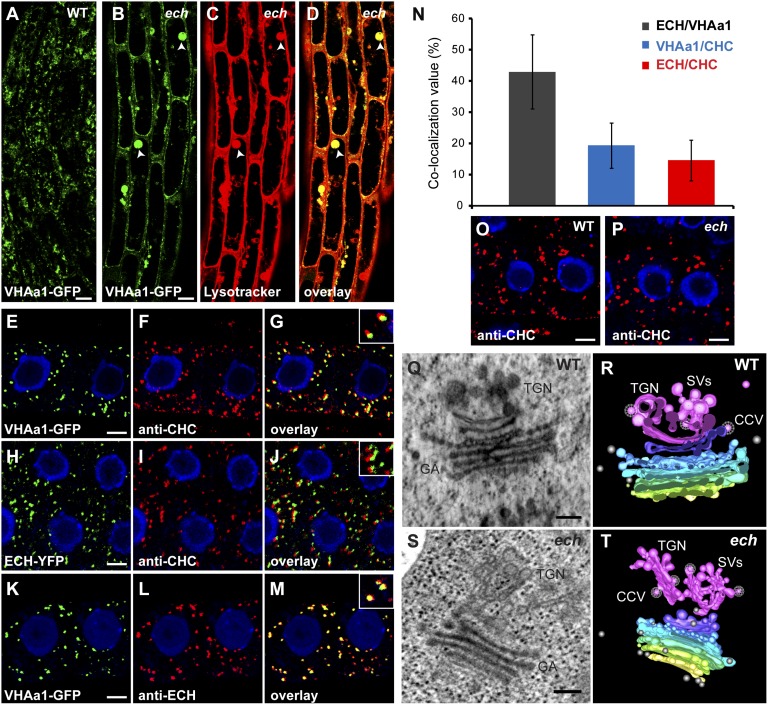
The plant hormone indole-acetic acid (auxin) is essential for many aspects of plant development. Auxin-mediated growth regulation typically involves the establishment of an auxin concentration gradient mediated by polarly localized auxin transporters. The localization of auxin carriers and their amount at the plasma membrane are controlled by membrane trafficking processes such as secretion, endocytosis, and recycling. In contrast to endocytosis or recycling, how the secretory pathway mediates the localization of auxin carriers is not well understood. In this study we have used the differential cell elongation process during apical hook development to elucidate the mechanisms underlying the post-Golgi trafficking of auxin carriers in Arabidopsis. We show that differential cell elongation during apical hook development is defective in Arabidopsis mutant echidna (ech). ECH protein is required for the trans-Golgi network (TGN)-mediated trafficking of the auxin influx carrier AUX1 to the plasma membrane. In contrast, ech mutation only marginally perturbs the trafficking of the highly related auxin influx carrier LIKE-AUX1-3 or the auxin efflux carrier PIN-FORMED-3, both also involved in hook development. Electron tomography reveals that the trafficking defects in ech mutant are associated with the perturbation of secretory vesicle genesis from the TGN. Our results identify differential mechanisms for the post-Golgi trafficking of de novo-synthesized auxin carriers to plasma membrane from the TGN and reveal how trafficking of auxin influx carriers mediates the control of differential cell elongation in apical hook development.

Keywords: IAA; morphogenesis; sorting.


Conflict of interest statement

The authors declare no conflict of interest.

Figures

Fig. 1.
Fig. 1.
ECHIDNA is involved in the apical hook maintenance phase and in the ethylene and auxin response. (A) WT and ech mutant dark-grown seedlings during distinct stages of apical hook development. (BD) Kinematic analyses of apical hook angles show that (B) compared with WT, ech mutant is defective in maintenance phase. (C) Compared with untreated WT, 10 µM ACC treatment exaggerates the formation phase. (D) ech mutant treated with 10 µM ACC does not result in exaggerated hook. (E) In WT, DR5::GUS is detected in the concave side of apical hook (arrowheads) and in cotyledons (arrows). (G) In WT, DR5::ER-GFP tissue localization pattern is restricted to epidermal (e) cells of the concave side of the hook (arrowhead). In ech mutant, DR5::GUS (F) and DR5::ER-GFP (H) are visualized in cotyledons (arrows) but not in apical hook (arrowheads). (I and J) Transmission picture of G and H, respectively. (Scale bars in EH, 50 µm).
Fig. 2.
Fig. 2.
The auxin influx carrier AUX1 genetically interacts with ECH and is mislocalized in the ech mutant. (AD) In WT hook region (black box in transmission picture in A) AUX1-YFP (B) and ECH–YFP (C) are localized in the epidermal (e) cell layer whereas PIN3–GFP (D) is localized in the cortical (c) and epidermal (e) cell layers. (E) Kinematic analyses of hook angles of aux1-21 and pin3-4 mutants seedlings untreated or treated with 10 µM ACC. (F) Kinematic analyses of hook angles of WT, aux1-21, and ech single mutants and ech;aux1-21 double mutant. (GN) As compared with WT (G and K), ech hook epidermal cells accumulate AUX1–YFP (H) in intracellular spherical compartments that colabel with Lysotracker Red (I and J; arrowheads in HJ), whereas PIN3–GFP (L) displays only a faint signal in these Lysotracker Red-positive compartments (M and N; arrowheads in LN). (OR) Confocal pictures of AUX1–YFP (O and P) and PIN3–GFP (Q and R) fluorescence in apical hook epidermal cells acquired under the same acquisition settings between WT (O and Q) and ech (P and R). (S) Plasma membrane fluorescence intensities quantification from experiments in OR. (Scale bars, 5 µm in GN, and 10 µm in OR.)
Fig. 3.
Fig. 3.
FRAP-monitored deposition of AUX1 to the plasma membrane in the ech mutant background or upon concA. Apical hook epidermal cells (14 cells from n = 7 individual seedlings) expressing AUX1–YFP (AC) were imaged before photobleaching (pre), after photobleaching (post), and during recovery after photobleaching at indicated time (over 180 min) in WT (A), ech mutant (B), and upon pretreatment with 10 µM concA for 1.5 h before photobleaching (C). (D) Recovery of AUX1–YFP in WT (A and D) is highly different from recovery of AUX1–YFP in the ech mutant (B and D). (E) ConcA pretreatment does not result in statistical difference in recovery curve of WT seedlings expressing AUX1–YFP (A, C, and E). (All scale bars, 5 µm.)
Fig. 4.
Fig. 4.
ECHIDNA acts at SV/VHAa1 sites rather than at CHC sites of TGN. (AD) In apical hook epidermal cells, compared with the WT (A) VHA-a1–GFP is mislocalized to vacuolar-like structures (arrowheads) in ech mutant (B), which colocalized strongly (arrowheads) with Lysotracker Red (C and D). (EN) In roots, VHA-a1–GFP-labeled structures (E) or ECH–YFP-positive compartments (H) are often associated with structures recognized by anti-CHC (F and I) but rarely colocalize (G and J; see the magnification in the top right corner box). (KM) VHA-a1–GFP compartments (K) and anti–ECH-positive structures (L) are found to strongly colocalize together (M; see the magnification in the top right corner box). (N) Quantification histogram of colocalization analyzed in EM. (O and P) Immunolocalization of anti–CHC-labeled compartments in the WT (O) and the ech mutant (P). (QT) Electron tomography of WT (Q and R) and ech mutant (S and T) roots. (Q and S) Still images of WT (Q) and ech mutant (S). GA, Golgi apparatus. (R and T) Models of WT (R) and ech mutant (T) tomograms. The cis-citernae of the Golgi apparatus is labeled in yellow, medial-citernae are labeled in gradient from green to blue, the trans-citernae is labeled in purple, and the TGN is highlighted in pink. SVs, in pink, are budding from the TGN. CCVs are represented with a white meshwork over the vesicle. Free vesicles are labeled in gray. (Scale bars, 5 µm in AM, 5 μm in O and P, and 200 nm in QT.)

References

    1. Okada K, Ueda J, Komaki M-K, Bell C-J, Shimura Y. Requirement of the auxin polar transport system in early stages of Arabidopsis floral bud formation. Plant Cell. 1991;3(7):677–684. - PMC - PubMed
    1. Sabatini S, et al. An auxin-dependent distal organizer of pattern and polarity in the Arabidopsis root. Cell. 1999;99(5):463–472. - PubMed
    1. Benková E, et al. Local, efflux-dependent auxin gradients as a common module for plant organ formation. Cell. 2003;115(5):591–602. - PubMed
    1. Friml J, et al. Efflux-dependent auxin gradients establish the apical-basal axis of Arabidopsis. Nature. 2003;426(6963):147–153. - PubMed
    1. Grieneisen V-A, Xu J, Marée A-F, Hogeweg P, Scheres B. Auxin transport is sufficient to generate a maximum and gradient guiding root growth. Nature. 2007;449(7165):1008–1013. - PubMed

Publication types

MeSH terms

LinkOut - more resources