. 2013 Oct 1;110(40):E3820-9.
doi: 10.1073/pnas.1308419110. Epub 2013 Sep 16.

Actin-related protein2/3 complex regulates tight junctions and terminal differentiation to promote epidermal barrier formation

Affiliations

Actin-related protein2/3 complex regulates tight junctions and terminal differentiation to promote epidermal barrier formation

Kang Zhou et al. Proc Natl Acad Sci U S A. .

Abstract

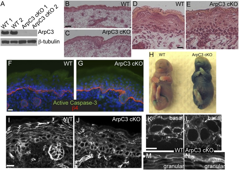
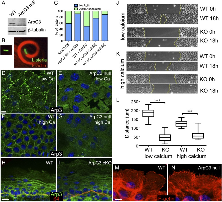
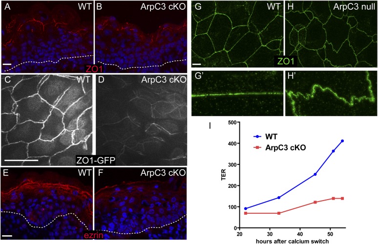
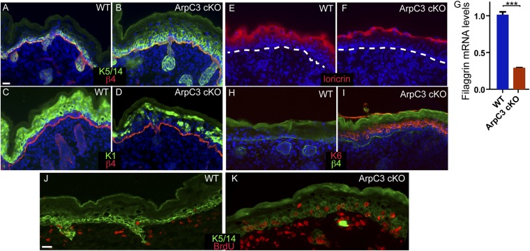
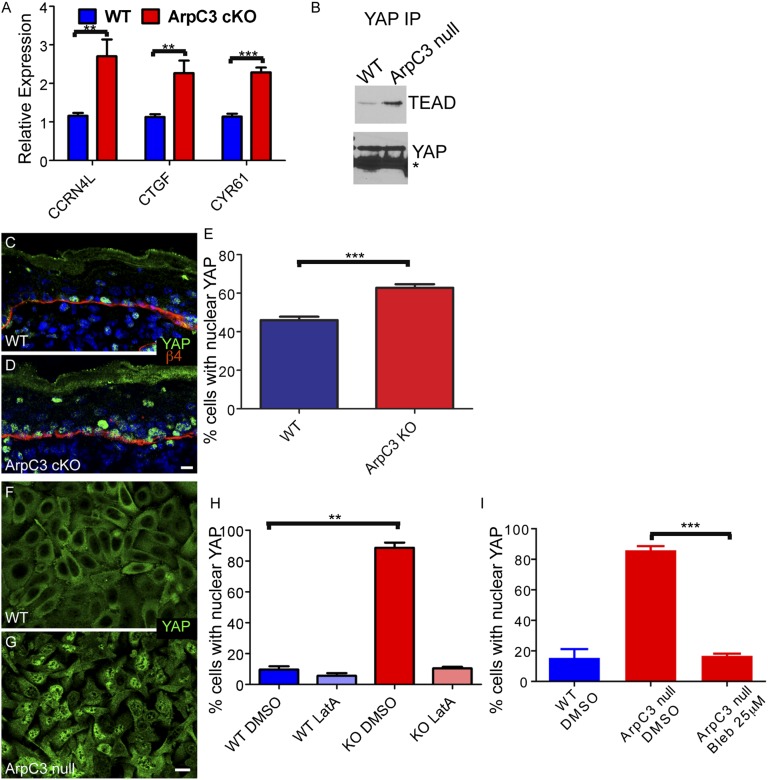
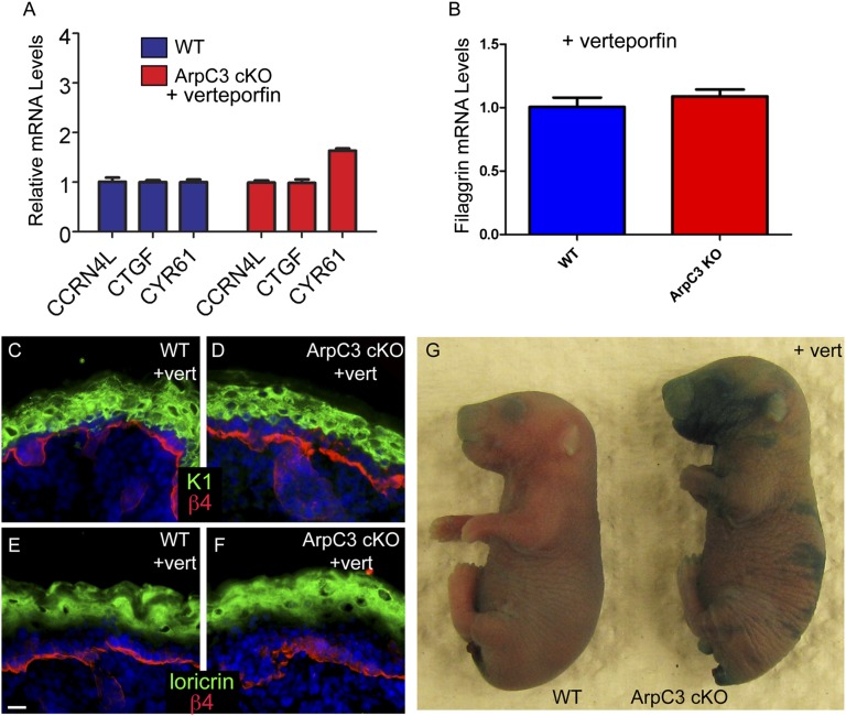
The epidermis provides an essential seal from the external environment and retains fluids within the body. To form an effective barrier, cells in the epidermis must form tight junctions and terminally differentiate into cornified envelopes. Here, we demonstrate that the branched actin nucleator, the actin-related protein (Arp)2/3 complex, is unexpectedly required for both these activities. Loss of the ArpC3 subunit of the Arp2/3 complex resulted in minimal changes in the morphogenesis and architecture of this stratified squamous epithelium, but resulted in profound defects in its physiology. Mutant embryos did not develop an effective barrier to the external environment and died within hours of birth. We discovered two underlying causes for these effects. First, ArpC3 was essential for robust assembly and function of tight junctions, specialized cell-cell adhesions that restrict water loss in the epidermis. Second, there were defects in differentiation of the epidermis and the production of cornified envelopes, structures essential for barrier activity. Underlying this defect, we found that YAP was inappropriately active not only in the ArpC3 mutant tissue, but also in cultured cells. Inhibition of YAP activity rescued the differentiation and barrier defects caused by loss of ArpC3. These results demonstrate previously unappreciated roles for the Arp2/3 complex and highlight the functions of branched actin networks in a complex tissue.


Conflict of interest statement

The authors declare no conflict of interest.

Figures

Fig. 1.
Fig. 1.
Loss of ArpC3 in the epidermis results in barrier defects but only subtle changes in architecture and F-actin organization. (A) Western blot of ArpC3 and β-tubulin in lysates prepared from two WT and two ArpC3 cKO epidermises. (BE) H&E-stained tissue sections from WT and ArpC3 cKO skin. (F and G) Immunofluorescence for active caspase 3 (green) and β4-integrin (red) in WT and ArpC3 cKO skin. β4-Integrin marks the junction between the dermis and the epidermis. (H) X-Gal barrier penetration assay in E18.5 WT and ArpC3 cKO embryos. The blue color indicates loss of barrier activity. (IN) Rhodamine–phalloidin staining of F-actin structures in WT and ArpC3 cKO epidermis as indicated. (Scale bars: B and C, 50 µm; D, E, I, and J, 20 µm; 10 µm in F, G, K, and L.)
Fig. 2.
Fig. 2.
Loss of ArpC3 results in defects in Arp2/3 complex activity and localization. (A) Western blot of ArpC3 and β-tubulin in lysates prepared from WT and ArpC3-null keratinocytes. (B) Examples of Listeria (green) that have not assembled actin around themselves (Left) or that have assembled F-actin (red; Right). (C) Quantification of Listeria actin assembly in control and ArpC3-null cells (ArpC3 fl/fl + AdCre) and in WT cells treated with the Arp2/3 inhibitor CK-636 (P < 0.0001 for ArpC3 fl/fl vs. ArpC3 fl/fl + AdCre and WT + DMSO vs. WT + CK-636 40 µM). (D and E) Arp3 localization in WT and ArpC3-null keratinocytes grown in low calcium containing media. (F and G) Arp3 localization in WT and ArpC3-null keratinocytes grown in 1.2 mM calcium-containing media. (H and I) Immunofluorescence of Arp3 (green) and β4-integrin (red) in WT and ArpC3 cKO epidermis. (J) Images of WT and ArpC3-null keratinocytes migrating into scratch wounds under low calcium conditions. (K) Images of WT and ArpC3-null keratinocytes migrating into scratch wounds in the presence of 1.2 mM calcium. (L) Quantification of migration rates of WT and ArpC3-null cells in low- and high-calcium conditions (P < 0.0001 for WT vs. KO under high- and low-calcium conditions). (M and N) F-actin organization at the leading edge of WT and ArpC3-null keratinocytes grown in 1.2 mM calcium-containing media. (Scale bars: 10 µm.)
Fig. 3.
Fig. 3.
Tight junction defects upon loss of ArpC3. (A and B) Immunofluorescence localization of the tight junction protein ZO-1 (red) in WT and ArpC3 cKO epidermis. The dotted line marks the basement membrane. (C and D) Epifluorescence of ZO1-GFP knock-in mice in the granular layer of the epidermis of WT and ArpC3 cKO embryos at E17.5. (E and F) Localization of ezrin (red) in the epidermis of WT and ArpC3 cKO epidermis. (G and H) ZO-1 organization in cultured WT and ArpC3-null keratinocytes. G’ and H’ highlight the difference in the linearity of the junctions. G’ and H’ are magnified views of areas of G and H, respectively. (I) Transepithelial resistance measurements for WT and ArpC3-null cells after a calcium switch. (Scale bars: A, B, and EI, 10 µm; C and D, 50 µm.)
Fig. 4.
Fig. 4.
Differentiation defects in ArpC3 cKO epidermis. (A and B) Immunofluorescence localization of K5/14 (green) and β4-integrin (red) in WT and ArpC3 cKO epidermis. (C and D) Localization of K1 (green) in the epidermis of WT and ArpC3 cKO embryos. (E and F) Loricrin protein localization (red) in WT and ArpC3 cKO epidermis. The dashed line indicates the basement membrane. (G) Quantitative PCR (qPCR) analysis of levels of filaggrin mRNA in WT and ArpC3-null epidermis (P < 0.0001). (H and I) K6 (red) expression in WT and ArpC3 cKO epidermis. (J and K) BrdU-incorporating cells (red) were visualized by anti-BrdU staining of WT and ArpC3 cKO skin sections. Dams were injected with BrdU 1 h before euthanasia and collection of embryos.
Fig. 5.
Fig. 5.
YAP activation upon loss of ArpC3. (A) qPCR analysis of the levels of three YAP target genes: CCRN4L, CTGF, and CYR61. RNA was collected from E18.5 embryos. P values for the differences are 0.0029, 0.0033, and <0.0001, respectively. (B) Cell extracts from WT or ArpC3-null cells were immunoprecipitated with anti-YAP antibodies. Bound proteins were examined by Western blot analysis. (C and D) Immunofluorescence localization of YAP (green) in WT and ArpC3 cKO epidermis. (E) Quantification of the number of basal cells with nuclear YAP in WT and ArpC3 cKO embryos. The ArpC3 cKO had a significant increase in cells with nuclear YAP (P = 0.0005, from n = 3 embryos and n > 250 cells counted per embryo). (F and G) Localization of YAP in confluent WT and ArpC3-null keratinocytes. (H) Quantification of cells with nuclear YAP grown under the indicated conditions. ArpC3-null cells had a significant increase in cells with nuclear YAP (P = 0.004, n > 300 cells from each of two independent experiments), and this effect was lost after disruption of F-actin with latrunculin-A. (I) Quantification of cells with nuclear YAP after treatment with the myosin II inhibitor blebbistatin. The inhibitor-treated ArpC3-null cells had a significant decrease in cells with nuclear YAP (P = 0.0009, n > 300 cells from each of two independent experiments). (Scale bars: 10 µm.)
Fig. 6.
Fig. 6.
YAP inhibition rescues terminal differentiation defects in the ArpC3 cKO epidermis. (A) qPCR analysis of three YAP target genes—CCRN4l, CTGF, and CYR61—in E18.5 epidermis from WT and ArpC3 embryos that were treated with verteporfin at E17.5 (P = 0.44, P = 0.42, and P = 0.003, respectively). (B) qPCR analysis of the levels of filaggrin mRNA in WT and ArpC3 cKO after verteporfin treatment. (C and D) Immunofluorescence localization of K1 (green) in WT and ArpC3 cKO epidermis after verteporfin treatment. (E and F) Localization of loricrin (green) in WT and ArpC3 cKO epidermis. (G) Barrier assay performed on E18.5 WT and ArpC3 cKO embryos that were treated with verteporfin at E17.5.

References

    1. Firat-Karalar EN, Welch MD. New mechanisms and functions of actin nucleation. Curr Opin Cell Biol. 2011;23(1):4–13. - PMC - PubMed
    1. Rotty JD, Wu C, Bear JE. New insights into the regulation and cellular functions of the ARP2/3 complex. Nat Rev Mol Cell Biol. 2013;14(1):7–12. - PubMed
    1. Pollard TD. Regulation of actin filament assembly by Arp2/3 complex and formins. Annu Rev Biophys Biomol Struct. 2007;36:451–477. - PubMed
    1. Robinson RC, et al. Crystal structure of Arp2/3 complex. Science. 2001;294(5547):1679–1684. - PubMed
    1. Volkmann N, et al. Structure of Arp2/3 complex in its activated state and in actin filament branch junctions. Science. 2001;293(5539):2456–2459. - PubMed

Publication types

MeSH terms

LinkOut - more resources