. 2013 Oct 15;191(8):4317-25.
doi: 10.4049/jimmunol.1301465. Epub 2013 Sep 16.

IL-33 markedly activates murine eosinophils by an NF-κB-dependent mechanism differentially dependent upon an IL-4-driven autoinflammatory loop

Affiliations

IL-33 markedly activates murine eosinophils by an NF-κB-dependent mechanism differentially dependent upon an IL-4-driven autoinflammatory loop

Carine Bouffi et al. J Immunol. .

Abstract

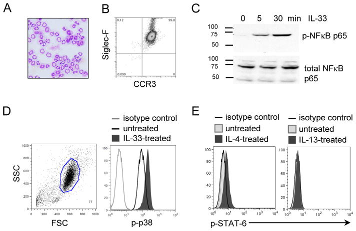
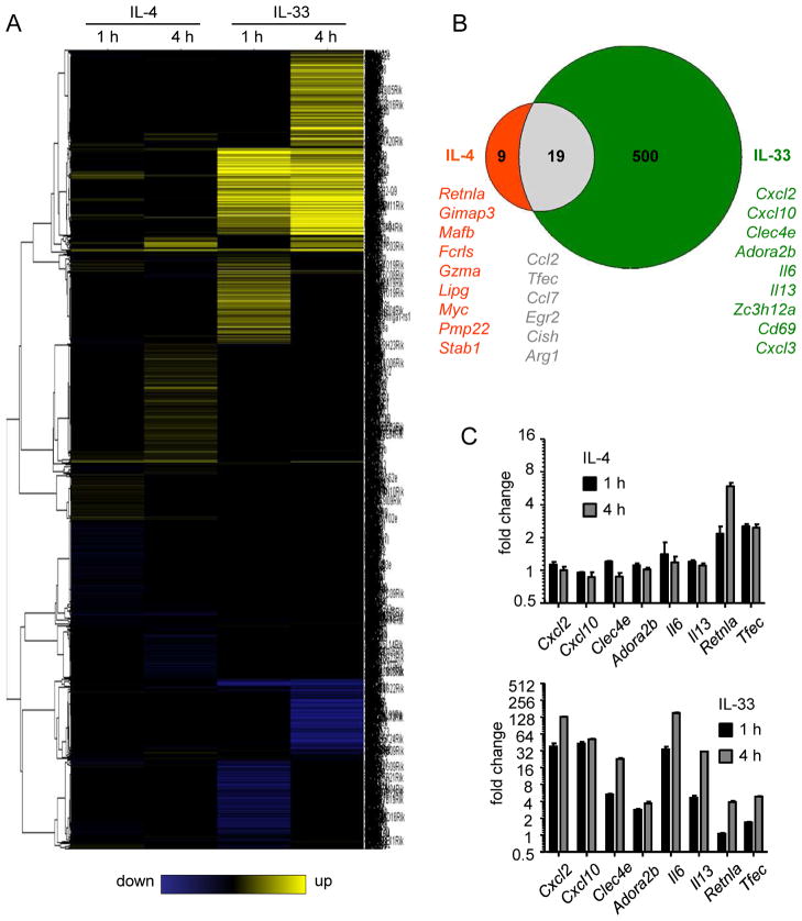
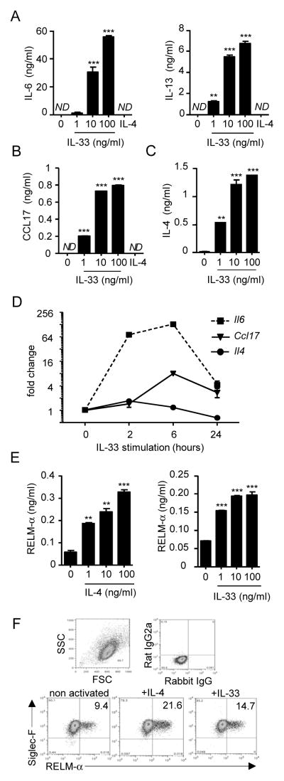
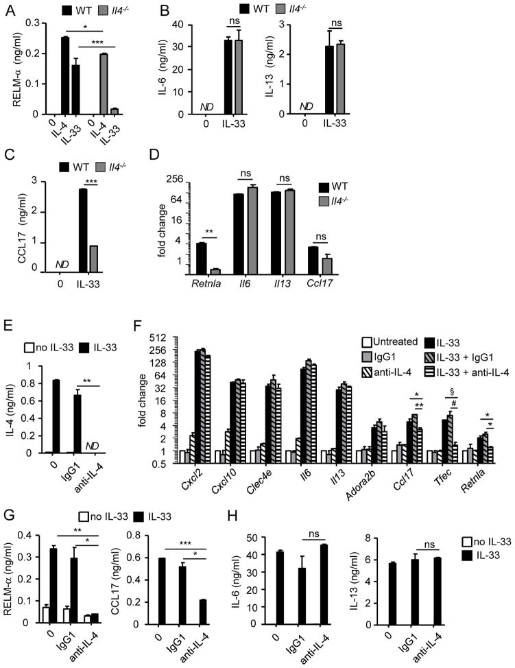
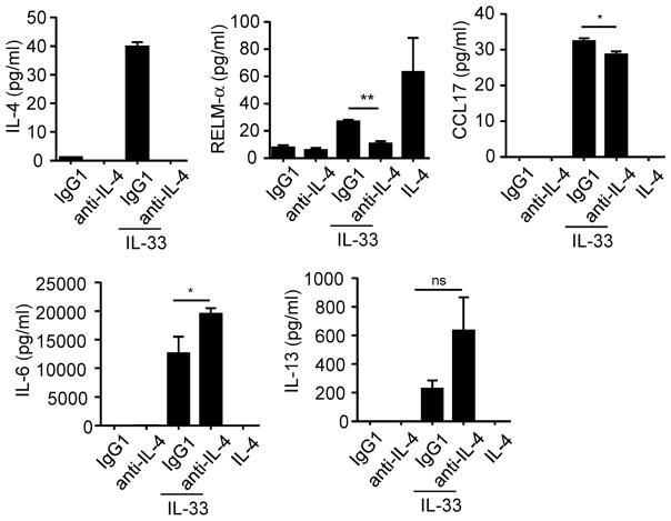
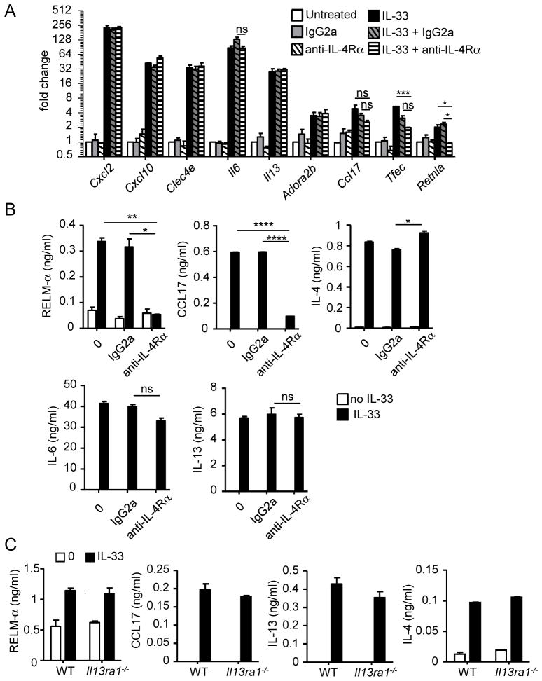
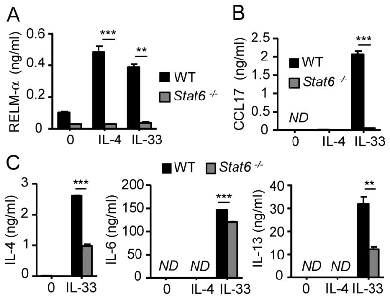
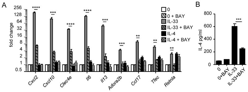
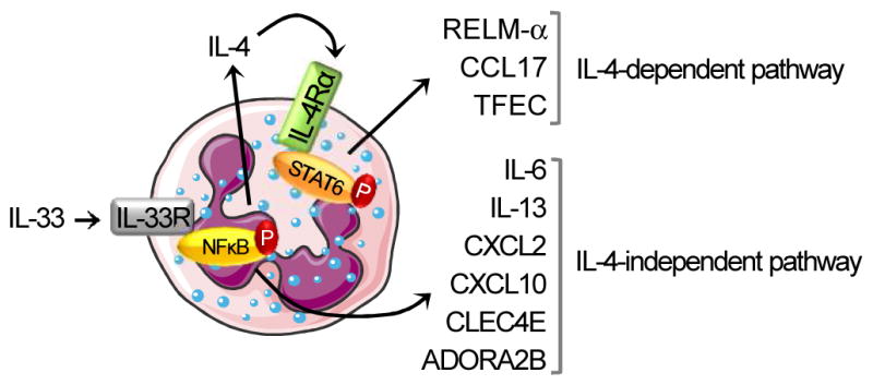
Eosinophils are major effector cells in type 2 inflammatory responses and become activated in response to IL-4 and IL-33, yet the molecular mechanisms and cooperative interaction between these cytokines remain unclear. Our objective was to investigate the molecular mechanism and cooperation of IL-4 and IL-33 in eosinophil activation. Eosinophils derived from bone marrow or isolated from Il5-transgenic mice were activated in the presence of IL-4 or IL-33 for 1 or 4 h, and the transcriptome was analyzed by RNA sequencing. The candidate genes were validated by quantitative PCR and ELISA. We demonstrated that murine-cultured eosinophils respond to IL-4 and IL-33 by phosphorylation of STAT-6 and NF-κB, respectively. RNA sequence analysis of murine-cultured eosinophils indicated that IL-33 induced 519 genes, whereas IL-4 induced only 28 genes, including 19 IL-33-regulated genes. Interestingly, IL-33 induced eosinophil activation via two distinct mechanisms, IL-4 independent and IL-4 secretion/autostimulation dependent. Anti-IL-4 or anti-IL-4Rα Ab-treated cultured and mature eosinophils, as well as Il4- or Stat6-deficient cultured eosinophils, had attenuated protein secretion of a subset of IL-33-induced genes, including Retnla and Ccl17. Additionally, IL-33 induced the rapid release of preformed IL-4 protein from eosinophils by a NF-κB-dependent mechanism. However, the induction of most IL-33-regulated transcripts (e.g., Il6 and Il13) was IL-4 independent and blocked by NF-κB inhibition. In conclusion, we have identified a novel activation pathway in murine eosinophils that is induced by IL-33 and differentially dependent upon an IL-4 auto-amplification loop.


Figures

Figure 1
Figure 1. Bone marrow-derived eosinophils respond to IL-33 and IL-4 but not IL-13
Hematoxylin/eosin staining of bone marrow-derived eosinophils at Day 14 of culture (magnification x400) (A). Characterization of eosinophils by the co-expression of Siglec-F and CCR3, using flow cytometry analysis (B). Western blot of p-NFκB and NFκB from eosinophils (4x10⁶/ml) treated with 100 ng/ml of IL-33 for 5 or 30 minutes (C). Flow cytometry of intracellular staining of p-p38 or p-STAT-6 from eosinophils (4×10⁶/ml) treated with 100 ng/ml of IL-33 for 5 minutes (D) or 100ng/ml of IL-4 or IL-13 for 30 minutes (E). The results shown are representative of 3 independent experiments.
Figure 2
Figure 2. Transcriptome analysis in eosinophils activated by IL-33 or IL-4
Eosinophils were treated for 1 or 4 hours with IL-4 or IL-33 at 10 ng/ml. RNA was sequenced by RNA-sequencing technology. Heatmap represents the genes differentially regulated by IL-4 and IL-33 after 1 and 4 hours of exposure (A). Venn diagram represents the number of genes (with a fold change ≥ 2 in either direction) differentially regulated by IL-4 (red), IL-33 (green), or both (grey) (B). Validation by qRT-PCR analysis of genes identified by RNA sequencing as upregulated in eosinophils activated by IL-4 or IL-33 at 1 or 4 hours (C). Bars represent the mean of 2 wells and the error bars represent the SEM values. Data are representative of 3 independent experiments.
Figure 3
Figure 3. Effect of IL-33 and IL-4 on cytokine/chemokine expression by eosinophils
Eosinophils (4×10⁶/ml) were activated for 24 hours in the presence of the indicated concentrations of IL-33 or IL-4 (IL-4 at 100 ng/ml for A and B). Supernatants were collected and IL-6 and IL-13 (A), CCL17(B), IL-4 (C), and RELM-α (E) were measured by ELISA. qRT-PCR analysis represents the kinetics of Il6, Ccl17, and Il4 induction by IL-33 in eosinophils (D). Bars (A-C, E) or symbols (D) represent the mean of 2 wells, and error bars represent the SEM values. Flow cytometry on eosinophils after overnight incubation with the different cytokines at 100 ng/ml to evaluate SiglecF+/RELM-α+ cells (F). The percentage of cells in each quadrant is indicated (F). Data are representative of 3 independent experiments. ND, not detected. **p < 0.01, ***p < 0.001.
Figure 4
Figure 4. IL-33 requires the IL-4 autocrine loop to induce RELM-αand CCL17
Wild-type (WT) or Il4−/− eosinophils (4×10⁶/ml) treated for 24 hours with IL-4 (10 ng/ml, A) or IL-33 (10 ng/ml, A–D). RELM-α, CCL17, IL-6, and IL-13 levels in the supernatants were determined by ELISA (A–C). qRT-PCR from wild-type (WT) or Il4−/− eosinophils (4×10⁶/ml) incubated for 4 hours in the presence of IL-33 (10 ng/ml); graph represents the fold change of gene expression in IL-33-treated eosinophils compare to untreated (D). Eosinophils (4×10⁶/ml) were activated for 4 hours (F) or 24 hours (E, G and H) by IL-33 (10 ng/ml) in the absence or presence of IgG1 (isotype control) or anti-IL-4 antibody (10 μg/ml). Gene expression of Cxcl2, Cxcl10, Clec4e, Il6, Il13, Adora2b, Ccl17, Tfec, and Retnla was analyzed by qRT-PCR (F). The levels of IL-4 (E), RELM-αand CCL17 (G), and IL-6 and IL-13 (H) in the supernatants were measured by ELISA. Bars represent the mean of 2 wells and the error bars represent the SEM values. Data are representative of 3 independent experiments. ND, not detected; ns, not significant. *p < 0.05, **p < 0.01, ***p < 0.001, § p=0.055 and # p=0.083.
Figure 5
Figure 5. Mature eosinophils respond to IL-33 and require IL-4 to induce RELM-αand CCL17
Eosinophils from Il5-transgenic mice were activated 24 hours with IL-33 in the presence of anti-IL-4 antibody or isotype control (IgG1). IL-4, RELM-α, CCL17, IL-6 and IL-13 released in the supernatants were measured by ELISA. *p < 0.05, **p<0.001, ns, not significant.
Figure 6
Figure 6. IL-4Rα, but not IL-13Rα1, is involved in IL-33-induced eosinophil activation
Eosinophils (4×10⁶/ml) activated for 4 hours (A) or 24 hours (B) by IL-33 (10 ng/ml) in the absence or presence of IgG2a (isotype control) or anti-IL-4Rαantibody, at 10 μg/ml. Gene expressions of Cxcl2, Cxcl10, Clec4e, Il6, Il13, Adora2b, Ccl17, Tfec, and Retnla were analyzed by qRT-PCR (A). The levels of RELM-α, CCL17, IL-4, IL-6, and IL-13 (B) in the supernatants were measured by ELISA. Wild-type (WT) or Il13ra1−/− eosinophils (4×10⁶/ml) were treated for 24 hours in the presence of IL-33 (10ng/ml), and the levels of RELM-α, CCL17, IL-13, and IL-4 in the supernatants were measured by ELISA (C). Bars represent the mean of 2 wells and the error bars represent the SEM values. Data are representative of 3 independent experiments. ND, not detected; ns, not significant. *p < 0.05, **p < 0.01, ****p < 0.0001.
Figure 7
Figure 7. Role of STAT-6 in eosinophil activation
Wild-type (WT) and Stat6-deficient eosinophils were activated with IL-33 (100 ng/ml) or IL-4 (100 ng/ml). RELM-α (A), CCL17 (B), IL-4, IL-6, and IL-13 (C) released in the supernatants were measured by ELISA. Bars represent the mean of 2 wells and the error bars represent the SEM values. Data are representative of 3 independent experiments. ND, not detected. **p < 0.01, ***p < 0.001.
Figure 8
Figure 8. Effect of NFκB inhibitor in the regulation in IL-33-induced gene expression
Eosinophils (4×10⁶/ml) were pretreated for 1 hour with NFκB inhibitor (BAY11-7082 [BAY]) at 5 μM and then cultured for 4 hours in the presence of IL-33 (AB) or IL-4 (A) at 10 ng/ml. Gene expression of Cxcl2, Cxcl10, Clec4e, Il6, Il13, Adora2b, Ccl17, Tfec, and Retnla were analyzed by qRT-PCR (A), and IL-4 in the supernatant was measured by ELISA (B). Bars represent the mean of 2 wells and the error bars represent the SEM values. Data are representative of 3 independent experiments. **p < 0.01, ***p < 0.001, ****p < 0.0001 (A–B).
Figure 9
Figure 9. Proposed model summarizing our results
After binding to its receptor, IL-33 induces eosinophil activation, as measured by gene transcription and release of several mediators, through the phosphorylation of NFκB. Il6, Il13, Cxcl2, Cxcl10, and Clec4e are directly induced by IL-33 (IL-4-independent pathway). Although IL-4 transcription is not increased after IL-33 treatment, the protein release of preformed IL-4 is dependent on the IL-33/ST2 signaling pathway. Retnla, Ccl17 and Tfec induction by IL-33 is indirect and requires the activation of the IL-4/IL4Rα pathway (IL-4-dependent pathway) via the phosphorylation of STAT-6. This figure was produced using Servier Medical Art (www.servier.com).

References

    1. Funakoshi-Tago M, Tago K, Hayakawa M, Tominaga S, Ohshio T, Sonoda Y, Kasahara T. TRAF6 is a critical signal transducer in IL-33 signaling pathway. Cell Signal. 2008;20:1679–1686. - PubMed
    1. Schmitz J, Owyang A, Oldham E, Song Y, Murphy E, McClanahan TK, Zurawski G, Moshrefi M, Qin J, Li X, Gorman DM, Bazan JF, Kastelein RA. IL-33, an interleukin-1-like cytokine that signals via the IL-1 receptor-related protein ST2 and induces T helper type 2-associated cytokines. Immunity. 2005;23:479–490. - PubMed
    1. Allakhverdi Z, Smith DE, Comeau MR, Delespesse G. Cutting edge: The ST2 ligand IL-33 potently activates and drives maturation of human mast cells. J Immunol. 2007;179:2051–2054. - PubMed
    1. Pecaric-Petkovic T, Didichenko SA, Kaempfer S, Spiegl N, Dahinden CA. Human basophils and eosinophils are the direct target leukocytes of the novel IL-1 family member IL-33. Blood. 2009;113:1526–1534. - PMC - PubMed
    1. Kurowska-Stolarska M, Stolarski B, Kewin P, Murphy G, Corrigan CJ, Ying S, Pitman N, Mirchandani A, Rana B, van Rooijen N, Shepherd M, McSharry C, McInnes IB, Xu D, Liew FY. IL-33 amplifies the polarization of alternatively activated macrophages that contribute to airway inflammation. J Immunol. 2009;183:6469–6477. - PubMed

Publication types

MeSH terms