. 2013 Oct 15;191(8):4095-102.
doi: 10.4049/jimmunol.1301079. Epub 2013 Sep 16.

Cholera toxin directly enhances IL-17A production from human CD4+ T cells

Affiliations

Cholera toxin directly enhances IL-17A production from human CD4+ T cells

Hsing-Chuan Tsai et al. J Immunol. .

Abstract

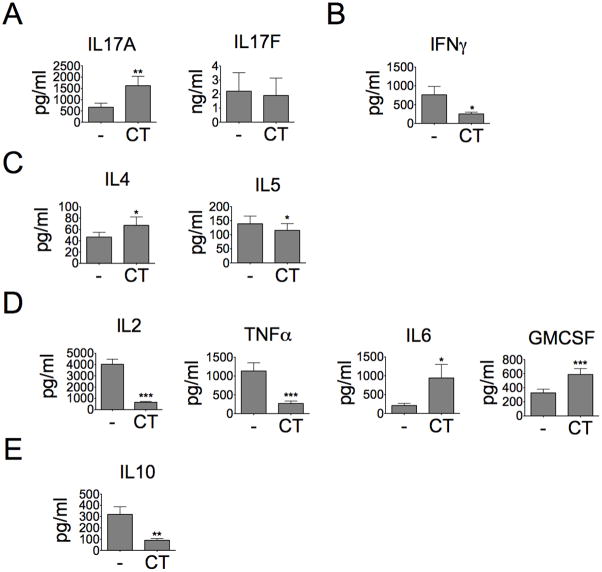
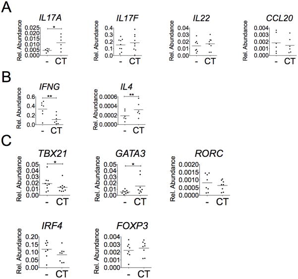
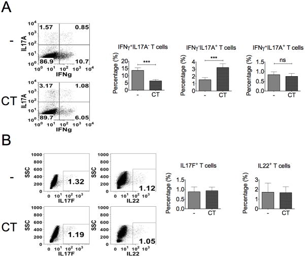
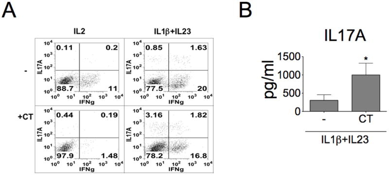
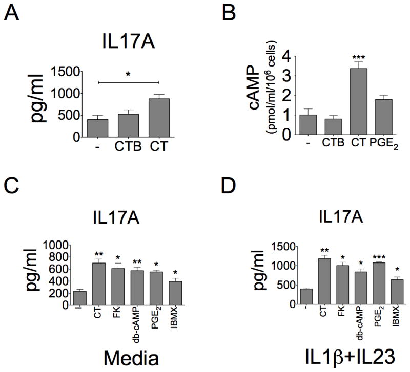
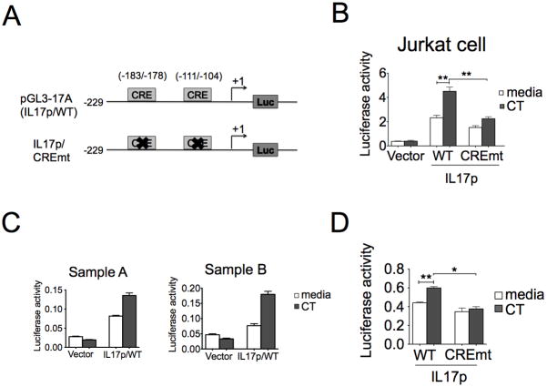
The significance of Th17 cells and IL-17A signaling in host defense and disease development has been demonstrated in various infection and autoimmune models. Additionally, the generation of Th17 cells is highly influenced by microbes. However, the specific bacterial components capable of shaping Th17 responses have not been well defined. The goals of this study were to understand how a bacterial toxin, cholera toxin (CT), modulates Th17-dominated response in isolated human CD4(+) T cells, and what are the mechanisms associated with this modulation. CD4(+) cells isolated from human peripheral blood were treated with CT. The levels of cytokine production and specific Th cell responses were determined by ELISA, Luminex assay, and flow cytometry. Along with the decreased production of other proinflammatory cytokines (IFN-γ, TNF-α, and IL-2), we found that CT could directly enhance the IL-17A production through a cAMP-dependent pathway. This enhancement is specific for IL-17A but not for IL-17F, IL-22, and CCL20. Interestingly, CT could increase IL-17A production only from Th17-committed cells, such as CCR6(+)CD4(+) T cells and in vitro-differentiated Th17 cells. Furthermore, we also demonstrated that this direct effect occurs at a transcriptional level because CT stimulates the reporter activity in Jurkat and primary CD4(+) T cells transfected with the IL-17A promoter-reporter construct. This study shows that CT has the capacity to directly shape Th17 responses in the absence of APCs. Our findings highlight the potentials of bacterial toxins in the regulation of human Th17 responses.


Conflict of interest statement

Conflict of interest declaration: The authors have no direct financial interest or relationship to the subject matter of this report.

Figures

Figure 1
Figure 1. CT directly induces IL-17A production but decreases Th1 cytokines from stimulated CD4+ T cells
Total CD4+ T cells were stimulated with or without CT (10ng/ml) for three days under CD3/CD28 activation (10 cells per bead). A, Culture supernatant was analyzed for IL-17A and IL-17F protein production by ELISA (n=7, *p<0.05). (B) Th1 cytokines; (C) Th2 cytokines, (D) pro-inflammatory cytokines; (E) anti-inflammatory cytokines, were detected by using the kit of Invitrogen Human Cytokine 10-plex panel (n≥7, *p<0.05).
Figure 2
Figure 2. CT increases IL17A, IL4, and GATA3 mRNA expression but decreases IFNG and TBX21 expression
CD4+ T cells were treated with or without CT (10ng/ml) for 2 days under CD3/CD28 activation and total RNAs of cells were collected. The expression of (A) Th17-associated genes, (B) Th1&Th2-associated genes, and (C) transcription factors were examined by SyBr Green real-time PCR assay (n≥6, *p<0.05, **p<0.01).
Figure 3
Figure 3. The percentage of IL-17A+ T cells is increased by the treatment of CT
CD4+ T cells were cultured with or without CT for 4 days and re-stimulated with TPA (50ng/ml) and ionomycin (1μM) for 5 hours. Intracellular staining for IFN-γ, IL-17A, IL-17F, and IL-22 expression in T cells was examined by flow cytometry. A, Data represent the percentage of IFNγ +IL-17A, IFNγIL-17A+ and IFNγ+IL-17A+ cells. B, Data represent the total percentage of IL17F+ and IL-22+ cells (n≥3, ***p<0.001).
Figure 4
Figure 4. CT only enhances IL-17A production from differentiated Th17 subset
Cells were sorted with CCR6 and CXCR3 surface marker to enriched Th1 (CXCR3+CCR6), Th17 (CXCR3CCR6+), and Th1/17 (CXCR+CCR6+). A, The purity (upper panels) and cytokine profile (lower panels) of sorted T cells were determined by using FACScan. Cells were activated with CD3/CD28 beads for three days, re-stimulated with TPA (50ng/ml) and ionomycin (1μM), and then stained with anti-IL17A and anti-IFNγ antibodies. Data represent one from three experiments. B, The IL-17A levels in cultured supernatant were examined after 3-day activation in the absence or presence of CT (n=3, *p<0.05, **p<0.01).
Figure 5
Figure 5. CT promotes Th17 differentiation from naïve T cells under Th17-polarizing condition
A, Naïve CD4+ T cells were isolated from human peripheral blood and differentiated under IL-2 alone or Th17-polarizing culture condition (IL-1β + IL-23 co-treatment) in the presence or absence of CT for 6 days and then rested in IL-2 (10 ng/ml) supplemented condition for additional 3 more days. Cells were then re-stimulated with TPA and ionomycin, and stained with anti-IL-17A and anti-IFNγ antibodies. Data represent one from three experiments. B, Effects of CT on IL-17A production from naïve CD4+ T cells cultured under Th17-polarizing culture condition (IL-1β + IL-23 co-treatment). These differentiated cells were re-stimulated with anti-CD3 antibody for 2 days. The IL-17A production in cultured supernatants was examined (n=4, *p<0.05).
Figure 6
Figure 6. Enhancing IL-17A production by cAMP-stimulating agents
A, T cells were stimulated with CD3/CD28 beads and treated with CT (10ng/ml) or CTB (10ng/ml). IL-17A production was examined in cultured supernatants from media, CT- or CTB-treated cells (n=5, ***p<0.001). B, Intracellular cAMP levels were examined in cell lysate from T cells by ELISA after one-hour treatment of CTB (10ng/ml), CT (10ng/ml) or PGE2 (1μM) (n=3, ***p<0.001). C–D, CD4+ T cells were treated with Forskolin (FK, 3μM), dibutyl-cAMP (db-cAMP, 100μM), IBMX (100μM) and PGE2 (1μM) for 3 days under (C) Th0 or (D) Th17-polarizing conditions. The IL-17A production in culture supernatants was assayed by ELISA (n=3, *p<0.05, **p<0.01, ***p<0.001, compared to “-” group). The “-” group means that cells were not treated by any cAMP agents.
Figure 7
Figure 7. CT directly enhances the IL-17A promoter activity dependent of cis-element CRE
A, A schematic representation of the hIL17A promoter depicting cAMP-responsive element motifs used for wild type and mutated promoter constructs. B, Jurkat cells were transfected with indicated plasmids (vector, IL17p/WT, and CREmt) and pRL-TK using lipofectamine 2000. One day after transfection, transfected cells were treated with CT for four hours under the stimulation of TPA and ionomycin and the luciferase activity were measured. Luciferase activity was displayed as relative activity normalized with Renilla luciferase activity. Data represent three independent experiments (n=3, **p<0.01). C–D, Primary T cells were stimulated for 16–18 hours and nucleofected individually with indicated plasmids (vector, IL17p/WT, and CREmt) and pRL-CMV. After 6hr recovery, cells were treated with CT for extra 18 hours under CD3/28 activation. Cell lysate were examined by luciferase reporter assay after the restimulation with TPA and ionomycin for four hours (D, n=3, *p<0.05, **p<0.01).
Figure 8
Figure 8. IL-12 modulates the effect of CT on T cells
Cells were treated with the combination of CT (10 ng/ml) and IL-12 (10 ng/ml) for three days and then re-stimulated with TPA and ionomycin. A, Re-stimulated T cells were stained with anti-IL-17A and anti-IFNγ antibodies. Data shown represent three independent experiments with similar results. B, IL-17A production was assessed by ELISA (n=6, *p< 0.05).

Similar articles

Cited by

References

    1. Wilson NJ, Boniface K, Chan JR, McKenzie BS, Blumenschein WM, Mattson JD, Basham B, Smith K, Chen T, Morel F, Lecron JC, Kastelein RA, Cua DJ, McClanahan TK, Bowman EP, de Waal Malefyt R. Development, cytokine profile and function of human interleukin 17-producing helper T cells. Nature immunology. 2007;8:950–957. - PubMed
    1. Dong C. TH17 cells in development: an updated view of their molecular identity and genetic programming. Nature reviews Immunology. 2008;8:337–348. - PubMed
    1. Hirota K, Yoshitomi H, Hashimoto M, Maeda S, Teradaira S, Sugimoto N, Yamaguchi T, Nomura T, Ito H, Nakamura T, Sakaguchi N, Sakaguchi S. Preferential recruitment of CCR6-expressing Th17 cells to inflamed joints via CCL20 in rheumatoid arthritis and its animal model. The Journal of experimental medicine. 2007;204:2803–2812. - PMC - PubMed
    1. El-Behi M, Ciric B, Dai H, Yan Y, Cullimore M, Safavi F, Zhang GX, Dittel BN, Rostami A. The encephalitogenicity of T(H)17 cells is dependent on IL-1- and IL-23-induced production of the cytokine GM-CSF. Nature immunology. 2011;12:568–575. - PMC - PubMed
    1. Gaffen SL. Recent advances in the IL-17 cytokine family. Current opinion in immunology. 2011;23:613–619. - PMC - PubMed

Publication types