

Two distinct types of the inhibition of vasculogenesis by different species of charged particles
- PMID: 24044765
- PMCID: PMC3856512
- DOI: 10.1186/2045-824X-5-16
Two distinct types of the inhibition of vasculogenesis by different species of charged particles
Abstract
Background: Charged particle radiation is known to be more biologically effective than photon radiation. One example of this is the inhibition of the formation of human blood vessels. This effect is an important factor influencing human health and is relevant to space travel as well as to cancer radiotherapy. We have previously shown that ion particles with a high energy deposition, or linear energy transfer (LET) are more than four times more effective at disrupting mature vessel tissue models than particles with a lower LET. For vasculogenesis however, the relative biological effectiveness between particles is the same. This unexpected result prompted us to investigate whether the inhibition of vasculogenesis was occurring by distinct mechanisms.
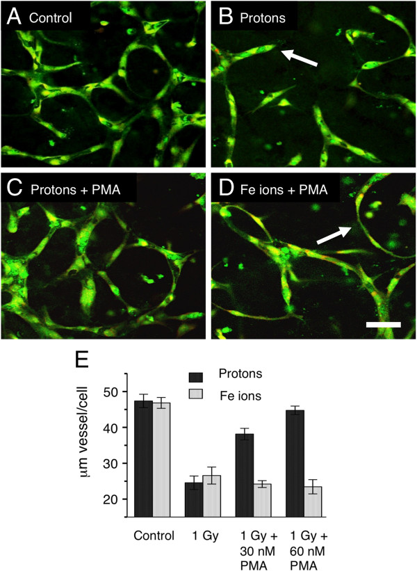
Methods: Using 3-Dimensional human vessel models, we developed assays that determine at what stage angiogenesis is inhibited. Vessel morphology, the presence of motile tip structures, and changes in the matrix architecture were assessed. To confirm that the mechanisms are distinct, stimulation of Protein Kinase C (PKC) with phorbol ester (PMA) was employed to selectively restore vessel formation in cultures where early motile tip activity was inhibited.
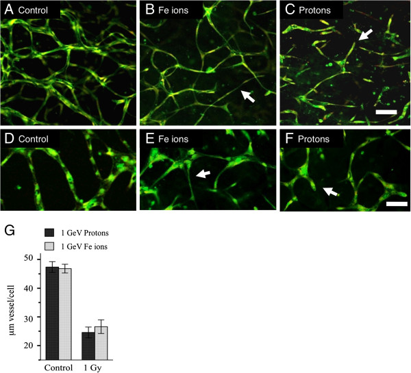
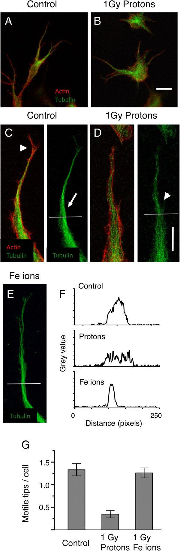
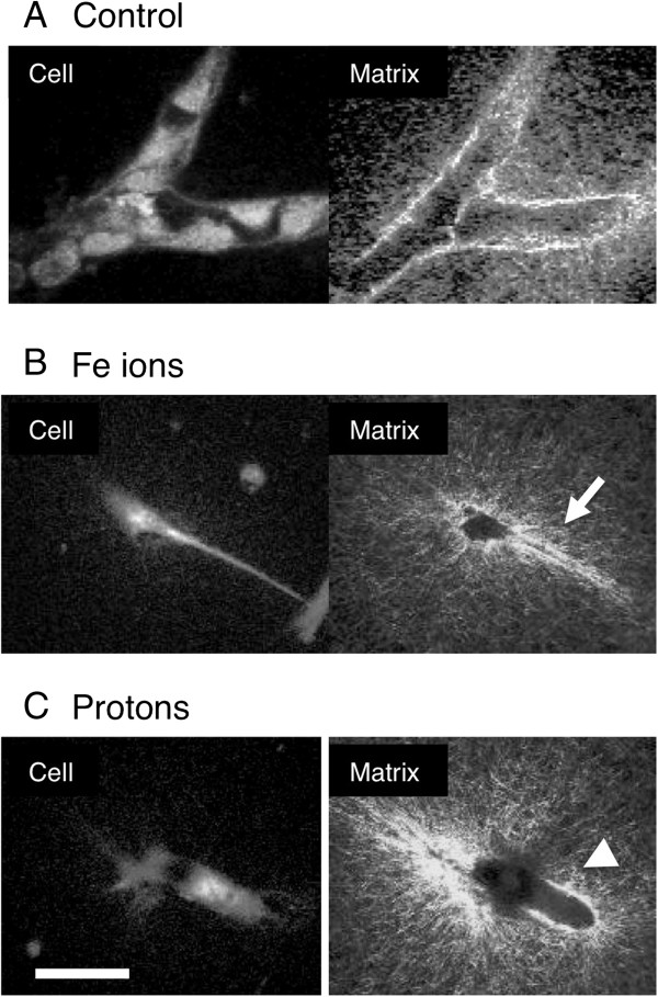
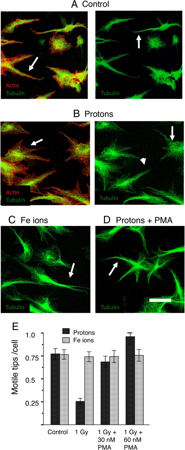
Results: Endothelial cells in 3-D culture exposed to low LET protons failed to make connections with other cells but eventually developed a central lumen. Conversely, cells exposed to high LET Fe charged particles extended cellular processes and made connections to other cells but did not develop a central lumen. The microtubule and actin cytoskeletons indicated that motility at the extending tips of endothelial cells is inhibited by low LET but not high LET particles. Actin-rich protrusive structures that contain bundled microtubules showed a 65% decrease when exposed to low LET particles but not high LET particles, with commensurate changes in the matrix architecture. Stimulation of PKC with PMA restored tip motility and capillary formation in low but not high LET particle treated cultures.
Conclusion: Low LET charged particles inhibit the early stages of vasculogenesis when tip cells have motile protrusive structures and are creating pioneer guidance tunnels through the matrix. High LET charged particles do not affect the early stages of vasculogenesis but they do affect the later stages when the endothelial cells migrate to form tubes.
Figures
Similar articles
-
Radiobiology with heavy charged particles: a historical review.Phys Med. 1998 Jul;14 Suppl 1:1-19. Phys Med. 1998. PMID: 11542635
-
Radiogenomics.Med Phys. 2018 Nov;45(11):e1111-e1122. doi: 10.1002/mp.13064. Med Phys. 2018. PMID: 30421807 Review.
-
Biological characterization of low-energy ions with high-energy deposition on human cells.Radiat Res. 2014 Sep;182(3):282-91. doi: 10.1667/RR13747.1. Epub 2014 Aug 6. Radiat Res. 2014. PMID: 25098728
-
LET-Dependent Low Dose and Synergistic Inhibition of Human Angiogenesis by Charged Particles: Validation of miRNAs that Drive Inhibition.iScience. 2020 Nov 25;23(12):101771. doi: 10.1016/j.isci.2020.101771. eCollection 2020 Dec 18. iScience. 2020. PMID: 33376971 Free PMC article.
-
DNA Damage Clustering after Ionizing Radiation and Consequences in the Processing of Chromatin Breaks.Molecules. 2022 Feb 24;27(5):1540. doi: 10.3390/molecules27051540. Molecules. 2022. PMID: 35268641 Free PMC article. Review.
Cited by
-
The effects of radiation on angiogenesis.Vasc Cell. 2013 Oct 26;5(1):19. doi: 10.1186/2045-824X-5-19. Vasc Cell. 2013. PMID: 24160185 Free PMC article.
-
Heart in space: effect of the extraterrestrial environment on the cardiovascular system.Nat Rev Cardiol. 2018 Mar;15(3):167-180. doi: 10.1038/nrcardio.2017.157. Epub 2017 Oct 20. Nat Rev Cardiol. 2018. PMID: 29053152 Review.
-
Differential Impact of Single-Dose Fe Ion and X-Ray Irradiation on Endothelial Cell Transcriptomic and Proteomic Responses.Front Pharmacol. 2017 Sep 22;8:570. doi: 10.3389/fphar.2017.00570. eCollection 2017. Front Pharmacol. 2017. PMID: 28993729 Free PMC article.
-
Microgravity × Radiation: A Space Mechanobiology Approach Toward Cardiovascular Function and Disease.Front Cell Dev Biol. 2021 Oct 29;9:750775. doi: 10.3389/fcell.2021.750775. eCollection 2021. Front Cell Dev Biol. 2021. PMID: 34778261 Free PMC article. Review.
-
Biological Effects of Space Radiation and Development of Effective Countermeasures.Life Sci Space Res (Amst). 2014 Apr 1;1:10-43. doi: 10.1016/j.lssr.2014.02.004. Life Sci Space Res (Amst). 2014. PMID: 25258703 Free PMC article.
References
LinkOut - more resources
Full Text Sources
Other Literature Sources
