. 2013 Dec;34(37):9486-95.
doi: 10.1016/j.biomaterials.2013.08.077. Epub 2013 Sep 14.

The independent roles of mechanical, structural and adhesion characteristics of 3D hydrogels on the regulation of cancer invasion and dissemination

Affiliations

The independent roles of mechanical, structural and adhesion characteristics of 3D hydrogels on the regulation of cancer invasion and dissemination

Jennifer N Beck et al. Biomaterials. 2013 Dec.

Abstract

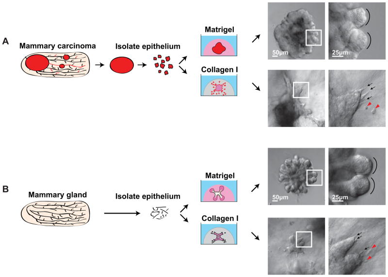
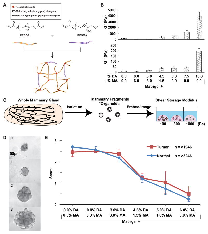
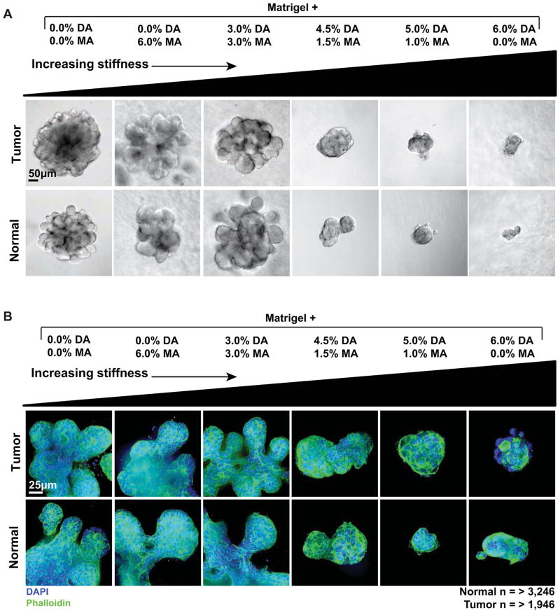
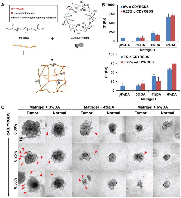
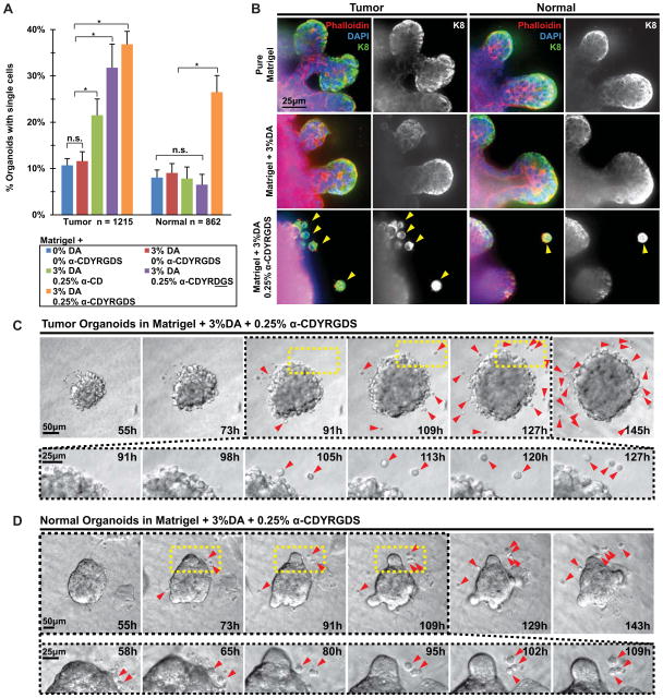
Metastasis begins with the escape, or dissemination, of cancer cells from the primary tumor. We recently demonstrated that tumors preferentially disseminate into collagen I and not into basement membrane protein gels (Matrigel). In this study, we used synthetic polymer systems to define material properties that could induce dissemination into Matrigel. We first specifically varied rigidity by varying the crosslinking density of poly(ethylene glycol) (PEG) networks within Matrigel scaffolds. Increased microenvironmental rigidity limited epithelial growth but did not promote dissemination. We next incorporated adhesive signals into the PEG network using peptide-conjugated cyclodextrin (α-CDYRGDS) rings. The α-CDYRGDS rings threaded along the PEG polymers, enabling independent control of matrix mechanics, adhesive peptide composition, and adhesive density. Adhesive PEG networks induced dissemination of normal and malignant mammary epithelial cells at intermediate values of adhesion and rigidity. Our data reveal that microenvironmental signals can induce dissemination of normal and malignant epithelial cells without requiring the fibrillar structure of collagen I or containing collagen I-specific adhesion sequences. Finally, the nanobiomaterials and assays developed in this study are generally useful both in 3D culture of primary mammalian tissues and in the systematic evaluation of the specific role of mechanical and adhesive inputs on 3D tumor growth, invasion, and dissemination.

Keywords: Breast cancer; Cell adhesion; Dissemination; Invasion; Mechanical properties; Poly(ethylene) glycol hydrogels.


Figures

Fig. 1
Fig. 1. Primary epithelial organoids as a model of tumor microenvironment induced dissemination
(A) Mouse mammary carcinomas were surgically isolated and then epithelial organoids were derived from the carcinoma through a combination of mechanical disruption and enzymatic digestion. (B) Normal epithelial organoids were similarly derived from normal mouse mammary glands. In both cases 1000s of organoids are recovered and so organoids from the same mouse were embedded in a series of microenvironments. Both normal and tumor organoids exhibited restrained epithelial growth in Matrigel and protrusive, disseminative growth in collagen I. DIC images show the organoid morphology in the different conditions. Black arrows highlight cellular protrusions, red arrowheads mark disseminated cells, black arcs highlight the rounded epithelial edge of mammary buds.
Fig. 2
Fig. 2. PEG-Matrigel composite hydrogels enable specific control of rigidity in 3D microenvironments
(A) Diagram describing the polymer system used to increase the rigidity of Matrigel. Poly(ethylene glycol) diacrylate (PEGDA) can crosslink PEG and increase the stiffness of the hydrogel whereas poly(ethylene glycol) monoacrylate (PEGMA) cannot crosslink and so limits the rigidity of the hydrogel. The total concentration of PEGDA+PEGMA was held constant at 6% so that the mechanics were varied independently of Matrigel or PEG concentration. (B) Parallel plate rheometer measurements of the shear storage modulus (G′) and shear loss modulus (G″) of a series of PEG-Matrigel composite hydrogels. Matrigel is the base polymer into which varying amounts of PEGDA and PEGMA were dissolved and photocrosslinked. Increasing concentrations of PEGDA correlated with increased rigidity of the hydrogel. (C) Schematic illustrating organoid extraction, isolation and embedding in multiple microenvironments of varying rigidity. (D) Phenotypic scores given to quantify growth of organoids. 0 = no growth, 1 = cyst-like structure, 2 = branching initiated, 3 = fully branched. Scale bar = 50 μm. (E) Graph of the trend in organoid development in microenvironments of varying rigidity, scored after 7 days of culture. Both tumor and normal displayed a negative correlation between rigidity and organoid development. Error bars = standard error of the mean.
Fig. 3
Fig. 3. Negative correlation between microenvironmental rigidity and epithelial morphogenesis
(A) DIC (scale bar = 50 μm) and (B) confocal (scale bar = 25 μm) images showing representative growth of tumor and normal organoids in microenvironments of increasing rigidity. The concentration of ECM ligands and the concentration of PEG were both held constant. Normal and tumor organoids were larger and more developed in the less rigid microenvironments. Blue = DAPI DNA stain, marking nuclei. Green = Phalloidin, marking F-actin.
Fig. 4
Fig. 4. Adhesive PEG-Matrigel composite hydrogels induce dissemination of epithelial cells
(A) We incorporated cyclodextrin rings with adhesive peptides (α-CDYRGDS) into the PEG networks to enable independent control over polymer mechanics and adhesivity. (B) Shear storage modulus (G′) and shear loss modulus (G″) measurements of PEG-Matrigel composite hydrogels revealed progressively increased rigidity with increased PEG concentration. The presence of α-CDYRGDS had a comparatively small effect on matrix mechanics. (C) Normal and tumor organoids were explanted into a range of microenvironments with different mechanics and adhesivity. We inferred cellular dissemination from the presence of single cells adjacent to organoids. Microenvironments with lower rigidity and intermediate concentrations of adhesive peptides displayed maximal dissemination (3%DA and 0.25% CDYRGDS) in both tumor and normal conditions. Scale bar = 50 μm
Fig. 5
Fig. 5. Normal and tumor organoids disseminate epithelial cells in response to synthetic microenvironmental signals
(A) Quantification of the fraction of organoids with adjacent single cells in each microenvironmental condition. Normal n = 862, tumor n = 1215 organoids counted. Statistical significance was tested using a generalized linear model to enable us to isolate the variation in single cell abundance from any possible batch effects in the 3 independent experiments. Conditions that were significantly different (P<0.05) are indicated. (B) Confocal images of tumor and normal organoids grown in Matrigel, Matrigel + 3% DA, and Matrigel + 3% DA,0.25% CDYRGDS. The single cells adjacent to epithelial organoids stained positive for antibodies targeting epithelial-specific cytokeratin 8, confirming their epithelial identity. Scale bar = 25 μm. We directly observed single cells disseminating from both tumor (C) and normal (D) organoids embedded within Matrigel + 3% DA,0.25% CDYRGDS using time-lapse DIC microscopy. Scale bar = 50 μm and 25 μm. The magnified portion shows more detail of dissemination as well as proliferation of disseminated cells.

References

    1. Sternlicht MD. Key stages in mammary gland development: the cues that regulate ductal branching morphogenesis. Breast Cancer Res. 2006;8(1):201. - PMC - PubMed
    1. Polyak K. Molecular markers for the diagnosis and management of ductal carcinoma in situ. J Natl Cancer Inst Monogr. 2010;2010(41):210–3. - PMC - PubMed
    1. Bogenrieder T, Herlyn M. Axis of evil: molecular mechanisms of cancer metastasis. Oncogene. 2003;22(42):6524–36. - PubMed
    1. Condeelis J, Singer RH, Segall JE. The great escape: when cancer cells hijack the genes for chemotaxis and motility. Annu Rev Cell Dev Biol. 2005;21:695–718. - PubMed
    1. Jeanes A, Gottardi CJ, Yap AS. Cadherins and cancer: how does cadherin dysfunction promote tumor progression? Oncogene. 2008;27(55):6920–9. - PMC - PubMed

Publication types

LinkOut - more resources