. 2013 Nov 15;19(22):6183-92.
doi: 10.1158/1078-0432.CCR-12-3904. Epub 2013 Sep 17.

Efficacy of BET bromodomain inhibition in Kras-mutant non-small cell lung cancer

Affiliations

Efficacy of BET bromodomain inhibition in Kras-mutant non-small cell lung cancer

Takeshi Shimamura et al. Clin Cancer Res. .

Abstract

Purpose: Amplification of MYC is one of the most common genetic alterations in lung cancer, contributing to a myriad of phenotypes associated with growth, invasion, and drug resistance. Murine genetics has established both the centrality of somatic alterations of Kras in lung cancer, as well as the dependency of mutant Kras tumors on MYC function. Unfortunately, drug-like small-molecule inhibitors of KRAS and MYC have yet to be realized. The recent discovery, in hematologic malignancies, that bromodomain and extra-terminal (BET) bromodomain inhibition impairs MYC expression and MYC transcriptional function established the rationale of targeting KRAS-driven non-small cell lung cancer (NSCLC) with BET inhibition.

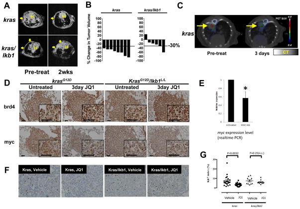
Experimental design: We performed functional assays to evaluate the effects of JQ1 in genetically defined NSCLC cell lines harboring KRAS and/or LKB1 mutations. Furthermore, we evaluated JQ1 in transgenic mouse lung cancer models expressing mutant kras or concurrent mutant kras and lkb1. Effects of bromodomain inhibition on transcriptional pathways were explored and validated by expression analysis.

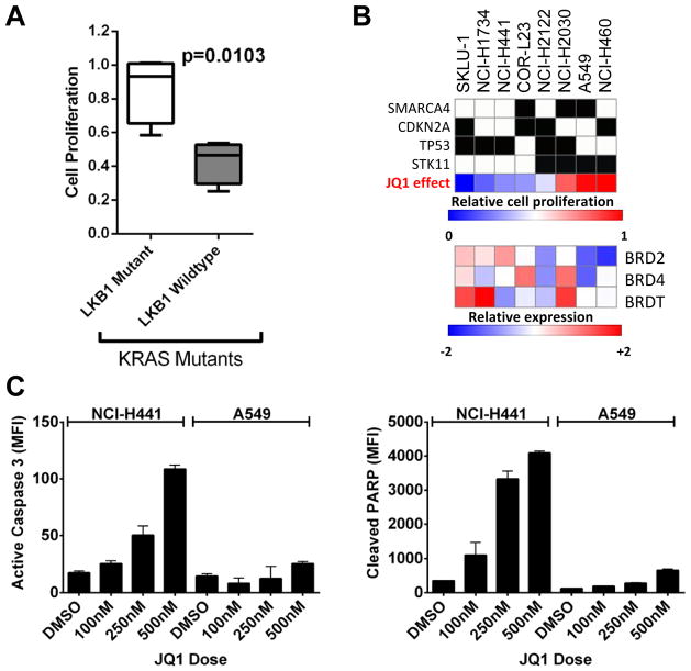
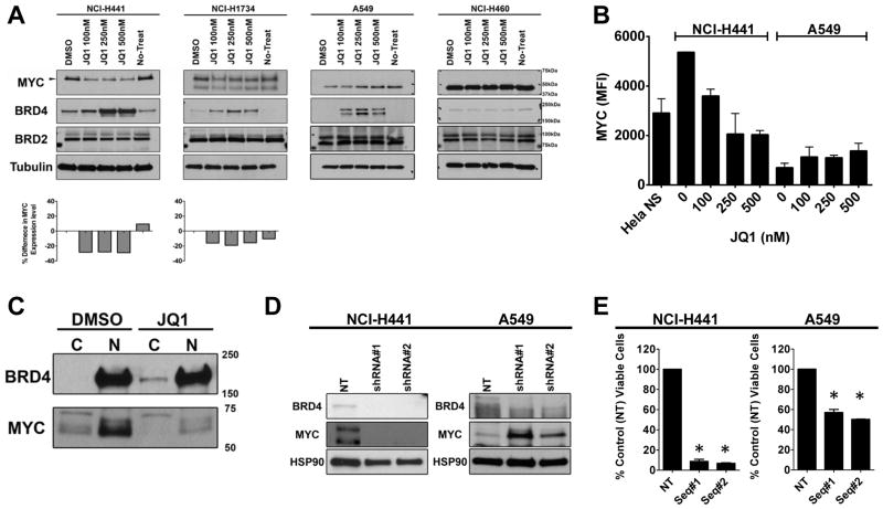
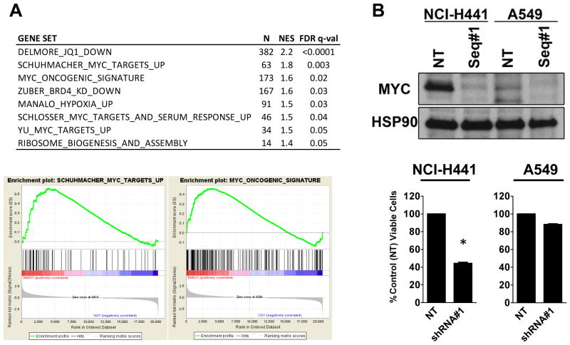
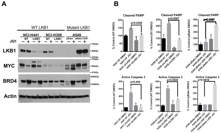
Results: Although JQ1 is broadly active in NSCLC cells, activity of JQ1 in mutant KRAS NSCLC is abrogated by concurrent alteration or genetic knockdown of LKB1. In sensitive NSCLC models, JQ1 treatment results in the coordinate downregulation of the MYC-dependent transcriptional program. We found that JQ1 treatment produces significant tumor regression in mutant kras mice. As predicted, tumors from mutant kras and lkb1 mice did not respond to JQ1.

Conclusion: Bromodomain inhibition comprises a promising therapeutic strategy for KRAS-mutant NSCLC with wild-type LKB1, via inhibition of MYC function. Clinical studies of BET bromodomain inhibitors in aggressive NSCLC will be actively pursued. Clin Cancer Res; 19(22); 6183-92. ©2013 AACR.


Conflict of interest statement

On behalf of all authors on this manuscript, we disclose the following conflicts of interest. All authors have completed separate conflict of interest forms, and we have no additional conflicts of interest to report.

Kwok-Kin Wong

Conflict of interest disclosure statement:

Drug-like BET bromodomain inhibitors created by Drs. Bradner and Qi have been licensed by the Dana-Farber Cancer Institute to Tensha Therapeutics (Cambridge, MA) for therapeutic development.

Consultant/Advisory Board

Drs. Bradner (Major) and Qi (Minor) consult for Tensha Therapeutics to contribute to the clinical translation of drug-like BET bromodomain inhibitors.

Dr. Kimmelman is a consultant for Forma Therapeutics (Minor).

Ownership

Dr. Bradner and the Dana-Farber Cancer Institute have been allocated equity (minority) in Tensha Therapeutics.

Takeshi Shimamura, Zhao Chen, Margaret Soucheray, Julian Carretero, Eiki Kikuchi, Jeremy H. Tchaicha, Yandi Gao, Katherine A. Cheng, Travis J. Cohoon, Esra Akbay, Andrew L. Kung and Kwok-Kin Wong

No conflicts of interest

Figures

Figure 1
Figure 1. JQ1 treatment induces apoptosis in NSCLC cells with mutant KRAS but not KRAS/LKB1 mutant cells
(A) NSCLC cells with KRAS/LKB1 mutations (Left, Open box) proliferate faster than NSCLC cells with KRAS mutation and wild-type LKB1 (Right, grey box) in the presence of JQ1. The proliferation rates of cells treated with 2.5 μM JQ1 were compared to that of cells in DMSO. Proliferation rates of the 24 cell lines tested are listed in Supplementary Fig. 1B. (B) Growth retardation by JQ1 treatment and genotypic characteristics of NSCLC cells harboring KRAS mutation. Percent growth with JQ1 in blue indicates small or no growth inhibition, while red indicates growth despite exposure to JQ1. In the heat map showing the expression level of BRD2, 4, and T, low relative expressions are represented by blue and high expression is indicated by red. (C) Exponentially growing NCI-H441 (KRASmut/LKB1WT) cells or A549 (KRASmut/LKB1mut) cells were treated with DMSO or indicated doses of JQ1 for 48 hours. Cells were harvested for Luminex-based active Caspase 3 (Left) and cleaved PARP (Right) assay for the detection of apoptosis. Dose dependent induction of activated Caspase-3 and cleaved PARP is evident in H441 cells but not in A549 cells. The results are from two independent experiments run with duplicate samples. Bars, S.D.
Figure 2
Figure 2. JQ1 promotes depletion of MYC in NSCLC cells with mutant KRAS
(A) A panel of exponentially growing NSCLC cells (NCI-H441 and NCI-H1734 cells are with KRASmut/LKB1WT and A549 and NCI-H460 cells are with KRASmut/LKB1mut) were treated with indicated doses of JQ1 for 48 hr and lysates were prepared for Western blotting with the indicated antibodies and demonstrate dose dependent accumulation of BRD4. Note that JQ1 induced depletion of MYC (57kDa) is evident in KRASmut/LKB1WT cells. MYC (57kDa) was not detectable in KRASmut/LKB1mut cells with the film exposure time. (B) NCI-H441 or A549 cells were treated with DMSO or indicated doses of JQ1 for 48 hours. Cells were harvested for a Luminex assay to quantify the MYC abundance. Units are mean fluorescent intensity (MFI) and the number is proportional to the amount of protein in the cell. The results represent an average of two independent assays run in duplicate samples. (C) Exponentially growing NCI-H441 cells (KRASmut/LKB1WT) were treated with 500 nM JQ1 or vehicle for 48 hours and lysates were harvested. The cytosolic (C) and nuclear (N) fractions were prepared and the samples were subjected to Western blotting with the indicated antibodies, demonstrating the accumulation of BRD4 takes place in the cytosol and MYC depletion takes place in the nucleus. (D) A representative Western blot showing NCI-H441 or A549 cells transduced with lentiviruses encoding two shRNA sequences targeting BRD4 or a shRNA sequence control. Western blots were performed 5 days post-transduction; in each case, the control construct did not reduce expression of BRD4. In H441 cells, BRD4 knockdown promoted the decrease of MYC expression while BRD4 knockdown in A549 resulted in an increase of MYC expression. (E) Viability assays were performed 7 days after infection, and viability was normalized to cells infected with a lentivirus encoding a non-targeting shRNA. Columns represent the average of triplicate samples. The viability for H441 and A549 cells was significantly (*) reduced by BRD4 knockdown with the most profound reduction being observed in H441 cells. Results represent an average of two independent assays. Bars, SD.
Figure 3
Figure 3. JQ1 suppresses the MYC transcriptional program and depletion of MYC phenocopies the JQ1 effect in H441 KRASmut/LKB1WT NSCLC cells
(A) Table of gene sets enriched among genes downregulated by JQ1-treatment in NCI-H441 cells. N: the number of genes in gene set, NES: the normalized enrichment score, and FDR q-val: statistical significance of FDR test. MYC gene sets are significantly downregulated in JQ1-treated NCI-H441 cells. (B) NCI-H441 or A549 cells were transduced with lentiviruses encoding two different sequences of shRNAs targeting MYC or non-target shRNA (control). Western blots were performed 5 days post-transduction; in each case, the control construct did not reduce expression of MYC. In H441 cells, MYC knockdown promoted the significant (*, p < 0.01) decrease in cell viability while MYC depletion in A549 demonstrates no growth retardation. Bars, SD.
Figure 4
Figure 4. The loss of LKB1 renders mutant KRAS NSCLC cells resistant to JQ1
(A) Two paired LKB1 and control (non-target, NT) knockdown cells were generated in KRASmut/LKB1WT NSCLC cell lines, H358 (H358LKB1 and H358NT) and H441 (H441LKB1 and H441NT). Additionally, wild-type LKB1 was ectopically expressed in KRASmut/LKB1mut A549 cell line (A549 pBabe-LKB1). A549 cells were transduced with retrovirus coding for pBabe vector as a paired control (A549 pBabe). The paired cell lines were challenged with DMSO (−) or JQ1 (+) for 48 hours and lysates were subjected to Western blotting with the indicated antibodies. Note that JQ1-induced MYC depletion is modest in the mutant KRAS NSCLC cells with the loss of LKB1. (B) Exponentially growing paired NCI-H441 or NCI-H358 or A549 cells were treated with DMSO or JQ1 (500 nM) for 48 hours. Cells were harvested for Luminex-based active Caspase-3 (bottom) and cleaved PARP (top) assay for the detection of apoptosis. The induction of apoptosis markers is significantly more in cells with LKB1 than in LKB1 deficient cells. The results are from two independent experiments run with duplicate samples. Bars, S.D.
Figure 5
Figure 5. JQ1 promotes BRD4 accumulation and MYC depletion in a mutant kras murine lung cancer model
(A) Mice harboring mutant kras or kras/lkb1 were subjected to MRI to establish measurable tumor burden. Next, mice were treated daily with JQ1 (50 mg/kg) by intraperitoneal injection. Two weeks post-treatment, the mice were imaged to evaluate tumor burden. Arrows indicate tumors; H indicates Heart. (B) Volumetric measurements of tumor burden in kras and kras/lkb1 mice after two weeks of JQ1 treatment. kras mice exhibited an objective response (> 30%) to JQ1 treatment while kras/lkb1 tumors do not respond as favorably to JQ1-treatment. (C) Representative FDG-PET/CT images of kras mice at baseline and 3 days post-treatment. Baseline and post-treatment PET images are depicted with identical scales. The colored FDG-PET images are superimposed with the grey-scale cross-sectional CT images. (D) Immunohistochemical (IHC) staining of lung tumors shows JQ1-treatments are more efficacious in downregulating myc expression in mutant kras mice than in mutant kras/lkb1 mice. IHC for brd4 (Top), and myc (Bottom) of lung tumors from kras or kras/lkb1 mice. Mice were treated with 3 doses of JQ1 over 3 days. The third dose was given 3 hours prior to euthanasia, and lung sections were stained with indicated antibodies. Photos shown are representative fields in each group in low and high magnification. Scale bars measure 50 μm. (E) Quantitative RT-PCR analysis of total myc transcript isolated from untreated and JQ1-treated mutant kras mice tumors shows that effects on myc mRNA expression are significantly (p < 0.05) with evident decreases in tumor nodules harvested from kras mice treated with JQ1 for 3 days. Each sample was analyzed in triplicate for quantification of both total myc and β-actin transcripts. The endogenous mouse myc level from untreated mice was arbitrarily designated as 1. Data were analyzed by relative quantitation using the ΔΔCt method with normalization to β-actin. Error bars, S.D. (F) Representative pictures of Ki-67 IHC staining of lung tumors from mutant kras and kras/lkb1 mutant mice untreated or treated with JQ1. (G) Scoring of Ki-67 positive cells shows significant (p = 0.0032, Student’s t-test) reduction in Ki-67 positive cells in mutant kras tumors treated with JQ1 for 3 days. Five areas of each slide were counted (n = 4 each for mutant kras treated and untreated; n = 2 each for kras/lkb1 mutant treated and untreated). Error bars represent S.D.

References

    1. Pylayeva-Gupta Y, Grabocka E, Bar-Sagi D. RAS oncogenes: weaving a tumorigenic web. Nat Rev Cancer. 2011;11:761–74. - PMC - PubMed
    1. Johnson L, Mercer K, Greenbaum D, Bronson RT, Crowley D, Tuveson DA, et al. Somatic activation of the K-ras oncogene causes early onset lung cancer in mice. Nature. 2001;410:1111–6. - PubMed
    1. Luo J, Emanuele MJ, Li D, Creighton CJ, Schlabach MR, Westbrook TF, et al. A genome-wide RNAi screen identifies multiple synthetic lethal interactions with the Ras oncogene. Cell. 2009;137:835–48. - PMC - PubMed
    1. Singh A, Greninger P, Rhodes D, Koopman L, Violette S, Bardeesy N, et al. A gene expression signature associated with “K-Ras addiction” reveals regulators of EMT and tumor cell survival. Cancer Cell. 2009;15:489–500. - PMC - PubMed
    1. Malumbres M, Barbacid M. RAS oncogenes: the first 30 years. Nat Rev Cancer. 2003;3:459–65. - PubMed

Publication types

MeSH terms