. 2014 Jan;34(1):34-42.
doi: 10.1038/jcbfm.2013.161. Epub 2013 Sep 18.

Cerebral arterial bolus arrival time is prolonged in multiple sclerosis and associated with disability

Affiliations

Cerebral arterial bolus arrival time is prolonged in multiple sclerosis and associated with disability

David Paling et al. J Cereb Blood Flow Metab. 2014 Jan.

Abstract

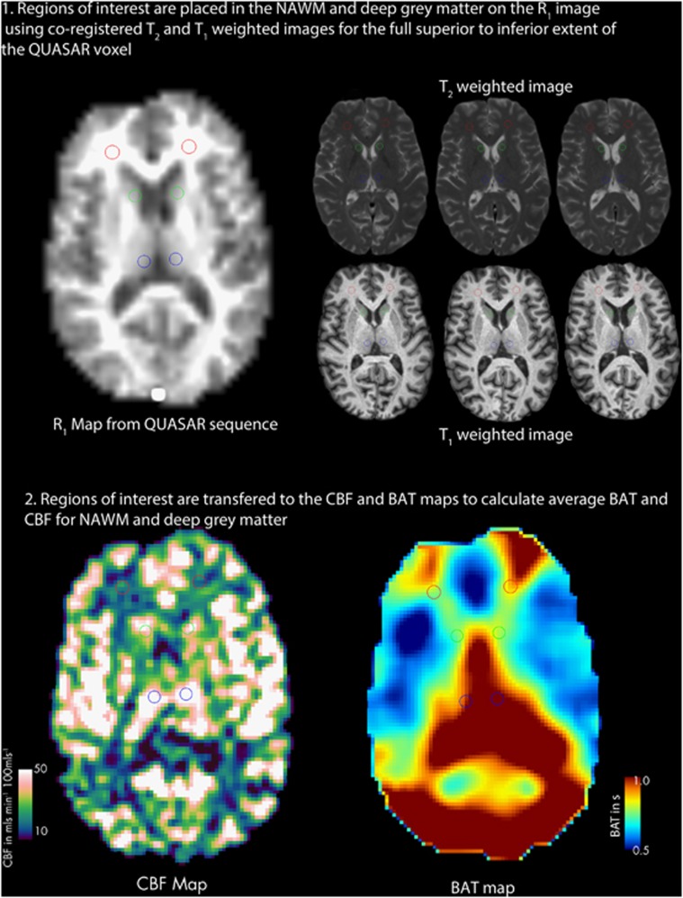
Alterations in the overall cerebral hemodynamics have been reported in multiple sclerosis (MS); however, their cause and significance is unknown. While potential venous causes have been examined, arterial causes have not. In this study, a multiple delay time arterial spin labeling magnetic resonance imaging sequence at 3T was used to quantify the arterial hemodynamic parameter bolus arrival time (BAT) and cerebral blood flow (CBF) in normal-appearing white matter (NAWM) and deep gray matter in 33 controls and 35 patients with relapsing-remitting MS. Bolus arrival time was prolonged in MS in NAWM (1.0±0.2 versus 0.9±0.2 seconds, P=0.031) and deep gray matter (0.90±0.18 versus 0.80±0.14 seconds, P=0.001) and CBF was increased in NAWM (14±4 versus 10±2 mL/100 g/min, P=0.001). Prolonged BAT in NAWM (P=0.042) and deep gray matter (P=0.01) were associated with higher expanded disability status score. This study demonstrates alteration in cerebral arterial hemodynamics in MS. One possible cause may be widespread inflammation. Bolus arrival time was longer in patients with greater disability independent of atrophy and T2 lesion load, suggesting alterations in cerebral arterial hemodynamics may be a marker of clinically relevant pathology.


Figures

Figure 1
Figure 1
Region of interest (ROI) placement. Regions of interest were placed on the R1 map from the arterial spin labeling (ASL) sequence using co-registered T1-weighted and T2-weighted scans. The image shows two of the 14 ROI placed in the white matter (red), and four of the six regions of interest placed in caudate (green) and thalamus (blue) Key: NAWM, normal-appearing white matter; CBF, cerebral blood flow; BAT, bolus arrival time.
Figure 2
Figure 2
Group-averaged maps of bolus arrival time (BAT) in controls (A) and patients with multiple sclerosis (B) registered to Montreal Neurological Institute space at three different levels (template shown for reference in panel C. Note that prolongations of BAT are seen in the border zone territories between the anterior and middle cerebral artery territories (at 1 o'clock and 11 o'clock with respect to the map) and the middle and posterior cerebral artery territories (at 4 o'clock and 8 o'clock with respect to the map). Global increase in BAT is seen in patients with multiple sclerosis (B) as compared with controls (A).
Figure 3
Figure 3
Group-averaged maps of cerebral blood flow (CBF) in controls (A) and patients with multiple sclerosis (B) registered to Montreal Neurological Institute space at three different levels (template shown for reference in panel C. A reduction in CBF can be seen in deep gray matter in patients with multiple sclerosis (B) when compared with controls (A). The increase in white matter CBF in patients with multiple sclerosis is harder to see because of the windowing chosen.
Figure 4
Figure 4
Box plot of cerebral blood flow (CBF) and bolus arrival time (BAT) in deep gray matter and normal-appearing white matter (NAWM) in controls and patients with multiple sclerosis (MS). Differences are assessed using linear regression with co-variates of gender, disease duration, and brain parenchymal fraction (BPF). In NAWM, significant increase in CBF (A) with prolongation of BAT (B) is seen in patients with MS (first row). In deep gray matter, decrease in CBF is not significantly different once BPF is added as a co-variate (C) but BAT is significantly prolonged (D) in patients with MS (second row).
Figure 5
Figure 5
Averaged plots of Δ M in deep gray matter (A) and normal-appearing white matter (B), in patients with MS (red) and controls (blue). In MS, deep gray matter Δ M rises later and to a lower peak compared with controls, indicating prolonged bolus arrival time (BAT) and lower cerebral blood flow (CBF). In normal-appearing white matter, Δ M rises later but to a higher peak compared with controls, indicating prolonged BAT but higher CBF. Note that the scale of the y-axis (Δ M) in the two charts is different, with the reduced scale in the normal-appearing white matter compared with deep gray matter indicating a comparative reduction in signal-to-noise in this region.
Figure 6
Figure 6
Scatter plots of expanded disability status score (EDSS) plotted against bolus arrival time (BAT) in the normal-appearing white matter (NAWM) (A) and deep gray matter (B). Significance of association was assessed using linear regression with co-variates of gender, disease duration, T2 lesion load, and brain parenchymal fraction. Longer BAT in NAWM and deep gray matter were associated with higher EDSS score.

References

    1. Kutzelnigg A, Lucchinetti CF, Stadelmann C, Bruck W, Rauschka H, Bergmann M, et al. Cortical demyelination and diffuse white matter injury in multiple sclerosis. Brain. 2005;128:2705–2712. - PubMed
    1. Zeis T, Graumann U, Reynolds R, Schaeren-Wiemers N. Normal-appearing white matter in multiple sclerosis is in a subtle balance between inflammation and neuroprotection. Brain. 2008;131:288–303. - PubMed
    1. Frischer JM, Bramow S, Dal-Bianco A, Lucchinetti CF, Rauschka H, Schmidbauer M, et al. The relation between inflammation and neurodegeneration in multiple sclerosis brains. Brain. 2009;132:1175–1189. - PMC - PubMed
    1. Howell OW, Rundle JL, Garg A, Komada M, Brophy PJ, Reynolds R. Activated microglia mediate axoglial disruption that contributes to axonal injury in multiple sclerosis. J Neurol Neurosurg Psychiatr. 2010;69:1017–1033. - PMC - PubMed
    1. Soon D, Tozer DJ, Altmann DR, Tofts PS, Miller DH. Quantification of subtle blood-brain barrier disruption in non-enhancing lesions in multiple sclerosis: a study of disease and lesion subtypes. Mult Scler. 2007;13:884–894. - PubMed

Publication types