. 2014 Jan;34(1):72-80.
doi: 10.1038/jcbfm.2013.163. Epub 2013 Sep 18.

Progesterone attenuates hemorrhagic transformation after delayed tPA treatment in an experimental model of stroke in rats: involvement of the VEGF-MMP pathway

Affiliations

Progesterone attenuates hemorrhagic transformation after delayed tPA treatment in an experimental model of stroke in rats: involvement of the VEGF-MMP pathway

Soonmi Won et al. J Cereb Blood Flow Metab. 2014 Jan.

Abstract

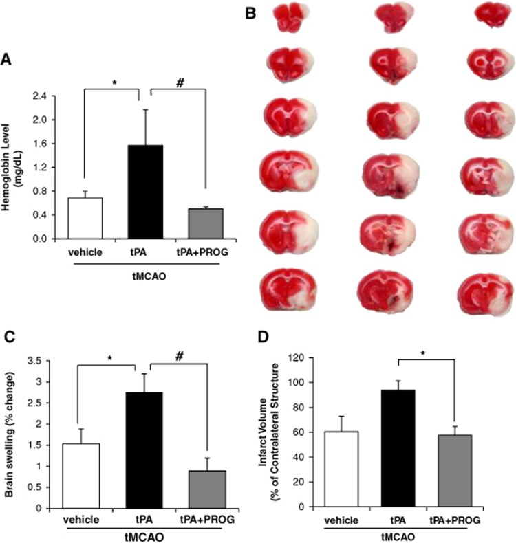
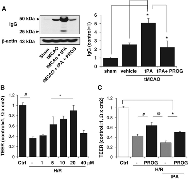
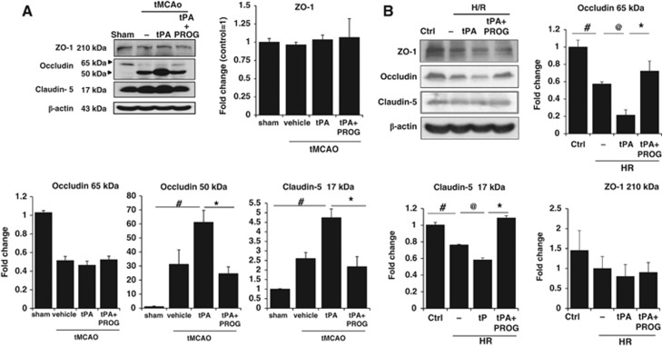
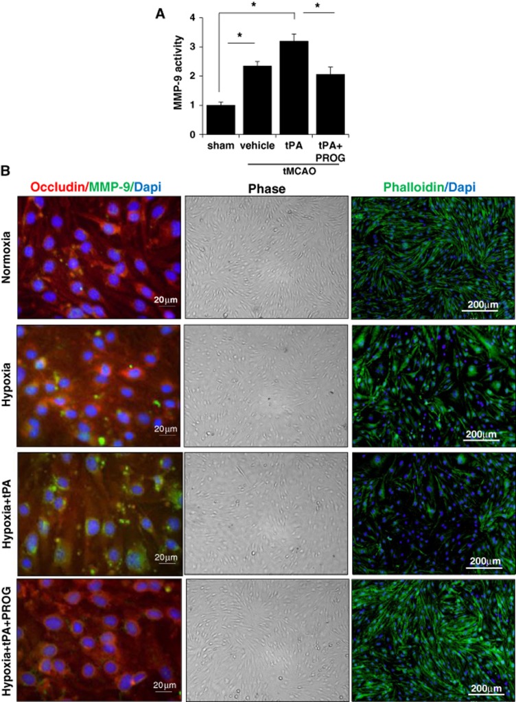
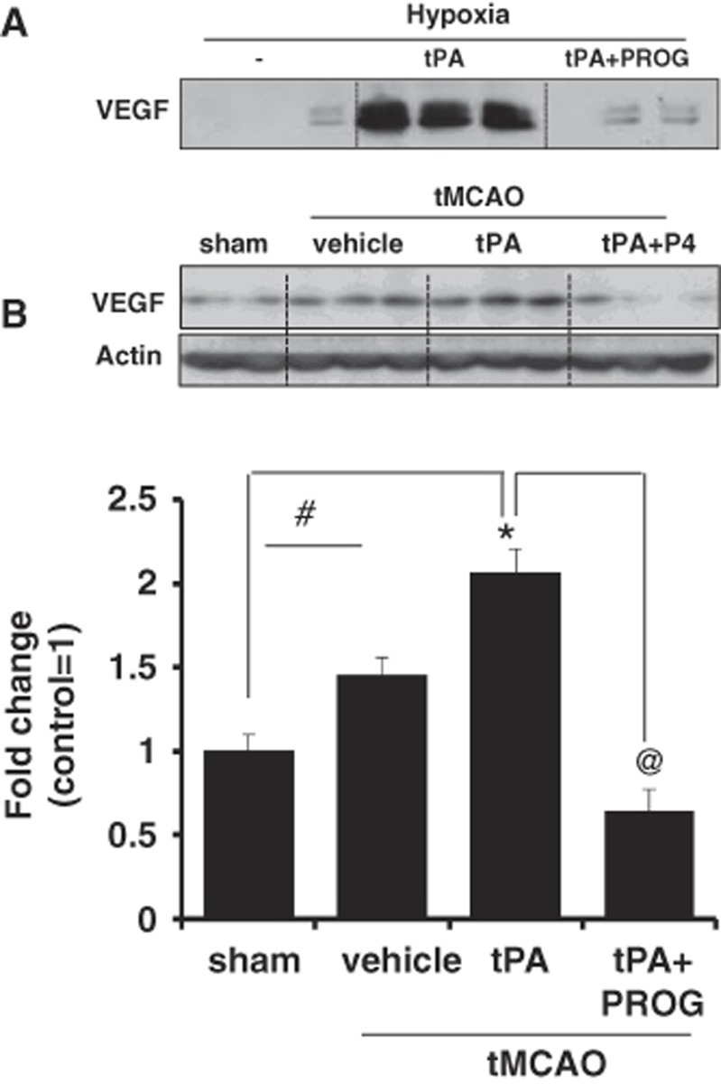
Tissue plasminogen activator (tPA) is the only FDA-approved treatment for acute stroke, but its use remains limited. Progesterone (PROG) has shown neuroprotection in ischemia, but before clinical testing, we must determine how it affects hemorrhagic transformation in tPA-treated ischemic rats. Male Sprague-Dawley rats underwent middle cerebral artery occlusion with reperfusion at 4.5 hours and tPA treatment at 4.5 hours, or PROG treatment intraperitoneally at 2 hours followed by subcutaneous injection at 6 hours post occlusion. Rats were killed at 24 hours and brains evaluated for cerebral hemorrhage, swelling, blood-brain barrier (BBB) permeability, and levels of matrix metalloproteinase-9 (MMP-9), vascular endothelial growth factor level (VEGF), and tight junction (TJ) proteins. We also evaluated PROG's efficacy in preventing tPA-induced impairment of transendothelial electrical resistance (TEER) and TJ proteins under hypoxia/reoxygenation in the endothelial cells. Delayed tPA treatment induced significant hemorrhagic conversion and brain swelling. Treatment with PROG plus tPA ameliorated hemorrhage, hemispheric swelling, BBB permeability, MMP-9 induction, and VEGF levels compared with controls. Progesterone treatment significantly prevented tPA-induced decrease in TEER and expression of occludin and claudin-5, and attenuated VEGF levels in culture media subjected to hypoxia. The study concluded that PROG may extend the time window for tPA administration in ischemic stroke and reduce hemorrhagic conversion.


Figures

Figure 1
Figure 1
Progesterone reduces delayed tissue plasminogen activator (tPA)-induced increase in edema, hemorrhage, and infarct volume after transient middle cerebral artery occlusion (tMCAO). (A) Quantitative analysis of cerebral hemorrhage volume with spectrophotometric assay. (B) Representative coronal sections of 2,3,5-triphenyltetrazolium chloride-stained brain sections of rats from the transient ischemia group (vehicle, left), tPA group (center), and tPA plus progesterone group (right). (C) Quantitative analysis of brain swelling 24 hours after ischemic onset. (D) Quantitative analysis of infarct volume in saline (n=6), tPA (n=6), and tPA+PROG (progesterone) (n=6). *P<0.05 versus saline, #P<0.05 versus tPA plus PROG. Data are expressed as means±s.e.m.
Figure 2
Figure 2
Effect of progesterone (PROG) on blood–brain barrier (BBB) disruption after transient middle cerebral artery occlusion (tMCAO) with tissue plasminogen activator (tPA) in rats and on the transendothelial electrical resistance (TEER) in endothelial cell monolayers under hypoxia and reperfusion (H/R). (A) Western blot analysis measured the levels of immunoglobulin G (IgG). Combination therapy with tPA plus PROG prevented tPA-associated increase in IgG. (B) Transendothelial electrical resistance was decreased by H/R. The decrease was significantly prevented by pretreatment with 5, 10, and 20 μmol/L PROG. (C) Pretreatment with PROG (with observed maximum protective dose of 20 μmol/L) prevented tPA (20 μg/mL)-associated H/R-induced, reduction in TEER. #P<0.05 versus H/R plus PROG, @P<0.05 versus tPA alone. *P<0.05 versus tPA plus PROG (n=5–6). Data are expressed as means ±s.e.m.
Figure 3
Figure 3
Effects of progesterone (PROG) on tight junction proteins after ischemia onset with tissue plasminogen activator (tPA), and after hypoxia/reperfusion (H/R) in brain endothelial cells. (A) Immunoblot images and quantitative data showing that, after tPA treatment (5 mg/kg, intravenous) in transient middle cerebral artery occlusion (tMCAO) rats, occludin and claudin-5 levels were decreased in peri-infarct cortex compared with sham, and increased after treatment with PROG. (B) Immunoblot images and quantitative data showing that administration of tPA (20 μg/mL) with H/R markedly decreased occludin and claudin-5 levels, and PROG significantly prevented that decrease in vitro. Data are expressed as means ±s.e.m. #P<0.05 versus Control, @P<0.05 versus tPA, *P<0.05 versus tPA plus PROG (n=6–7).
Figure 4
Figure 4
Effects of progesterone (PROG) on metalloproteinase-9 (MMP-9) activity and actin cytoskeletal change. (A) Metalloproteinase-9 activity of the lysates from the control or ischemic cortices at 24 hours after ischemia measured by fluorometric assay using fluorescence resonance energy transfer peptides as substrates. Progesterone treatment significantly attenuated the tissue plasminogen activator (tPA)-induced increase in MMP-9 levels. #P<0.01 versus control group, *P<0.05 (n=6–7). (B) Triple immunofluorescent staining (left panel) of MMP-9 (green), occludin (red), and 4′,6-Diamidino-2-phenylindole (DAPI) (blue) after hypoxia/reperfusion (H/R) with tPA in brain endothelial cells. Digital photomicrographs (middle panel) of phase contrast images of confluent bEnd.3 cells under H/R with or without tPA in the presence or absence of 20 μmol/L PROG. Representative fluorescence microscopy images (right panel) of phalloidin staining (green) showing actin cytoskeleton reorganization under H/R and after tPA (20 μg/mL) application under H/R. The cells pretreated with 20 μmol/L PROG preserved the cell morphology and actin cytoskeletal component. tMCAO, transient middle cerebral artery occlusion.
Figure 5
Figure 5
Effect of progesterone (PROG) on vascular endothelial growth factor (VEGF) level. (A) Immunoblot images and quantitative data showing VEGF levels in cell culture media. Application of tissue plasminogen activator (tPA) (20 μg/mL) with hypoxia increased the levels of VEGF in the culture media. Equal volumes of media were loaded in each lane. Progesterone (20 μmol/L) prevented the increase of VEGF levels in the cell culture media. (B) Immunoblot images and quantitative data showing VEGF levels in the brain tissue. Vascular endothelial growth factor levels in the cortical area were significantly elevated in the ischemic group given tPA (5 mg/kg, intravenous) compared with the sham group. Progesterone treatment (8 mg/kg) significantly reduced VEGF levels in the cortical area after delayed tPA treatment. Data are expressed as mean ±s.e.m. *P<0.05 versus sham, #P<0.05 versus saline, and @P<0.05 versus PROG. tMCAO, transient middle cerebral artery occlusion.

References

    1. Hacke W, Kaste M, Bluhmki E, Brozman M, Dávalos A, Guidetti D, et al. Thrombolysis with alteplase 3 to 4.5 hours after acute ischemic stroke. N Engl J Med. 2008;359:1317–1329. - PubMed
    1. Donnan GA, Davis SM, Parsons MW, Ma H, Dewey HM, Howells DW. How to make better use of thrombolytic therapy in acute ischemic stroke. Nat Rev Neurol. 2011;7:400–409. - PubMed
    1. Wang X, Tsuji K, Lee SR, Ning M, Furie KL, Buchan AM, et al. Mechanisms of hemorrhagic transformation after tissue plasminogen activator reperfusion therapy for ischemic stroke. Stroke. 2004;35:2726–2730. - PubMed
    1. Su EJ, Fredriksson L, Geyer M, Folestad E, Cale J, Andrae J, et al. Activation of PDGF-CC by tissue plasminogen activator impairs blood-brain barrier integrity during ischemic stroke. Nat Med. 2008;14:731–737. - PMC - PubMed
    1. Sayeed I, Wali B, Stein DG. Progesterone inhibits ischemic brain injury in a rat model of permanent middle cerebral artery occlusion. Restor Neurol Neurosci. 2007;25:151–159. - PubMed

Publication types

MeSH terms

Substances

LinkOut - more resources