. 2013:4:2472.
doi: 10.1038/ncomms3472.

A peptide probe for targeted brown adipose tissue imaging

Affiliations

A peptide probe for targeted brown adipose tissue imaging

Ali Azhdarinia et al. Nat Commun. 2013.

Abstract

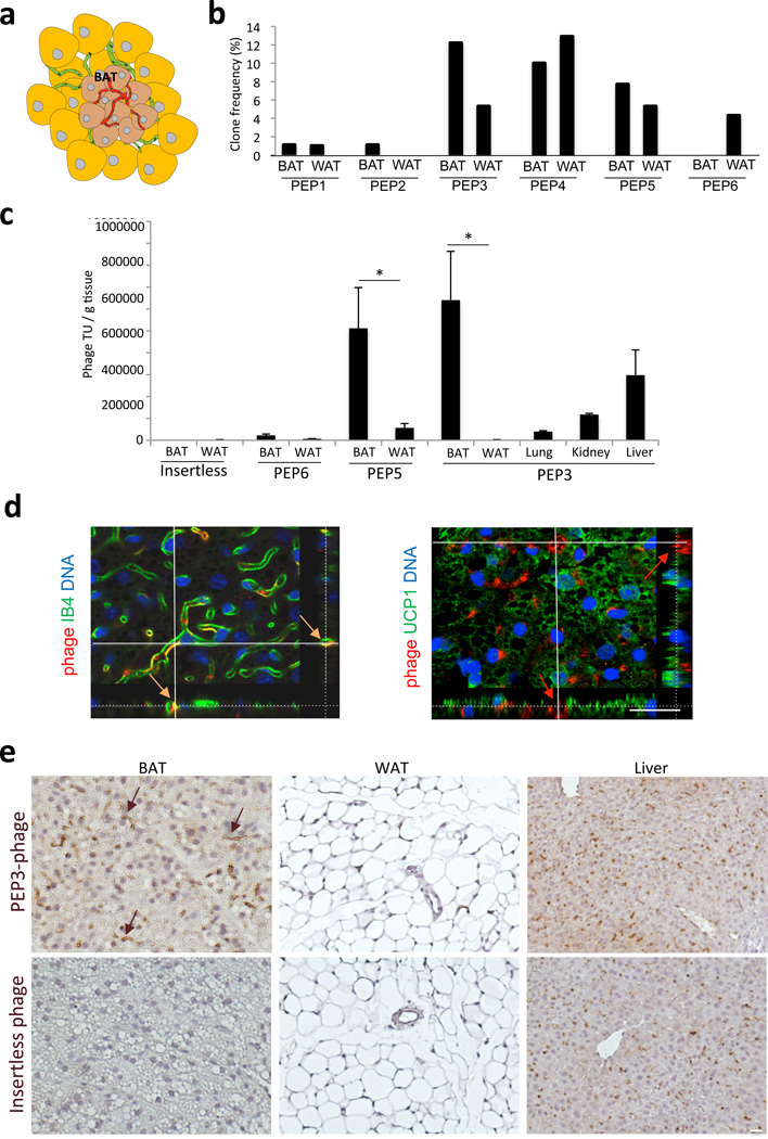
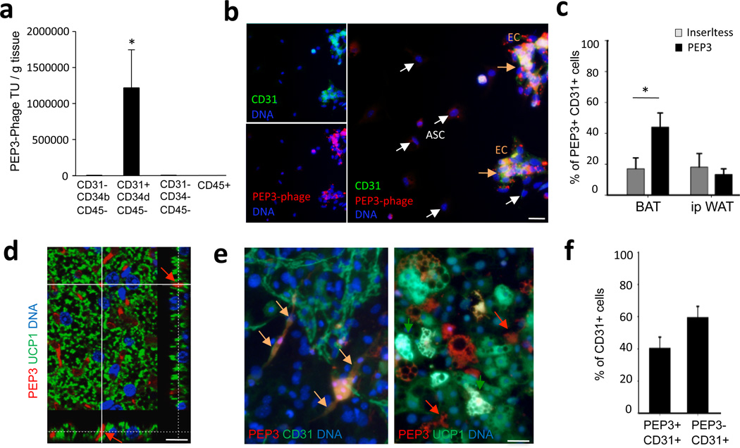
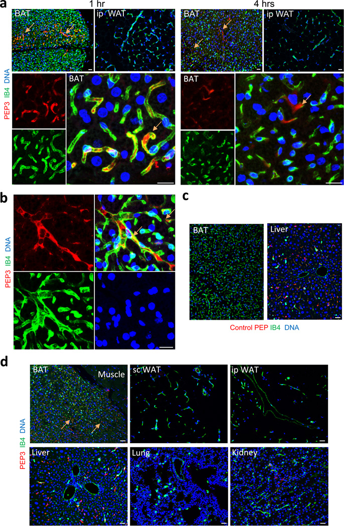
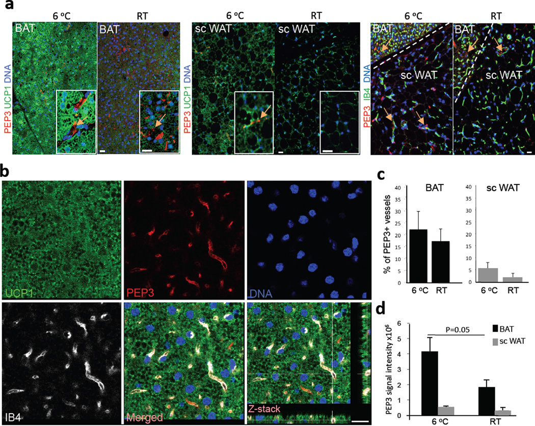
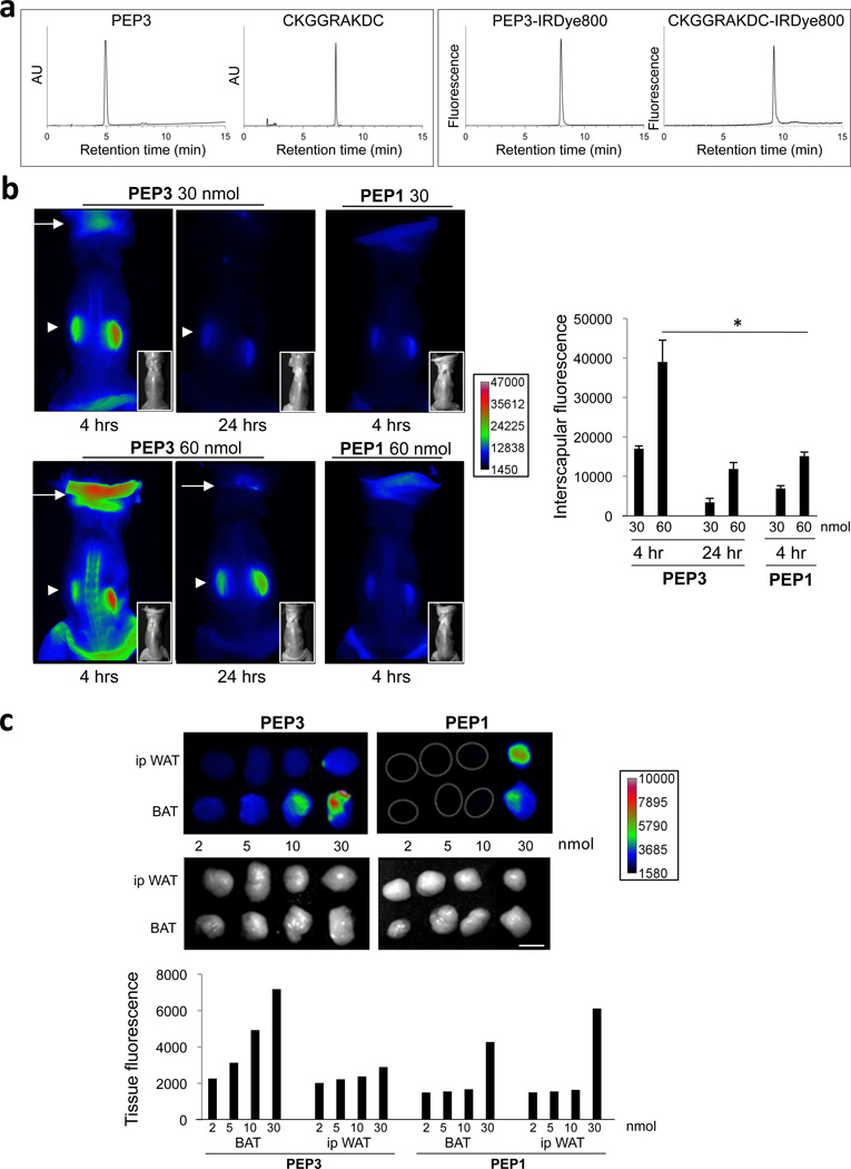
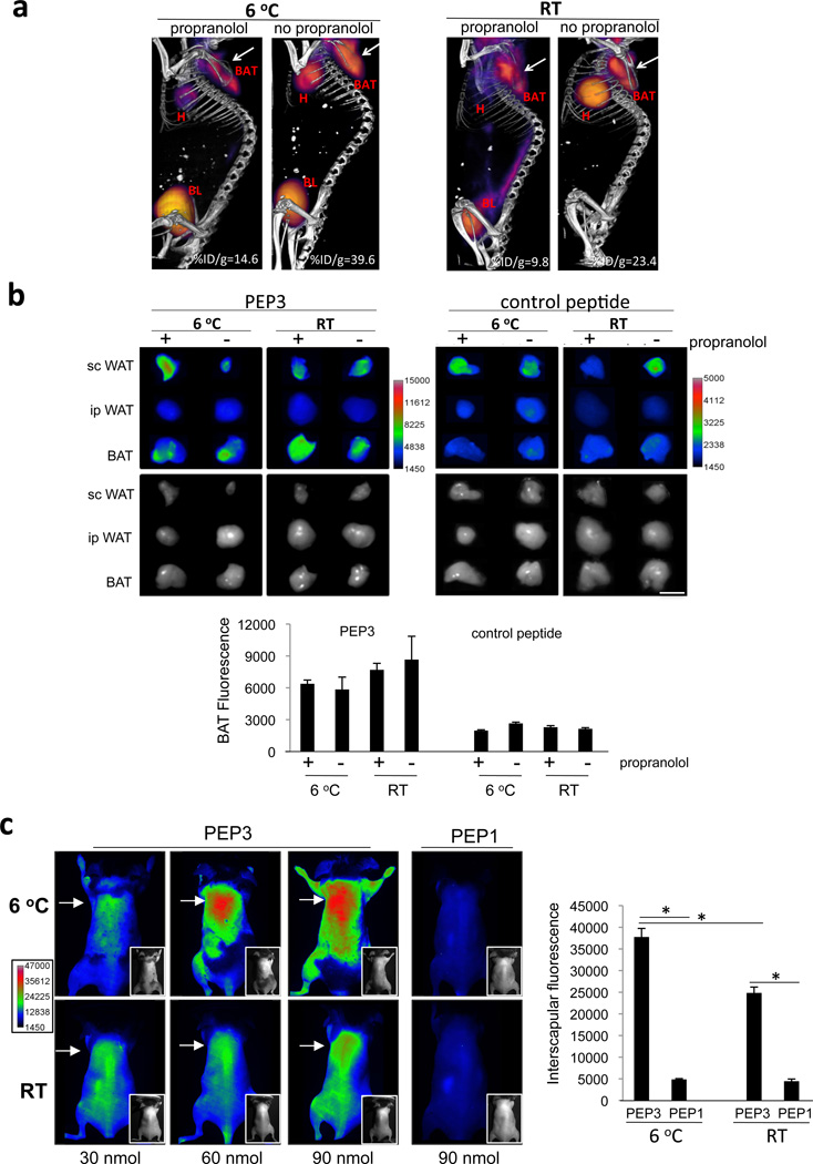
The presence of brown adipose tissue responsible for thermogenic energy dissipation has been revealed in adult humans and has high clinical importance. Owing to limitations of current methods for brown adipose tissue detection, analysing the abundance and localization of brown adipose tissue in the body has remained challenging. Here we screen a combinatorial peptide library in mice and characterize a peptide (with the sequence CPATAERPC) that selectively binds to the vascular endothelium of brown adipose tissue, but not of intraperitoneal white adipose tissue. We show that in addition to brown adipose tissue, this peptide probe also recognizes the vasculature of brown adipose tissue-like depots of subcutaneous white adipose tissue. Our results indicate that the CPATAERPC peptide localizes to brown adipose tissue even in the absence of sympathetic nervous system stimulation. Finally, we demonstrate that this probe can be used to identify brown adipose tissue depots in mice by whole-body near-infrared fluorescence imaging.


Figures

Figure 1
Figure 1. A screen for BAT-homing peptides
(a) The hypothesis: endothelium of BAT expresses specific surface receptors. (b) Comparison of recovery frequency for phage clones displaying peptides (PEP1 through PEP6) from BAT and WAT at round 3 of combinatorial library selection in mouse BAT. (c) Validation of BAT homing for selected phage-displayed peptides compared to peptide-free phage (insertless) through administration of 109 transforming units (TU) into mice. After 1 hr of circulation, PEP3-phage displays superior selectivity of accumulation in BAT but not in control organs. Error bars: s.e.m. * P<0.05 (Student’s t-Test); n=3 (different phage titration dilutions). (d) Anti-phage immunofluorescence (red) upon injection into a mouse revealed in BAT sections. Confocal Z-stack projections of median series demonstrate luminal localization of PEP3-phage in vessels of BAT revealed by IB4 labeling (green, left), but not to adipocytes expressing UCP1 (green, right). (e) Anti-phage immunohistochemistry (brown) showing that PEP3-phage homes to vessels of BAT (arrows), but not of ip WAT. Mouse injected with insertless phage has no BAT or WAT signal, and liver trapping of phage is comparable to trapping of PEP3-phage. Hematoxylin counterstaining: blue. Nuclei are blue. All scale bars: 20 µm.
Figure 2
Figure 2. PEP3 targets BAT endothelium
(a) 1 hr after PEP3-phage injection into a mouse, BAT cells were separated by FACS and recovery of TU (per gram of tissue ± s.e.m.; n=2 different phage titration dilutions) was graphed for CD31+CD34dimCD45-endothelial cells, CD31-CD34brightCD45- stromal cells, CD31-CD34-CD45-preadipocytes, and CD45+ leukocytes. (b) BAT-derived cells from mice injected with PEP3-phage or insertless phage were analyzed in adherent culture using anti-CD31 and anti-phage antibodies. Immunofluorescence reveals CD31+ endothelial cells (green) containing PEP3-phage (red speckles) in contrast to CD31- cells (ASC) showing only background autofluorescence. (c) Frequency of CD31+ cells from the mouse analyzed in (b) stained positive with anti-phage antibodies (mean + s.e.m., n=14). (d) Immunofluorescence analysis of BAT from a mouse iv-injected with 60 nmol of PEP3-Cy3 (red) demonstrating peptide accumulation in the vessels (arrow) but not in adipocytes expressing UCP1 (green). Confocal Z-stack projections demonstrate lack of green and red signal co-localization. (e) The stromal/vascular cells of mouse BAT subjected to brown adipogenesis induction in adherent culture were incubated with PEP3-Cy3 (5 µg/ml) and then fixed and subjected to anti-CD31 or anti-UCP1 immunofluorescence (green). Yellow arrows: bound PEP3 co-localized with endothelial networks (left). Red arrows: lack of bound PEP3 co-localization with UCP1-expressing adipocytes (green, right). (f) Adherent BAT-derived cells were incubated with PEP3-Cy3 (5 µg/ml). Plotted is the frequency of CD31+ cells bound with PEP3 (mean + s.e.m., n=12). Nuclei are blue. All scale bars: 20 µm. * P<0.05 (Student’s t-Test).
Figure 3
Figure 3. PEP3 localizes to BAT vasculature in cold- and warm-acclimated mice
Confocal fluorescence analysis of tissue sections from after iv-injection of 60 nmol of indicated Cy3-conjugated peptide. Endothelium is labeled with IB4 (green). Arrows indicate PEP3 localization in the vasculature of BAT. (a) PEP3 in BAT, but not ip WAT, of cold-acclimated mice 1 hr and 4 hr post-injection. High-magnification images (lower panels) demonstrate that peptide localization is exclusively intravascular/luminal (yellow). (b) PEP3 in BAT vasculature of warm-acclimated mice 1 hr post-injection. (c) Control WAT-homing peptide WAT7 detected in liver (non-endothelial) but not in BAT 1 hr post-injection into warm-acclimated mice (d) PEP3 detected in BAT, and occasionally in sc WAT, but not in ip WAT, muscle, or lungs, 1 hr post-injection into warm-acclimated mice. Occasional kidney and liver PEP3 trapping (non-endothelial) is comparable to that of the Control peptide. Nuclei are blue. All scale bars: 20 µm.
Figure 4
Figure 4. PEP3 localizes to vasculature of BAT and metabolically active sc WAT
(a) Confocal immunofluorescence analysis of tissue sections from cold (6 °C) or warm (RT) -acclimated mice 1 hr after iv-injection of PEP3-Cy3. Anti-UCP1 (green) immunofluorescence demonstrates UCP1 induction by cold in both BAT and WAT. Insets (high-magnification) show PEP3 vascular homing (arrow), to UCP1-expressing areas. IB4 colocalization shows PEP3-Cy3 presence in interscapular sc WAT adjacent to BAT. (b) Three-color confocal immunofluorescence analysis of BAT sections from a cold-acclimated mouse 1 hr after iv injection of PEP3-Cy3 showing co-localization of PEP3 (red) with an endothelial marker IB4 (white), but not with brown adipocyte-expressed UCP1 (green). Z-stack projections confirm colocalization of IB4 and PEP3. (c) Tissue sections (n=5) from cold- and warm-acclimated mice injected with PEP3-Cy3 were analyzed and the percentage of microvessels associated with PEP3-Cy3 signal (mean ± s.e.m.) was plotted. (d) Cumulative PEP3-Cy3 pixel intensity (mean ± s.e.m.) in sections (n=4) of indicated tissues was measured by Amira 5.4 (VSG) software. Nuclei are blue. All scale bars: 20 µm.
Figure 5
Figure 5. Biodistribution of PEP3 conjugated with a NIR fluorophore
(a) HPLC chromatograms of peptide probes. Left: UV detection of PEP3 and WAT-homing peptide CKGGRAKDC (containing two Dde protecting groups on internal Lys residues) at 280 nm. AU denotes relative absorption units. Right: fluorescence detection of PEP3-IRDye800, and CKGGRAKDC-IRDye800. (b) Whole-body NIR fluorescence imaging of cold-acclimated mice 4 and 24 hrs after iv administration of indicated doses of IRDye800-conjugated PEP3 or control PEP1. Arrows: interscapular signal; arrowheads: perirenal signal. Insets show black/white photographs of mice that had skin removed from the back for imaging. Right: Plotted data analysis from n=3 mice per group (mean ± s.e.m., *P<0.05, Student’s t-Test). (c) NIR fluorescence imaging of ip WAT and interscapular BAT isolated from cold-acclimated mice iv-injected with increasing doses of IRDye800-conjugated PEP3 or a control peptide (PEP1) after 1 hr of circulation. Scale shows fluorescence intensity. Black/white photographs of tissues are shown below. Scale bar: 5 mm. Graph: plotted quantitative data corresponding to NIR images.
Figure 6
Figure 6. Whole body NIR fluorescent imaging with a PEP3-directed probe
(a) Modulation of metabolism by warm-acclimation combined with propranolol treatment. Shown are PET/CT images of 6 °C- and RT-acclimated mice that were ip injected with propranolol where indicated. Note decreased FDG uptake by interscapular BAT (arrow) and heart (H) in propranolol-treated mice. FDG uptake in BAT is quantified as percent injected dose per gram (%ID/g). BL: bladder. (b) Tissues isolated from mice pre-conditioned as in (a) 1 hr after iv-injection with 100 nmol of IRDye800-conjugates of PEP3 or a control WAT-homing peptide CKGGRAKDC . NIR fluorescent imaging demonstrates accumulation of PEP3 in interscapular BAT and sc WAT, but not in ip WAT in all groups, while CKGGRAKDC accumulates in WAT but not in BAT. Black/white photographs of tissue are shown below. Scale bar: 5 mm. Graph: Plotted data analysis from n=3 mice per group (mean ± s.e.m.). (c) Whole-body NIR fluorescence imaging of cold (6 °C) or warm (RT) -acclimated mice 1 hr after iv administration of indicated doses of IRDye800-conjugated PEP3 or control PEP1. Arrow indicates interscapular area. Insets show black/white photographs of mice that were shaved for imaging (skin left on). Scale shows fluorescence intensity. Graph: Plotted data analysis from n=3 mice per group (mean ± s.e.m., *P<0.05, Student’s t-Test).

References

    1. Cannon B, Nedergaard J. Brown adipose tissue: function and physiological significance. Physiol. Rev. 2004;84:277–359. - PubMed
    1. Enerback S. The origins of brown adipose tissue. N. Engl. J. Med. 2009;360:2021–2023. - PubMed
    1. Tam CS, Lecoultre V, Ravussin E. Brown adipose tissue: mechanisms and potential therapeutic targets. Circulation. 2012;125:2782–2791. - PubMed
    1. Paidisetty S, Blodgett TM. Brown fat: atypical locations and appearances encountered in PET/CT. Am. J. Roentgenol. 2009;193:359–366. - PubMed
    1. Virtanen KA, et al. Functional brown adipose tissue in healthy adults. N. Engl. J. Med. 2009;360:1518–1525. - PubMed

Publication types