. 2013 Nov 1;288(44):31477-87.
doi: 10.1074/jbc.M113.514984. Epub 2013 Sep 17.

Dhrs3 protein attenuates retinoic acid signaling and is required for early embryonic patterning

Affiliations

Dhrs3 protein attenuates retinoic acid signaling and is required for early embryonic patterning

Richard Kin Ting Kam et al. J Biol Chem. .

Abstract

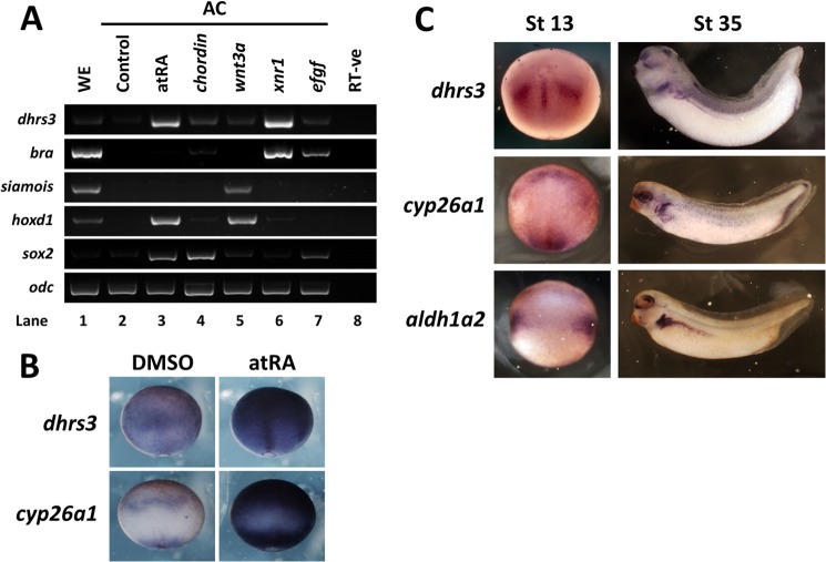
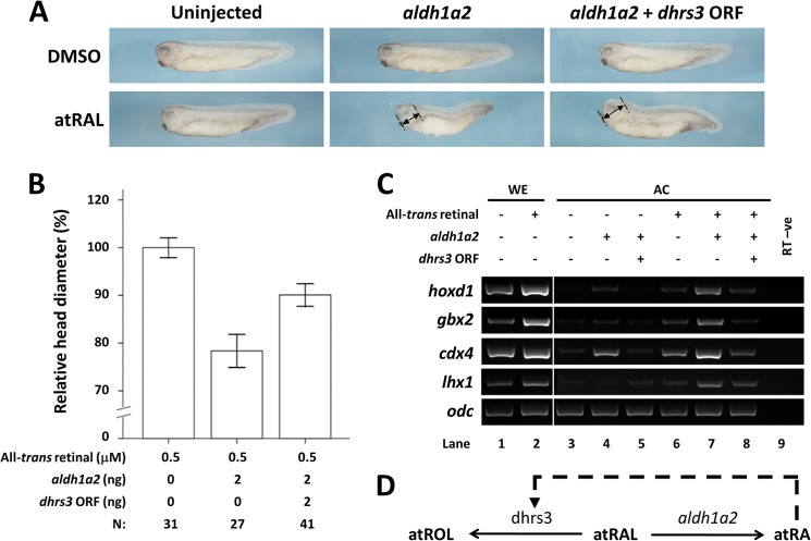
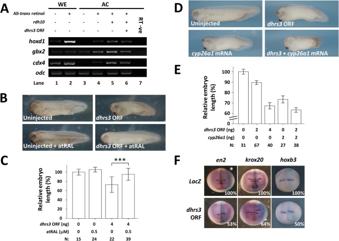
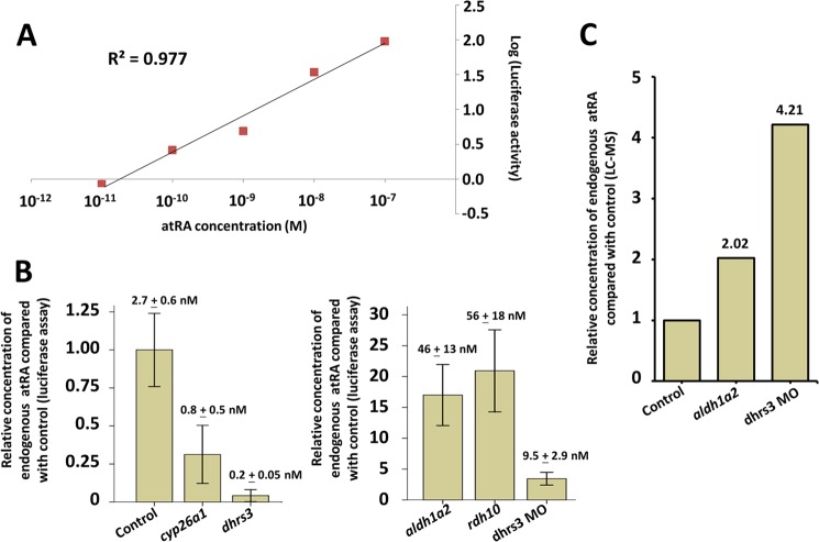
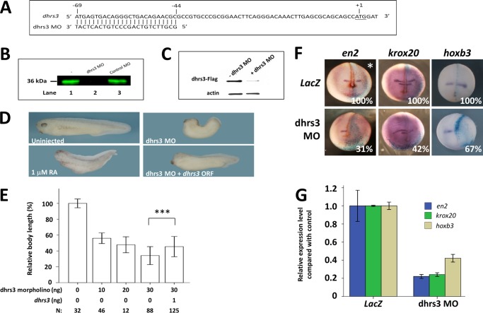
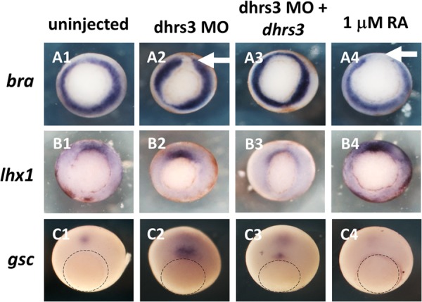
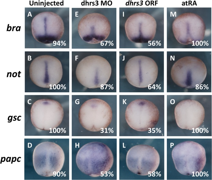
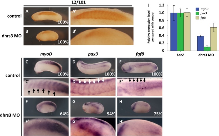
All-trans-retinoic acid (atRA) is an important morphogen involved in many developmental processes, including neural differentiation, body axis formation, and organogenesis. During early embryonic development, atRA is synthesized from all-trans-retinal (atRAL) in an irreversible reaction mainly catalyzed by retinal dehydrogenase 2 (aldh1a2), whereas atRAL is converted from all-trans-retinol via reversible oxidation by retinol dehydrogenases, members of the short-chain dehydrogenase/reductase family. atRA is degraded by cytochrome P450, family 26 (cyp26). We have previously identified a short-chain dehydrogenase/reductase 3 (dhrs3), which showed differential expression patterns in Xenopus embryos. We show here that the expression of dhrs3 was induced by atRA treatment and overexpression of Xenopus nodal related 1 (xnr1) in animal cap assay. Overexpression of dhrs3 enhanced the phenotype of excessive cyp26a1. In embryos overexpressing aldh1a2 or retinol dehydrogenase 10 (rdh10) in the presence of their respective substrates, Dhrs3 counteracted the action of Aldh1a2 or Rdh10, indicating that retinoic acid signaling is attenuated. Knockdown of Dhrs3 by antisense morpholino oligonucleotides resulted in a phenotype of shortened anteroposterior axis, reduced head structure, and perturbed somitogenesis, which were also found in embryos treated with an excess of atRA. Examination of the expression of brachyury, not, goosecoid, and papc indicated that convergent extension movement was defective in Dhrs3 morphants. Taken together, these studies suggest that dhrs3 participates in atRA metabolism by reducing atRAL levels and is required for proper anteroposterior axis formation, neuroectoderm patterning, and somitogenesis.

Keywords: Development; Embryo; Gene Regulation; Retinal Metabolism; Retinoid; dhrs3, Retinoic Acid, Embryonic Patterning, Xenopus.


Figures

FIGURE 1.
FIGURE 1.
dhrs3 expression is induced by atRA. A, expression of dhrs3 in whole embryos and animal caps was induced in response to a 4-h treatment of 1 μm atRA or xnr1 overexpression. WE, whole embryos; AC, animal cap. B, expression patterns of dhrs3 and cyp26a1 in stage 12 embryos in response to a 4-h treatment of 1 μm atRA. Dorsal view. C, expression patterns of dhrs3, cyp26a1, and aldh1a2, in stage 13 (dorsal view) and stage 35 embryos (lateral view).
FIGURE 2.
FIGURE 2.
Overexpression of dhrs3 counteracts the action of Aldh1a2. A, morphology of stage 35 embryos injected with 2 ng of aldh1a2 alone or in combination with 2 ng of dhrs3 at the two-cell stage, followed by treatment with either DMSO or 0.5 μm atRAL during gastrulation. Arrows indicate the measurement of the head diameter. B, quantification of the relative embryo head diameter of uninjected and injected embryos treated with atRAL. C, expression of atRA-responsive genes hoxd1, gbx2, cdx4, and lhx1 in animal caps overexpressing aldh1a2 alone or in combination with dhrs3, DMSO, or atRAL treatment. D, proposed role of dhrs3 in the atRA metabolism. Dashed arrow indicates the induced expression of dhrs3 by atRA. WE, whole embryos; AC, animal caps.
FIGURE 3.
FIGURE 3.
Overexpression of dhrs3 attenuates RA signaling. A, overexpression of dhrs3 counteracts the action of rdh10. Expression of atRA-responsive genes hoxd1, gbx2, and cdx4 in animal caps overexpressing rdh10 alone or in combination with dhrs3 in the presence of atROL. The expression of atRA-responsive genes was induced by ectopic rdh10, but this induction was suppressed by co-expression with dhrs3. WE, whole embryos; AC, animal caps. B and C, dhrs3 overexpression phenotypes were reversed by atRAL. Lateral view of stage 35 embryos showed phenotypes induced by dhrs3 overexpression, which were reversed by a nonteratogenic dose of 0.5 μm atRAL during gastrulation (B). Quantification of relative embryo length of embryos exposed to atRAL with or without dhrs3 overexpression (C). ***, analysis of variance, p < 0.0005. D and E, dhrs3 overexpression enhanced the phenotypes induced by ectopic cyp26a1. Lateral view of stage 35 embryos exhibited shortened trunk of AP axis when overexpressing dhrs3, cyp26a1, or both (D). Two-cell stage embryos were injected with 4 ng of dhrs3, 2 ng of cyp26a1 mRNA, or both. Quantification of relative embryo length is shown in E. F, overexpression of dhrs3 caused a posterior shift of the expression domain of en2, krox20, and hoxb3. At the four-cell stage, one dorsal blastomere of the embryo was injected with 4 ng of dhrs3 mRNA together with 100 pg of lacZ mRNA as lineage tracer. Injected side is on the right (asterisk). Overexpression of dhrs3 caused a posterior shift of the expression domain of the examined neural markers. Images of en2, krox20, and hoxb3 are the frontal view of the embryos. Percentages of embryos showing the phenotype is marked in the figure.
FIGURE 4.
FIGURE 4.
dhrs3 negatively regulates the synthesis of endogenous atRA. A, log-log plot showing the relationship between different concentrations of atRA standard applied to HEK293T cells and the respective luciferase activity. B, relative concentration of embryonic atRA under different treatment conditions compared with control as measured by cell-based luciferase assay. Overexpression of cyp26a1 and dhrs3 reduced the endogenous atRA level, whereas overexpression of aldh1a2, rdh10, or knockdown of dhrs3 strongly increased atRA concentration in the embryos. The measured concentration of atRA in cell culture is indicated in the plot as mean ± S.D. in nm. Readings were taken in triplicate. C, relative concentration of embryonic atRA in aldh1a2-overexpressing embryos and Dhrs3 knockdown embryos, as measured by LC-MS. Relative level of atRA is represented as fold change compared with control reading. Injection dose of each condition per embryo is as follows: 4 ng for cyp26a1; 4 ng for dhrs3 ORF; 30 ng for Dhrs3 MO; 4 ng for aldh1a2; 4 ng for rdh10.
FIGURE 5.
FIGURE 5.
dhrs3 morphants exhibit shortened axis and reduced head structure. A, 25-base-long morpholino was designed to target the 5′-UTR region of dhrs3. The start codon ATG is underlined. B, Western blot shows that Dhrs3 MO inhibited in vitro translation of C-terminal FLAG-tagged full-length dhrs3 (Dhrs3-FLAG). Dhrs3-FLAG contained the targeting sequence of Dhrs3 MO. C, Dhrs3 MO decreased translation of Dhrs3-FLAG in embryos. Embryos were first injected with dhrs3 mRNA with or without Dhrs3 MO, followed by protein extraction at stage 10. Anti-FLAG antibody was used to detect Dhrs3-FLAG, and endogenous actin was employed as the loading control. D, stage 35 embryos injected with 30 ng of Dhrs3 MO at the two-cell stage or treated with 1 μm atRA during gastrulation showed a shortened AP axis and reduced head structure compared with uninjected embryos. Co-injection of 1 ng of dhrs3 mRNA lacking 5′-UTR rescued the morphant phenotypes. E, quantification of the relative head diameter of uninjected and Dhrs3 MO-injected embryos. F, expression of en2, krox20, and hoxb3 in Dhrs3 morphants. At the four-cell stage, one dorsal blastomere was injected with 12.5 ng of Dhrs3 MO together with 100 pg of lacZ mRNA as the lineage tracer. Injected side is on the right (asterisk). Whole mount in situ hybridization was performed at stage 19. Expression of en2, krox20, and hoxb3 was localized in the anterior neuroectoderm. Knockdown of dhrs3 suppressed the expression of these three genes on the injected side (asterisk). Percentages of embryos showing the phenotype are marked in the figure. G, real time PCR indicated that expression level of en2, krox20, and hoxb3 at stage 19 was down-regulated by dhrs3 knockdown. ***, analysis of variance, p < 0.0005.
FIGURE 6.
FIGURE 6.
Expression patterns of mesoderm and organizer marker genes in Dhrs3 morphants and embryos treated with 1 μm atRA during gastrulation. Embryos were injected at the two-cell stage with either 30 ng of Dhrs3 MO or 30 ng of Dhrs3 MO plus 1 ng of dhrs3 mRNA or treated with 1 μm of atRA at gastrulation stages. Whole mount in situ hybridization was used to visualize mesoderm marker gene brachyury (bra) (A1–A4), organizer marker genes lhx1 (B1–B4), and goosecoid (gsc) (C1–C4). Expression of bra was disrupted in Dhrs3 morphants (A2, arrow) and in embryos treated with atRA (A4, arrow). Injection of 1 ng of dhrs3 mRNA rescued the disruption (A3). The expression intensity of lhx1 was elevated by Dhrs3 knockdown (B2) and atRA treatment (B4). Compared with the control embryos (C1), the expression domain of gsc was closer to the blastopore in Dhrs3 morphants (C2) and was abolished by atRA treatment (C4). Dotted line outlined the blastopore.
FIGURE 7.
FIGURE 7.
Defective convergent extension movement induced by knockdown of Dhrs3. Expression of mesoderm and notochord marker genes brachyury (bra), not, and goosecoid (gsc), and CE marker gene papc in control embryos (A–D), in response to dhrs3 knockdown (E–H), dhrs3 overexpression (I–L), or exogenous atRA (M–P). The expression domain of bra and not was shortened by Dhrs3 knockdown (E and F) and embryos treated with atRA (M and N). A similar but less severe shortening of bra expression domain was observed in embryos overexpressing dhrs3 (I). The not expression domain remained in normal shape, but the signal was less intensified in embryos overexpressing dhrs3 (J). The expression of gsc was suppressed by Dhrs3 knockdown (G) and atRA treatment (O) but was unaffected by dhrs3 overexpression (K). papc expression domain was more diffused in Dhrs3 morphants (H) and atRA treated embryos (P) when compared with control embryos (D). Percentages of embryos showing the phenotypes were marked in the figure. All embryos are dorsal view.
FIGURE 8.
FIGURE 8.
Knockdown of Dhrs3 interferes with somitogenesis. Dhrs3 MO (30 ng) was injected at the two-cell stage, and the morphants were collected at stage 28 for immunostaining and whole mount in situ hybridization analysis. A–B′, immunostaining with 12/101 antibody indicated that in Dhrs3 morphants the somites showed a diffused pattern and a reduction in immunoreactivity. C–H′, expression of somite marker genes myoD, pax3, and fgf8 on the dorsal side of the embryos showed the segmented expression pattern of somite units in the control embryos (C′–E′, arrow) was not observed in Dhrs3 morphants (F′–H′). I, real time PCR indicated that expression level of myoD, pax3, and fgf8 at stage 21 was down-regulated in Dhrs3 morphants.

Similar articles

Cited by

References

    1. Mic F. A., Haselbeck R. J., Cuenca A. E., Duester G. (2002) Novel retinoic acid generating activities in the neural tube and heart identified by conditional rescue of Raldh2 null mutant mice. Development 129, 2271–2282 - PMC - PubMed
    1. Niederreither K., Vermot J., Messaddeq N., Schuhbaur B., Chambon P., Dollé P. (2001) Embryonic retinoic acid synthesis is essential for heart morphogenesis in the mouse. Development 128, 1019–1031 - PubMed
    1. Ryckebusch L., Wang Z., Bertrand N., Lin S. C., Chi X., Schwartz R., Zaffran S., Niederreither K. (2008) Retinoic acid deficiency alters second heart field formation. Proc. Natl. Acad. Sci. U.S.A. 105, 2913–2918 - PMC - PubMed
    1. Chen F., Desai T. J., Qian J., Niederreither K., Lü J., Cardoso W. V. (2007) Inhibition of Tgfβ signaling by endogenous retinoic acid is essential for primary lung bud induction. Development 134, 2969–2979 - PubMed
    1. Esteban-Pretel G., Marín M. P., Renau-Piqueras J., Barber T., Timoneda J. (2010) Vitamin A deficiency alters rat lung alveolar basement membrane: reversibility by retinoic acid. J. Nutr. Biochem. 21, 227–236 - PubMed

Publication types

MeSH terms

LinkOut - more resources