. 2013 Nov 1;288(44):31842-52.
doi: 10.1074/jbc.M113.471318. Epub 2013 Sep 17.

Binding of the sialic acid-binding lectin, Siglec-9, to the membrane mucin, MUC1, induces recruitment of β-catenin and subsequent cell growth

Affiliations

Binding of the sialic acid-binding lectin, Siglec-9, to the membrane mucin, MUC1, induces recruitment of β-catenin and subsequent cell growth

Shuhei Tanida et al. J Biol Chem. .

Abstract

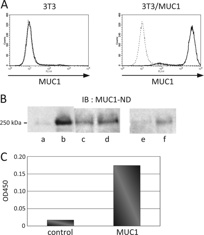
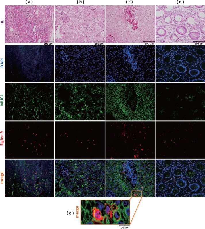
Because MUC1 carries a variety of sialoglycans that are possibly recognized by the siglec family, we examined MUC1-binding siglecs and found that Siglec-9 prominently bound to MUC1. An immunochemical study showed that Siglec-9-positive immune cells were associated with MUC1-positive cells in human colon, pancreas, and breast tumor tissues. We investigated whether or not this interaction has any functional implications for MUC1-expressing cells. When mouse 3T3 fibroblast cells and a human colon cancer cell line, HCT116, stably transfected with MUC1cDNA were ligated with recombinant soluble Siglec-9, β-catenin was recruited to the MUC1 C-terminal domain, which was enhanced on stimulation with soluble Siglec-9 in dose- and time-dependent manners. A co-culture model of MUC1-expressing cells and Siglec-9-expressing cells mimicking the interaction between MUC1-expressing malignant cells, and Siglec-9-expressing immune cells in a tumor microenvironment was designed. Brief co-incubation of Siglec-9-expressing HEK293 cells, but not mock HEK293 cells, with MUC1-expressing cells similarly enhanced the recruitment of β-catenin to the MUC1 C-terminal domain. In addition, treatment of MUC1-expressing cells with neuraminidase almost completely abolished the effect of Siglec-9 on MUC1-mediated signaling. The recruited β-catenin was thereafter transported to the nucleus, leading to cell growth. These findings suggest that Siglec-9 expressed on immune cells may play a role as a potential counterreceptor for MUC1 and that this signaling may be another MUC1-mediated pathway and function in parallel with a growth factor-dependent pathway.

Keywords: Lectin; Mucins; Signaling; Tumor Microenvironment; β-Catenin.


Figures

FIGURE 1.
FIGURE 1.
Binding of soluble recombinant Siglec-9 to MUC1. A, expression of MUC1 in 3T3/mock and 3T3/MUC1 cells was analyzed by flow cytometry after incubation with mouse anti-MUC1-ND antibodies (solid line) or control antibodies (broken line) and then with FITC-labeled goat anti-mouse IgG antibodies. B, recombinant Fc-tagged Siglec-3, -5, and -9, or His-tagged Siglec-1 was added to a lysate of 3T3/MUC1 cells. Siglec-binding proteins were pulled down using protein G-Sepharose (lanes a–d) or nickel-nitrilotriacetic acid-agarose (lanes e and f) and then subjected to SDS-PAGE followed by Western blotting (IB). MUC1-ND was detected as described under “Materials and Methods.” Lane a, control (protein G-Sepharose); lane b, Siglec-9; lane c, Siglec-3; lane d, Siglec-5; lane e, control (nickel-nitrilotriacetic acid-agarose); lane f, Siglec-1. C, MUC1-coated plate was prepared as described under “Materials and Methods.” The plate assay was performed in duplicate using Fc-tagged Siglec-9.
FIGURE 2.
FIGURE 2.
Distribution of Siglec-9-positive cells in tumor tissues. Sections of paraffin-embedded human pancreas (a), breast (b), and colon (c) tumor tissues, and nonmalignant colon tissues (d) were stained with H&E, DAPI (blue), and the combinations of rabbit anti-Siglec-9 antibodies and Alexa Fluor 594-labeled goat anti-rabbit IgG antibodies (red), and mouse anti-MUC1-ND antibodies and Alexa Fluor 488-labeled goat anti-mouse IgG antibodies (green) and then observed by microscopy. A magnified image in colon tumor tissues is shown in e.
FIGURE 3.
FIGURE 3.
Analyses of proteins co-immunoprecipitated with MUC1. A, expression of FGFR-3 and EGFR was analyzed by flow cytometry as described under “Materials and Methods.” Broken line, control IgG; solid line, rabbit anti-FGFR-3 antibodies or rabbit anti-EGFR antibodies. B, MUC1 (lanes a and b) and FGFR-3 (lanes c and d) were immunoprecipitated (IP) with mouse anti-MUC1-ND antibodies and rabbit anti-FGFR-3 antibodies, respectively, from a lysate of 3T3/MUC1 cells, and the immunoprecipitate was subjected to SDS-PAGE, followed by Western blotting and detection with anti-MUC1-ND antibodies (lanes a and c) and anti-FGFR-3 antibodies (lanes b and d). C and D, after treatment of 3T3/MUC1 cells with FGF (C) or sSiglec-9 (D), the same procedure was performed as described in B. Each lane contained a sample treated similarly to that in B. E, MUC1 (lanes a and b) and EGFR (lanes c and d) were immunoprecipitated with mouse anti-MUC1-ND antibodies and rabbit anti-EGFR antibodies, respectively, from a lysate of HCT116/MUC1 cells, and then subjected to SDS-PAGE. After Western blotting, MUC1-ND (lanes a and c) and EGFR (lanes b and d) were detected with the same antibodies as described above.
FIGURE 4.
FIGURE 4.
Recruitment of β-catenin to MUC1 on stimulation with sSiglec-9. A, 3T3/mock (lanes a and b), 3T3/MUC1 (lanes c–f), HCT116/MUC1 (lanes g and h), and A549 (lanes i and j) cells (1 × 10⁶ cells) were stimulated with or without sSiglec-9 for 20 min, and then MUC1-CD (lanes a–d, g–j) and β-catenin (lanes e and f) were immunoprecipitated (IP) with Armenian hamster anti-MUC1 Ab5 antibodies and mouse anti-β-catenin antibodies, respectively, from the cell lysates. MUC1-CD and β-catenin were detected with the same antibodies as described above after SDS-PAGE and Western blotting (IB). B, MUC1-CD was immunoprecipitated as described above from a lysate of 3T3/MUC1 cells (1 × 10⁶ cells) stimulated with sSiglec-9 for 0–40 min. After SDS-PAGE and Western blotting, co-immunoprecipitated β-catenin was detected. C, 3T3/MUC1 cells (1 × 10⁶ cells) were stimulated with sSiglec-9 (0–5 μg of protein/ml) for 20 min, and the same procedure was performed as described in B. D, the intensity of the band of β-catenin in B was estimated with ImageJ software. The level of β-catenin relative to that of MUC1-CD was compared. The value obtained in the experiment in which 3T3/MUC1 cells were treated with sSiglec-9 for 0 min was taken as 1 (mean ± S.D. (error bars), n = 5; *, p < 0.05). E, the level of β-catenin in C was estimated as described in D, and the intensity of β-catenin relative to that of MUC1-CD was compared. The value obtained in the experiment in which 3T3/MUC1 cells were treated without sSiglec-9 for 20 min was taken as 1. F, a lysate of 3T3/MUC1 cells (lane a) and immunoprecipitates with Armenian hamster anti-MUC1 Ab5 antibodies from lysates of 3T3/MUC1 cells (1 × 10⁶ cells) treated with (lane c) or without (lane b) sSiglec-9 for 20 min were subjected to SDS-PAGE, followed by Western blotting and detection with rabbit anti-c-Src, anti-Lck, and anti-Lyn antibodies. G, 3T3/MUC1 cells (1 × 10⁶ cells) were stimulated with sSiglec-9 in the presence (lane b) or absence (lane a) of PP2 (10 μm) for 20 min, and β-catenin co-immunoprecipitated with MUC1 was detected as described in A. H, the intensity of β-catenin in H was estimated as described in D, and the level of β-catenin relative to that of MUC1-CD was compared. The value obtained in the experiment in which 3T3/MUC1 cells were treated with sSiglec-9 in the absence of PP2 (10 μm) for 20 min was taken as 1 (mean ± S.D., n = 3; *, p < 0.01).
FIGURE 5.
FIGURE 5.
MUC1-mediated signaling in 3T3/MUC1 and HCT116/MUC1 cells co-incubated with Siglec-9-expressing cells and effect of neuraminidase treatment of MUC1-expressing cells. A, expression of Siglec-9 in HEK293/Siglec-9 and HEK293/mock cells was examined with a flow cyotmeter. Solid trace, mouse anti-Siglec-9 antibodies; broken trace, control antibodies. B, binding of MAM to the surface of 3T3/MUC1 and HCT116/MUC1 cells was analyzed before (thick solid trace) and after (thin solid trace) neuraminidase treatment with a flow cytometer as described under “Materials and Methods.” Control analysis (broken trace) was performed using intact cells without MAM. C, lysates of 3T3/MUC1 and HCT116/MUC1 cells (1 × 10⁶ cells) treated with or without neuraminidase were subjected to SDS-PAGE, followed by Western blotting and detection with mouse anti-MUC1-ND antibodies. D, binding of sSiglec-9 to 3T3/MUC1 and HCT116/MUC1 cells (1 × 10⁶ cells) was examined before (thick solid trace) after (thin solid trace) neuraminidase treatment. Control analysis (broken trace) was performed using intact cells without sSiglec-9. E, 3T3/MUC1 and HCT116/MUC1 cells (1 × 10⁶ cells) treated with or without neuraminidase were co-incubated with HEK293/Siglec-9 or HEK293/mock cells, and then recruitment of β-catenin to MUC1-CD was examined as described above. IP, immunoprecipitation; IB, Western blotting. F, the intensity of β-catenin in E was estimated as described in Fig. 4D, and the level of β-catenin relative to that of MUC1-CD was compared. The value obtained in the experiment in which 3T3/MUC1 and HCT116/MUC1 cells were stimulated with sSiglec-9 before neuraminidase treatment was taken as 1 (mean ± S.D. (error bars), n = 3; *, p < 0.05).
FIGURE 6.
FIGURE 6.
Down-regulation of β-catenin phosphorylation and enhancement of nuclear transport of β-catenin by treatment with sSiglec-9. A, 3T3/mock and 3T3/MUC1 cells (1 × 10⁶ cells) were stimulated with or without sSiglec-9 for 40 min. Phosphorylated β-catenin, β-catenin, and β-actin were detected with mouse anti-phosphorylated β-catenin, anti-β-catenin, and anti-β-actin antibodies, respectively, after SDS-PAGE and Western blotting (IB). B, the intensity of phosphorylated β-catenin in A was estimated as described in Fig. 4D. The level of phosphorylated β-catenin relative to that of β-catenin was compared. The value obtained in the experiment in which 3T3/MUC1 cells were not treated with sSiglec-9 was taken as 1 (mean ± S.D. (error bars), n = 4; *, p < 0.001). C, 3T3/MUC1 cells (1 × 10⁶ cells) were cultured in the presence or absence of sSiglec-9 for 1 h. The cells were immunostained with mouse anti-β-catenin antibodies as described under “Materials and Methods.” Red, propidium iodide; green, β-catenin. D, 3T3/MUC1 cells (1 × 10⁶ cells) were treated as described in C, and cell fractionation was performed as described under “Materials and Methods.” Cytosol and nuclear fractions were subjected to SDS-PAGE, followed by Western blotting and detection with mouse anti-histone 2B antibodies and anti-IκB-α antibodies. E, 3T3/MUC1 cells (1 × 10⁶ cells) were treated as described in A, and cell fractionation was performed. β-Catenin was immunoprecipitated (IP) from the nuclear fraction obtained from 3T3/MUC1 cells cultured in the presence (lane b) or absence (lane a) of sSiglec-9, and β-catenin and MUC1-CD were detected after SDS-PAGE and Western blotting as described above in Fig. 4A. F, distribution of MUC1-CD and β-catenin in the nucleus of 3T3/MUC1 cells treated with or without sSiglec-9 as described in C was observed using the combinations of mouse anti-β-catenin antibodies and Alexa Fluor 488-labeled goat anti-mouse IgG antibodies (green) and Armenian hamster anti-MUC1 Ab5 antibodies and DyLight 594 labeled goat anti-hamster IgG antibodies (red), respectively. G, 3T3/MUC1 cells (1 × 10⁶ cells) were stimulated with or without sSiglec-9 for 20 min and then cultured for 24 h. The cell lysates were subjected to SDS-PAGE, followed by Western blotting and detection with anti-c-myc antibodies. H, 3T3/MUC1 cells (2 × 10³ cells) were stimulated with (■) or without (♦) sSiglec-9 for 20 min and then cultured for 72 h. The level of cell proliferation was determined by means of the MTT assay (mean ± S.D., n = 5; *, p < 0.01).

Similar articles

Cited by

References

    1. Workman H. C., Sweeney C., Carraway K. L., 3rd (2009) The membrane mucin Muc4 inhibits apoptosis induced by multiple insults via ErbB2-dependent and ErbB2-independent mechanisms. Cancer Res. 7, 2845–2852 - PMC - PubMed
    1. Wesseling J., van der Valk S. W., Vos H. L., Sonnenberg A., Hilkens J. (1995) Episialin (MUC1) overexpression inhibits integrin-mediated cell adhesion to extracellular matrix components. J. Cell Biol. 129, 255–265 - PMC - PubMed
    1. Ligtenberg M. J., Kruijshaar L., Buijs F., van Meijer M., Litvinov S. V., Hilkens J. (1992) Cell-associated episialin is a complex containing two proteins derived from a common precursor. J. Biol. Chem. 267, 6171–6177 - PubMed
    1. Parry S., Silverman H. S., McDermott K., Willis A., Hollingsworth M. A., Harris A. (2001) Identification of MUC1 proteolytic cleavage sites in vivo. Biochem. Biophys. Res. Commun. 283, 715–720 - PubMed
    1. Siddiqui J., Abe M., Hayes D., Shani E., Yunis E., Kufe D. (1988) Isolation and sequencing of a cDNA coding for the human DF3 breast carcinoma-associated antigen. Proc. Natl. Acad. Sci. U.S.A. 85, 2320–2323 - PMC - PubMed

MeSH terms