. 2013 Nov 1;288(44):31423-36.
doi: 10.1074/jbc.M113.473736. Epub 2013 Sep 17.

The adaptor molecule signaling lymphocytic activation molecule (SLAM)-associated protein (SAP) is essential in mechanisms involving the Fyn tyrosine kinase for induction and progression of collagen-induced arthritis

Affiliations

The adaptor molecule signaling lymphocytic activation molecule (SLAM)-associated protein (SAP) is essential in mechanisms involving the Fyn tyrosine kinase for induction and progression of collagen-induced arthritis

Ming-Chao Zhong et al. J Biol Chem. .

Abstract

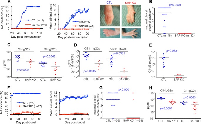
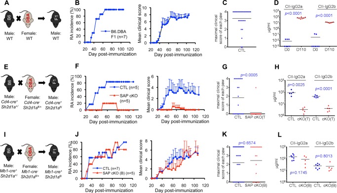
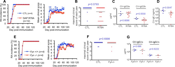
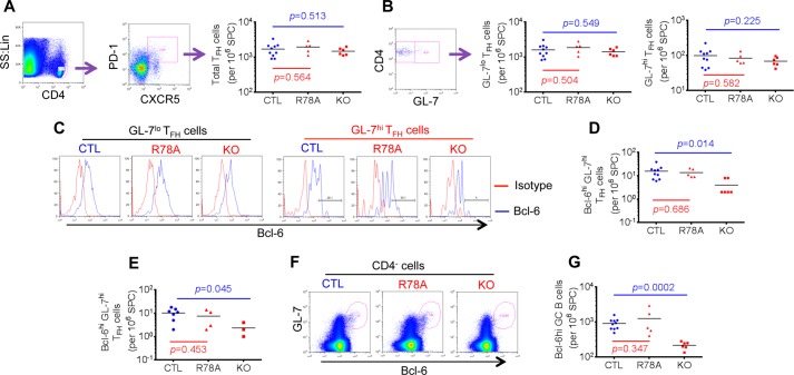
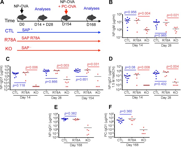
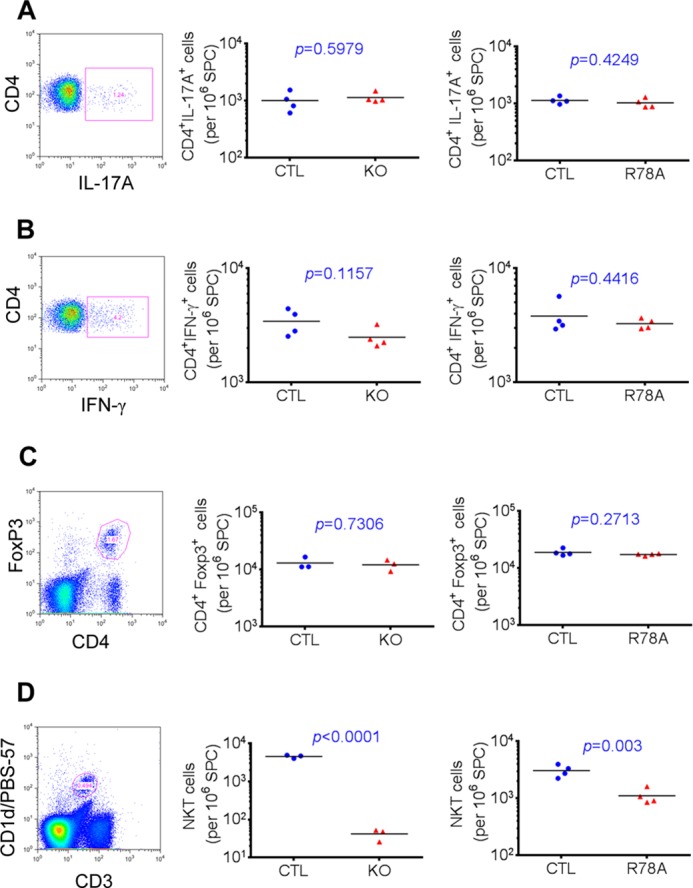
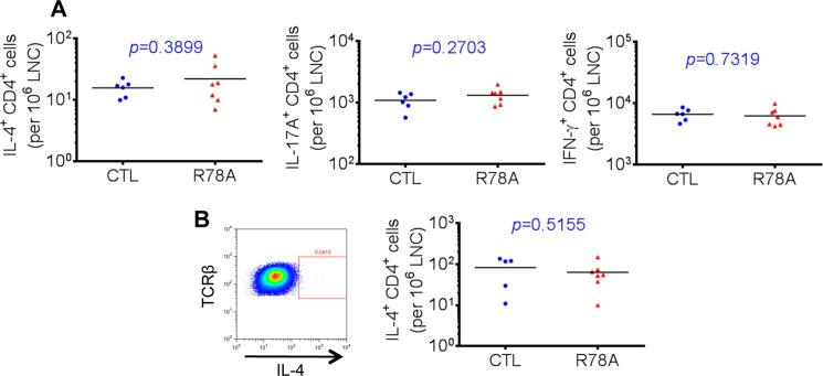
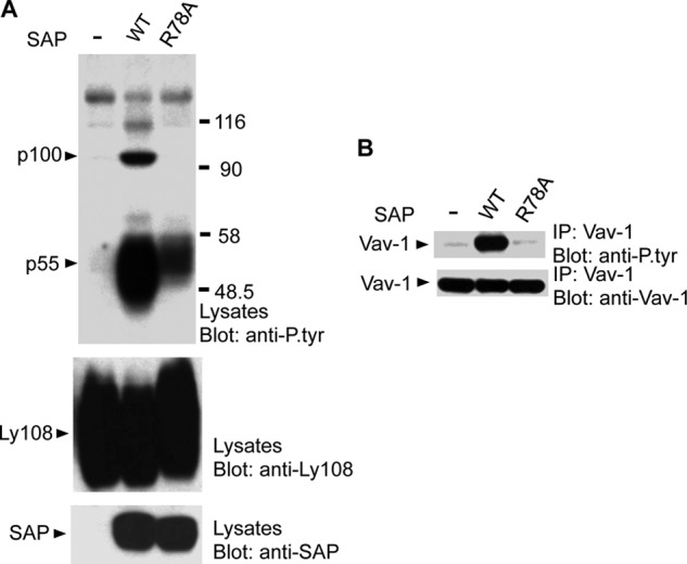
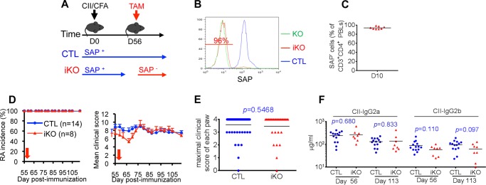
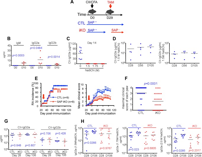
Signaling lymphocytic activation molecule-associated protein (SAP) is an Src homology 2 domain-only adaptor involved in multiple immune cell functions. It has also been linked to immunodeficiencies and autoimmune diseases, such as systemic lupus erythematosus. Here, we examined the role and mechanism of action of SAP in autoimmunity using a mouse model of autoimmune arthritis, collagen-induced arthritis (CIA). We found that SAP was essential for development of CIA in response to collagen immunization. It was also required for production of collagen-specific antibodies, which play a key role in disease pathogenesis. These effects required SAP expression in T cells, not in B cells. In mice immunized with a high dose of collagen, the activity of SAP was nearly independent of its ability to bind the protein tyrosine kinase Fyn and correlated with the capacity of SAP to promote full differentiation of follicular T helper (TFH) cells. However, with a lower dose of collagen, the role of SAP was more dependent on Fyn binding, suggesting that additional mechanisms other than TFH cell differentiation were involved. Further studies suggested that this might be due to a role of the SAP-Fyn interaction in natural killer T cell development through the ability of SAP-Fyn to promote Vav-1 activation. We also found that removal of SAP expression during progression of CIA attenuated disease severity. However, it had no effect on disease when CIA was clinically established. Together, these results indicate that SAP plays an essential role in CIA because of Fyn-independent and Fyn-dependent effects on TFH cells and, possibly, other T cell types.

Keywords: Adaptor Proteins; Autoimmune Diseases; Immunology; SH2 Domains; Src.


Figures

FIGURE 1.
FIGURE 1.
SAP deficiency fully protects against collagen-induced arthritis. A–E, mice (males, 8- to 10 weeks old) in the DBA/1J background were immunized with CII in complete Freund adjuvant containing M. tuberculosis as detailed under “Experimental Procedures.” The incidence of RA and the mean clinical score over time were determined by clinical examination (A). The number (n) of mice studied is shown in brackets. Error bars represent mean ± S.E. of the clinical score. Clinical scores were as follows: 0, no erythema and swelling; 1, erythema and mild swelling confined to the mid-foot (tarsals) or ankle joint; 2, erythema and mild swelling extending from the ankle to mid-foot; 3, erythema and moderate swelling extending from the ankle to the metatarsal joints; and 4, erythema and severe swelling encompassing the ankle, foot, and digits. The clinical score was the sum of the scores for the four paws. Photographs of representative animals (RA score of 3 for the control mouse and 0 for the SAP-deficient mouse) are shown on the right. B, maximal clinical score for each paw observed during the experiment period. Levels of antibodies against CII (C), high-affinity antibodies against the collagen fragment CB11 (measured by addition of 1.5 m NaSCN during ELISA (D), and IgE antibodies against CII (E) were measured 105 days after immunization as detailed under “Experimental Procedures.” Individual mice are depicted by individual symbols, and the horizontal line shows mean values. p values are also shown. F–H, mice were immunized a second time (boost) with CII 105 days after the first immunization. RA incidence and mean clinical score (F) and maximal clinical score for each paw (G) were determined. H, levels of antibodies against CII were measured 70 days after the boost. CTL, control mice; SAP KO, SAP-deficient mice.
FIGURE 2.
FIGURE 2.
SAP expression in T cells, not in B cells, is required for collagen-induced arthritis. A–D, F1 mice were generated from a cross between WT C57BL/6 and WT DBA/1J (B6.DBA, A) mice. Induction of RA (B and C) and anti-collagen (CII) antibodies (D) was monitored as detailed in Fig. 1. Antibody levels were measured 110 days after immunization. Individual mice are depicted by individual symbols, and the horizontal line shows mean values. p values are shown. E–H, mice expressing a conditional allele of the SAP-encoding gene (Sh2d1afl/, SAP cKO) were bred with mice expressing the Cre recombinase under the control of the CD4 promoter (Cd4-cre) (E). Induction of RA (F and G) and anti-CII antibodies (H) was monitored. Antibody levels were measured 28 days after immunization. I–L, Sh2d1afl/ mice were bred with mice expressing the Cre recombinase under the control of the MB1 promoter (Mb1-cre) (I). Induction of RA (J and K) and anti-CII antibodies (L) was monitored. Antibody levels were measured 28 days after immunization. CTL, control mice.
FIGURE 3.
FIGURE 3.
SAP-Fyn interaction is not required for CIA induction at the standard dose of collagen. A–D, SAP R78A mice. A, SAP R78A mice in DBA/1J background were immunized with 100 μg of CII. RA development was monitored as detailed in Fig. 1. B, the maximal clinical score for each paw is shown. CII-specific IgG2a and IgG2b (C) and IgE (D) were assayed 56 days after immunization. Individual mice are depicted by individual symbols, and the horizontal line shows mean values. p values are also shown. E–G, Fyn-deficient mice. Fyn-deficient mice in the DBA/1J background were immunized with 100 μg of CII. RA development (E) and maximal clinical score for each paw (F) were monitored. G, CII-specific IgG2a and IgG2b were measured 56 days after immunization. CTL, control mice.
FIGURE 4.
FIGURE 4.
SAP-Fyn interaction is critical for CIA induction at the low dose of collagen. SAP R78A mice in the DBA/1J background were immunized with 2 μg of CII. A, RA development was monitored as detailed in Fig. 1. B, maximal clinical score for each paw. CII-specific IgG2a (C), IgG2b (D), and IgE (E) were measured at the indicated times after immunization. Individual mice are depicted by individual symbols, and the horizontal line shows mean values. p values are also shown. CTL, control mice.
FIGURE 5.
FIGURE 5.
The SAP-Fyn interaction is not required for full TFH cell differentiation. A–D, mice were immunized with 100 μg of CII. Ten days later, splenocytes were isolated and analyzed by flow cytometry. A, numbers of TFH cells (defined as CD4+CXCR5+PD-1+ cells) in the spleen were determined. The gating strategy is shown on the left, whereas values for all mice are depicted by individual symbols on the right. The horizontal lines show mean values. p values are also shown. B, numbers of GC TFH cells (GL-7hiCD4+CXCR5+PD-1+ cells) were determined. The gating strategy is shown on the left, whereas numbers of GL-7lo and GL-7hi TFH cells for all mice are depicted on the right. C and D, Bcl-6 expression in GL-7lo and GL-7hi TFH cells was determined by intracellular staining. Histograms from representative mice are shown in C, and numbers of Bcl-6hiGL-7hi TFH cells for all mice are depicted in D. Bcl-6hi was defined as a mean fluorescence intensity of > 200. E, the experiment was performed as detailed in D, except that mice were immunized with 2 μg of CII, and analyses were performed 7 days after immunization. F and G, numbers of GC B cells (CD4GL-7hiBcl-6hi cells) in spleens of mice immunized with 100 μg of CII were determined. The gating strategy is shown in F, whereas values for individual mice are depicted by individual symbols in G. SS, side scatter; Lin, linear; SPC, spleen cells; CTL, control mice; R78A, SAP R78A mice; KO, SAP-deficient mice.
FIGURE 6.
FIGURE 6.
SAP-Fyn interaction is not required for maintenance or reactivation of TFH cell-dependent antibody responses. A, mice in the C57BL/6 background were immunized with a low dose (2 μg) of NP-OVA in the presence of alum. After 156 days, they were immunized again with low doses (2 μg each) of NP-OVA and PC-OVA. NP-specific IgM (B) and IgG1 (C) and high-affinity anti-NP IgG1 (measured by addition of 1.5 m NaSCN during ELISA, D) were measured at the indicated times after primary immunization with NP-OVA. Moreover, NP-specific IgG1 (E) and PC-specific IgG1 (F) were assayed 14 days (that is, on day 168) after the secondary immunization. Individual mice are depicted by individual symbols, and the horizontal line shows mean values. p values are shown. CTL, control mice; R78A, SAP R78A mice; KO, SAP-deficient mice.
FIGURE 7.
FIGURE 7.
SAP is not needed for TH7 cytokine production. Mice in the DBA/1J background were immunized with 100 μg of collagen II. On day 14, splenocytes were isolated and reactivated at 37 °C for 4 h in the presence of Golgi Plug, phorbol myristate acetate, and ionomycin. Relative numbers of CD4+ T cells producing IL-17A (A) and IFN-γ (B) were determined by intracellular staining. Isolated splenocytes were also analyzed by flow cytometry for detection of regulatory T cells (C), which are CD4+FoxP3+, and NK-T cells (D), which are CD3+CD1d/PBS-57+. In all cases, the gating strategy is depicted on the left. On the right, values for individual mice are depicted by individual symbols, and the horizontal line shows mean values. p values are also shown. SPC, spleen cells; CTL, control mice; KO, SAP-deficient mice; R78A, SAP R78A mice.
FIGURE 8.
FIGURE 8.
SAP-Fyn interaction is not needed for IL-4 production. Mice in the DBA/1J background were immunized with either a low dose (2 μg) or high dose (100 μg) of CII supplemented with 200 μg of M. tuberculosis in the hind footpads. On day 18, draining lymph node T cells were isolated and reactivated for 8 h at 37 °C in the presence of phorbol myristate acetate and ionomycin, followed by intracellular staining for cytokines. Golgi Stop was added during the last 6 h of stimulation. A, low dose of CII. Relative numbers of CD4+ T cells producing IL-4, IL-17A, and IFN-γ were determined by intracellular staining. B, high dose of CII. Relative numbers of CD4+ T cells producing IL-4 were determined by intracellular staining. Values for individual mice are depicted by individual symbols, and the horizontal line shows mean values. p values are also shown. CTL, control mice; R78A, SAP R78A mice.
FIGURE 9.
FIGURE 9.
SAP-Fyn association is required for Vav-1 tyrosine phosphorylation during homotypic cell-cell interactions. BI-141 T cell transfectants expressing Ly108 in the absence of SAP or in the presence of WT SAP or SAP R78A were seeded overnight at the same confluency to allow homotypic cell-cell interactions and Ly108 self-ligation and then lysed. A, lysates were immunoblotted with antibodies against phosphotyrosine, Ly108, or SAP. The positions of the predominant substrates showing augmented tyrosine phosphorylation in cells expressing WT SAP are shown on the left. B, lysates were immunoprecipitated (IP) with antibodies against Vav-1 and probed by immunoblotting with antibodies against phosphotyrosine or Vav-1.
FIGURE 10.
FIGURE 10.
SAP is not required for maintenance of collagen-induced arthritis. A, mice expressing the conditional (“inducible”) allele of the SAP-encoding gene (Sh2d1afl/, SAP iKO) were bred with mice expressing the TAM-responsive Cre recombinase under the control of the ubiquitin C promoter (UBC-creERT2). They were then immunized with CII and complete Freund adjuvant (CFA). After 56 days, SAP was deleted by treatment of mice with TAM. B, expression of SAP in representative mice was examined by intracellular staining and flow cytometry using peripheral blood lymphocytes gated on CD3+CD4+ cells. Green line, SAP-deficient mouse; red line, SAP iKO mouse; blue line, control (CTL) mouse. The percentage of SAP-negative cells in the SAP iKO mouse is indicated. C, percentages of SAP-negative CD3+CD4+ T cells in multiple SAP iKO mice treated with TAM. Analyses were performed 10 days after TAM treatment. Induction of RA (D and E) and anti-CII antibodies (F) was monitored as detailed in Fig. 1. The time of TAM treatment is shown by a red arrow. Values for individual mice are depicted by individual symbols, and the horizontal line shows mean values. p values are also shown.
FIGURE 11.
FIGURE 11.
SAP is required for progression of collagen-induced arthritis. A, this experiment was performed as detailed in Fig. 10, except that TAM treatment was initiated 29 days after immunization. B, anti-CII antibodies were measured 10 days (D10) after immunization of wild-type mice with CII when the clinical symptoms were not yet observed. C, high-affinity IgG2a antibodies against CII were measured on day 14 after CII injection using the chaotropic agent NaSCN. D, high-affinity IgG2a and IgG2b antibodies against CII were measured on the indicated days after CII injection. Induction of RA (E and F) and anti-CII antibodies (G) was monitored as detailed in Fig. 1. Time of TAM treatment is shown by a red arrow. Values for individual mice are depicted by individual symbols, and the horizontal line shows mean values. p values are also shown. H, the abundance of high-affinity anti-CII IgG2a was determined by performing ELISA in presence of progressively higher concentrations of NaSCN. CTL, control mice; iKO, inducible KO mice; CFA, complete Freund adjuvant.

Similar articles

Cited by

References

    1. Schwartzberg P. L., Mueller K. L., Qi H., Cannons J. L. (2009) SLAM receptors and SAP influence lymphocyte interactions, development and function. Nat. Rev. Immunol. 9, 39–46 - PubMed
    1. Veillette A., Dong Z., Latour S. (2007) Consequence of the SLAM-SAP signaling pathway in innate-like and conventional lymphocytes. Immunity 27, 698–710 - PubMed
    1. Calpe S., Wang N., Romero X., Berger S. B., Lanyi A., Engel P., Terhorst C. (2008) The SLAM and SAP gene families control innate and adaptive immune responses. Adv. Immunol. 97, 177–250 - PubMed
    1. Ma C. S., Nichols K. E., Tangye S. G. (2007) Regulation of cellular and humoral immune responses by the SLAM and SAP families of molecules. Annu. Rev. Immunol. 25, 337–379 - PubMed
    1. Kageyama R., Cannons J. L., Zhao F., Yusuf I., Lao C., Locci M., Schwartzberg P. L., Crotty S. (2012) The receptor Ly108 functions as a SAP adaptor-dependent on-off switch for T cell help to B cells and NKT cell development. Immunity 36, 986–1002 - PMC - PubMed

MeSH terms

Substances

LinkOut - more resources