Clinical Trial
. 2013 Oct 25;288(43):30956-68.
doi: 10.1074/jbc.M113.509893. Epub 2013 Sep 17.

Glycoepitopes of staphylococcal wall teichoic acid govern complement-mediated opsonophagocytosis via human serum antibody and mannose-binding lectin

Affiliations
Clinical Trial

Glycoepitopes of staphylococcal wall teichoic acid govern complement-mediated opsonophagocytosis via human serum antibody and mannose-binding lectin

Kenji Kurokawa et al. J Biol Chem. .

Abstract

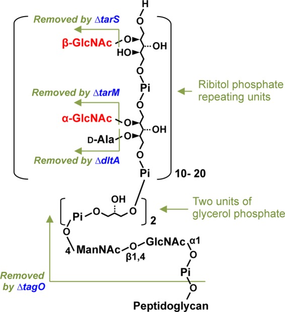
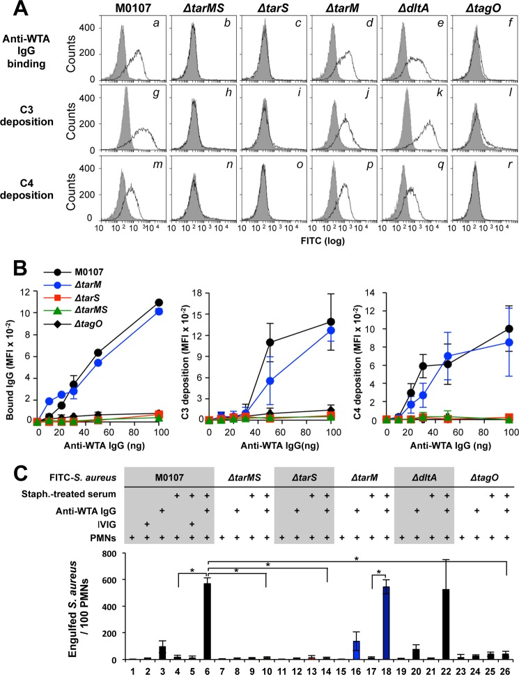
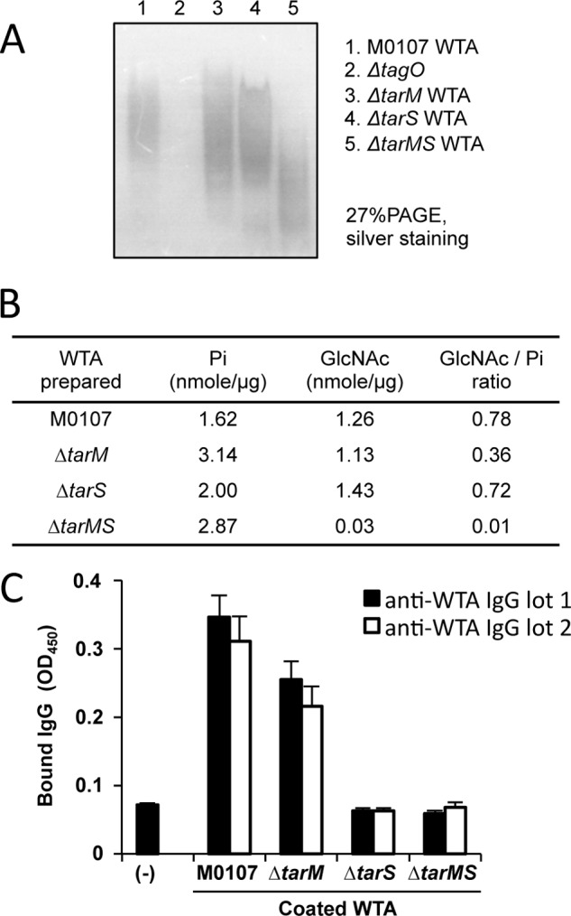
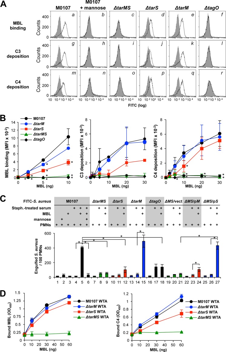
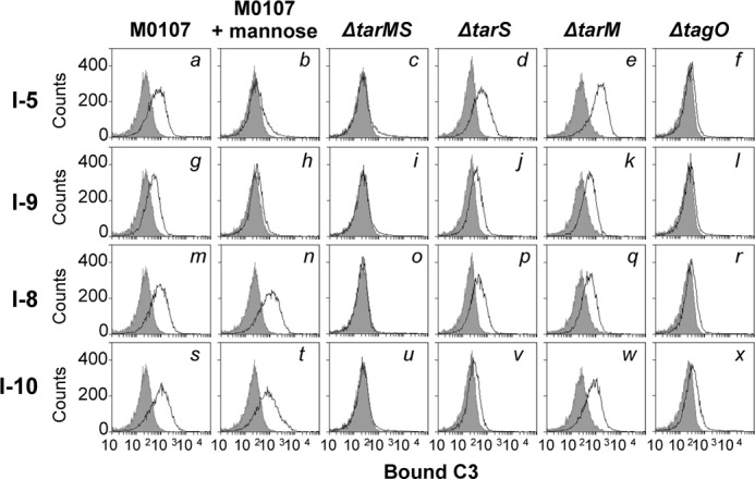
Serum antibodies and mannose-binding lectin (MBL) are important host defense factors for host adaptive and innate immunity, respectively. Antibodies and MBL also initiate the classical and lectin complement pathways, respectively, leading to opsonophagocytosis. We have shown previously that Staphylococcus aureus wall teichoic acid (WTA), a cell wall glycopolymer consisting of ribitol phosphate substituted with α- or β-O-N-acetyl-d-glucosamine (GlcNAc) and d-alanine, is recognized by MBL and serum anti-WTA IgG. However, the exact antigenic determinants to which anti-WTA antibodies or MBL bind have not been determined. To answer this question, several S. aureus mutants, such as α-GlcNAc glycosyltransferase-deficient S. aureus ΔtarM, β-GlcNAc glycosyltransferase-deficient ΔtarS, and ΔtarMS double mutant cells, were prepared from a laboratory and a community-associated methicillin-resistant S. aureus strain. Here, we describe the unexpected finding that β-GlcNAc WTA-deficient ΔtarS mutant cells (which have intact α-GlcNAc) escape from anti-WTA antibody-mediated opsonophagocytosis, whereas α-GlcNAc WTA-deficient ΔtarM mutant cells (which have intact β-GlcNAc) are efficiently engulfed by human leukocytes via anti-WTA IgG. Likewise, MBL binding in S. aureus cells was lost in the ΔtarMS double mutant but not in either single mutant. When we determined the serum concentrations of the anti-α- or anti-β-GlcNAc-specific WTA IgGs, anti-β-GlcNAc WTA-IgG was dominant in pooled human IgG fractions and in the intact sera of healthy adults and infants. These data demonstrate the importance of the WTA sugar conformation for human innate and adaptive immunity against S. aureus infection.

Keywords: Cell Wall; Complement System; Gram-positive Bacteria; Host Defense; Host-Pathogen Interactions; Innate Immunity; S. aureus.


Figures

FIGURE 1.
FIGURE 1.
Schematic structure of S. aureus WTA. WTA of S. aureus is composed of a short linkage unit connected to peptidoglycan, consisting of a ManNAc-GlcNAc disaccharide with two glycerol phosphates, followed by a longer chain of ribitol phosphate repeating units substituted with α- or β-GlcNAc and d-alanine. Changes by ΔtarS, ΔtarM, ΔdltA, or ΔtagO mutations on the WTA structure are indicated.
FIGURE 2.
FIGURE 2.
Anti-WTA IgG-mediated complement activation and opsonophagocytosis of S. aureus cells are dependent on β-GlcNAc WTA. A, in the top panels, ethanol-killed S. aureus mutant cells were incubated without (gray area) or with anti-WTA IgG (50 ng; black line) in 20 μl of buffer, and bound IgG was detected by flow cytometric analysis. The middle and bottom panels show the measurement of C3 and C4 depositions in 10% S. aureus-treated adult serum without (gray area) or with anti-WTA IgG (50 ng; black line) in 20 μl of buffer. C3 and C4 were detected by specific antibodies with flow cytometric analysis. B, anti-WTA IgG binding (left), C3 deposition (middle), and C4 deposition (right) on each S. aureus strain was examined as in A with respect to affinity-purified anti-WTA IgG concentrations. C, ethanol-killed Δspa (parent; columns 1–6) and the indicated isogenic mutant S. aureus cells were labeled with FITC (0.1 mm) and opsonized without or with S. aureus-treated serum (10%) in 20 μl of buffer. For descriptions of columns 1–26, see “Results.” Purified anti-WTA IgG (50 ng) or IVIG (50 ng) was simultaneously added, as indicated. Opsonized FITC-labeled S. aureus cells were incubated with human PMNs (1 × 10⁵ PMNs) at a multiplicity of infection of 25 in 40 μl of RPMI 1640 medium at 37 °C for 1 h. Phagocytosed S. aureus cells per 100 PMNs were counted under fluorescent phase-contrast microscopy. Data are represented as the means ± S.D. (error bars) of the results of three independent experiments. *, p < 0.05.
FIGURE 3.
FIGURE 3.
Biochemical characterization of purified WTAs and their affinities for purified anti-WTA IgG. A, purified WTAs (10 μg) from indicated strains were separated by PAGE (27%) and visualized by silver staining. B, amounts of phosphate, GlcNAc, and GlcNAc/inorganic phosphate (GlcNAc/Pi) ratio of purified WTA are shown. C, each purified WTA (5 nmol of phosphate) was used to coat a 96-well ELISA plate, and affinity-purified anti-WTA IgG (100 ng) was assayed for its binding ability to each coated WTA by ELISA. Two independently purified anti-WTA IgGs were used. Data are represented as the means ± S.D. (error bars) of the results of three independent experiments.
FIGURE 4.
FIGURE 4.
WTA GlcNAc dependence of MBL-mediated complement activation and opsonophagocytosis of S. aureus cells by PMNs. A, in the top panels, ethanol-killed S. aureus M0107 (Δspa) cells or their derivatives were incubated without (gray area) or with MBL (10 ng; black line) in 20 μl of buffer, and bound MBL was detected by flow cytometry analysis. In the middle and bottom panels, depositions of C3 and C4, respectively, onto S. aureus cells were carried out in 10% S. aureus-treated adult serum without (gray area) or with MBL (10 ng, black line) in 20 μl of buffer. C3 and C4 were detected by specific antibodies with FCM analysis. B, MBL binding (left), C3 deposition (middle), and C4 deposition (right) were examined as in A with respect to MBL concentrations. Mean fluorescent intensities on flow cytometry analyses were expressed on the vertical axis. C, ethanol-killed M0107 (Δspa, parent; columns 1–6) and mutant S. aureus cells were labeled with FITC (0.1 mm) and opsonized without or with S. aureus-treated serum (10%) in 20 μl of buffer. Purified MBL (10 ng) and mannose (100 mm) were simultaneously added as indicated. Opsonized FITC-labeled S. aureus cells were incubated with human PMNs (1 × 10⁵ PMNs) at a multiplicity of infection of 25 in 40 μl of RPMI 1640 medium at 37 °C for 1 h. Phagocytosed S. aureus cells per 100 PMNs were counted under fluorescent phase-contrast microscopy. Data are represented as the means ± S.D. (error bars) of the results of three independent experiments. *, p < 0.05. D, purified WTA was assayed for MBL binding and C4 deposition by ELISA. Left, each purified WTA (3 μg) was coated onto a 96-well ELISA plate, and purified MBL/MASP at the indicated amount was incubated at 4 °C for 2 h. Amounts of MBL bound to each coated WTA were determined by ELISA. Right, each purified WTA (3 μg) was coated onto a 96-well ELISA plate, and purified MBL/MASP at the indicated amount and C4 (100 ng) were incubated at 37 °C for 1 h, and C4 bound to each coated WTA was determined by ELISA. Data are represented as the means ± S.D. of results of three independent experiments.
FIGURE 5.
FIGURE 5.
The MBL-mediated lectin pathway and anti-WTA IgG-mediated classical complement pathway are activated GlcNAc-dependently in intact infant sera. Ethanol-killed S. aureus M0107 (Δspa) cells or its derivatives were incubated without (gray area) or with 5% of each intact infant serum (black line) in 20 μl of buffer at 37 °C for 1 h, and bound C3 was detected by flow cytometric analysis. Data are representative of at least three independent experiments.

Similar articles

Cited by

References

    1. Lowy F. D. (1998) Staphylococcus aureus infections. N. Engl. J. Med. 339, 520–532 - PubMed
    1. Weidenmaier C., Peschel A. (2008) Teichoic acids and related cell-wall glycopolymers in Gram-positive physiology and host interactions. Nat. Rev. Microbiol. 6, 276–287 - PubMed
    1. Swoboda J. G., Campbell J., Meredith T. C., Walker S. (2010) Wall teichoic acid function, biosynthesis, and inhibition. Chembiochem 11, 35–45 - PMC - PubMed
    1. Xia G., Kohler T., Peschel A. (2010) The wall teichoic acid and lipoteichoic acid polymers of Staphylococcus aureus. Int. J. Med. Microbiol. 300, 148–154 - PubMed
    1. Weidenmaier C., Kokai-Kun J. F., Kristian S. A., Chanturiya T., Kalbacher H., Gross M., Nicholson G., Neumeister B., Mond J. J., Peschel A. (2004) Role of teichoic acids in Staphylococcus aureus nasal colonization, a major risk factor in nosocomial infections. Nat. Med. 10, 243–245 - PubMed

Publication types

MeSH terms

LinkOut - more resources