

Epidermal growth factor (EGF)-enhanced vascular cell adhesion molecule-1 (VCAM-1) expression promotes macrophage and glioblastoma cell interaction and tumor cell invasion
- PMID: 24045955
- PMCID: PMC3814745
- DOI: 10.1074/jbc.M113.499020
Epidermal growth factor (EGF)-enhanced vascular cell adhesion molecule-1 (VCAM-1) expression promotes macrophage and glioblastoma cell interaction and tumor cell invasion
Erratum in
- J Biol Chem. 2014 Jul 4;289(27):18667
Abstract
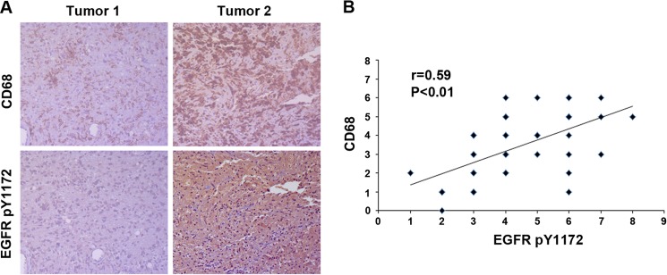
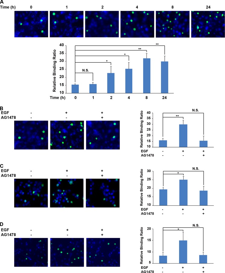
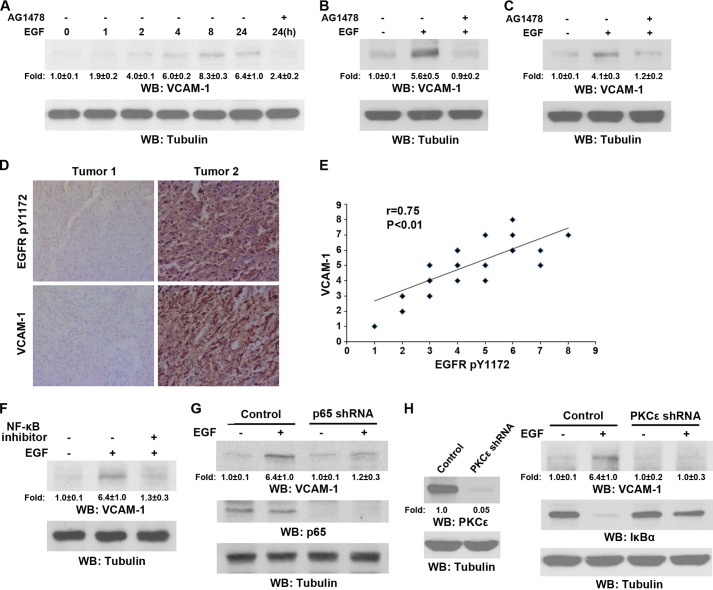
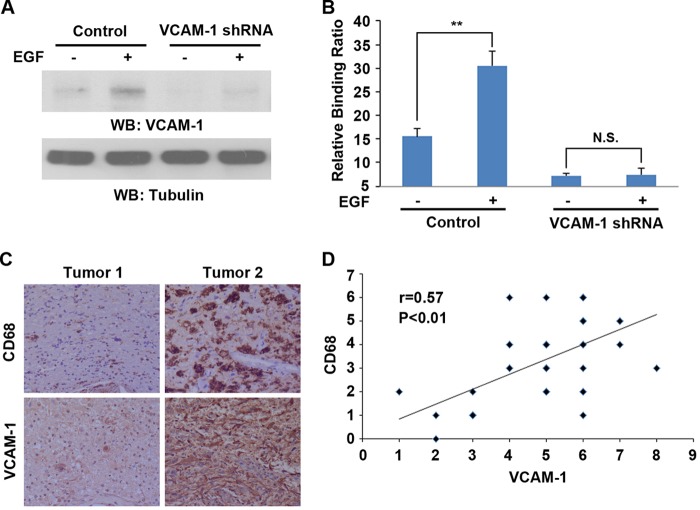
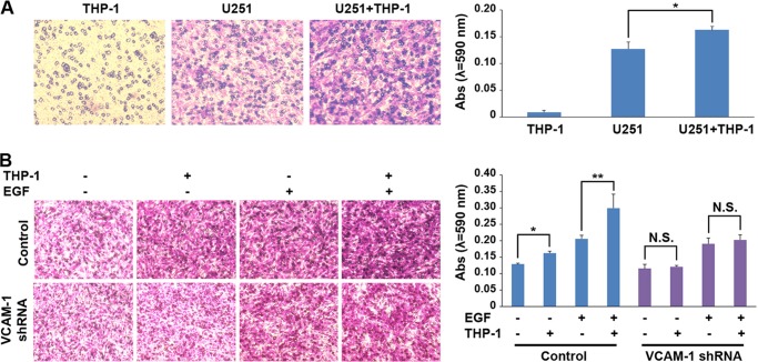
Activated EGF receptor (EGFR) signaling plays an instrumental role in glioblastoma (GBM) progression. However, how EGFR activation regulates the tumor microenvironment to promote GBM cell invasion remains to be clarified. Here, we demonstrate that the levels of EGFR activation in tumor cells correlated with the levels of macrophage infiltration in human GBM specimens. This was supported by our observation that EGFR activation enhanced the interaction between macrophages and GBM cells. In addition, EGF treatment induced up-regulation of vascular cell adhesion molecule-1 (VCAM-1) expression in a PKCε- and NF-κB-dependent manner. Depletion of VCAM-1 interrupted the binding of macrophages to GBM cells and inhibited EGF-induced and macrophage-promoted GBM cell invasion. These results demonstrate an instrumental role for EGF-induced up-regulation of VCAM-1 expression in EGFR activation-promoted macrophage-tumor cell interaction and tumor cell invasion and indicate that VCAM-1 is a potential molecular target for improving cancer therapy.
Keywords: Epidermal Growth Factor Receptor (EGFR); Glioblastoma; Invasion; Macrophages; Migration.
Figures
Similar articles
-
EGFR-induced and PKCε monoubiquitylation-dependent NF-κB activation upregulates PKM2 expression and promotes tumorigenesis.Mol Cell. 2012 Dec 14;48(5):771-84. doi: 10.1016/j.molcel.2012.09.028. Epub 2012 Nov 1. Mol Cell. 2012. PMID: 23123196 Free PMC article.
-
MiR-181b modulates EGFR-dependent VCAM-1 expression and monocyte adhesion in glioblastoma.Oncogene. 2017 Aug 31;36(35):5006-5022. doi: 10.1038/onc.2017.129. Epub 2017 May 1. Oncogene. 2017. PMID: 28459461
-
Regulatory effects of IL-1β in the interaction of GBM and tumor-associated monocyte through VCAM-1 and ICAM-1.Eur J Pharmacol. 2021 Aug 15;905:174216. doi: 10.1016/j.ejphar.2021.174216. Epub 2021 May 28. Eur J Pharmacol. 2021. PMID: 34058204
-
MicroRNAs involved in the EGFR pathway in glioblastoma.Biomed Pharmacother. 2021 Feb;134:111115. doi: 10.1016/j.biopha.2020.111115. Epub 2020 Dec 16. Biomed Pharmacother. 2021. PMID: 33341046 Review.
-
EGFR-dependent mechanisms in glioblastoma: towards a better therapeutic strategy.Cell Mol Life Sci. 2014 Sep;71(18):3465-88. doi: 10.1007/s00018-014-1608-1. Epub 2014 Mar 27. Cell Mol Life Sci. 2014. PMID: 24671641 Free PMC article. Review.
Cited by
-
WNT/β-catenin-suppressed FTO expression increases m6A of c-Myc mRNA to promote tumor cell glycolysis and tumorigenesis.Cell Death Dis. 2021 May 8;12(5):462. doi: 10.1038/s41419-021-03739-z. Cell Death Dis. 2021. PMID: 33966037 Free PMC article.
-
Combination treatment with oncolytic Vaccinia virus and cyclophosphamide results in synergistic antitumor effects in human lung adenocarcinoma bearing mice.J Transl Med. 2014 Jul 17;12:197. doi: 10.1186/1479-5876-12-197. J Transl Med. 2014. PMID: 25030093 Free PMC article.
-
Context-Dependent Glioblastoma-Macrophage/Microglia Symbiosis and Associated Mechanisms.Trends Immunol. 2021 Apr;42(4):280-292. doi: 10.1016/j.it.2021.02.004. Epub 2021 Mar 1. Trends Immunol. 2021. PMID: 33663953 Free PMC article. Review.
-
The Vitamin D status is associated with serum C-reactive protein and adhesion molecules in patients with renal cell carcinoma.Sci Rep. 2019 Nov 13;9(1):16719. doi: 10.1038/s41598-019-53395-9. Sci Rep. 2019. PMID: 31723229 Free PMC article.
-
Microglial SMAD4 regulated by microRNA-146a promotes migration of microglia which support tumor progression in a glioma environment.Oncotarget. 2018 May 18;9(38):24950-24969. doi: 10.18632/oncotarget.25116. eCollection 2018 May 18. Oncotarget. 2018. PMID: 29861845 Free PMC article.
References
-
- Clarke J., Butowski N., Chang S. (2010) Recent advances in therapy for glioblastoma. Arch. Neurol. 67, 279–283 - PubMed
-
- Libermann T. A., Razon N., Bartal A. D., Yarden Y., Schlessinger J., Soreq H. (1984) Expression of epidermal growth factor receptors in human brain tumors. Cancer Res. 44, 753–760 - PubMed
-
- Larysz D., Kula D., Kowal M., Rudnik A., Jarząb M., Blamek S., Bierzyńska-Macyszyn G., Kowalska M., Bażowski P., Jarząb B. (2011) Epidermal growth factor receptor gene expression in high grade gliomas. Folia Neuropathol. 49, 28–38 - PubMed
-
- Zhu Y., Parada L. F. (2002) The molecular and genetic basis of neurological tumours. Nat. Rev. Cancer 2, 616–626 - PubMed
Publication types
MeSH terms
Substances
Grants and funding
LinkOut - more resources
Full Text Sources
Other Literature Sources
Research Materials
Miscellaneous
