. 2013 Oct 21;54(10):6806-19.
doi: 10.1167/iovs.13-12551.

Gene expression signatures in tree shrew sclera in response to three myopiagenic conditions

Affiliations

Gene expression signatures in tree shrew sclera in response to three myopiagenic conditions

Lin Guo et al. Invest Ophthalmol Vis Sci. .

Abstract

Purpose: We compared gene expression signatures in tree shrew sclera produced by three different visual conditions that all produce ocular elongation and myopia: minus-lens wear, form deprivation, and dark treatment.

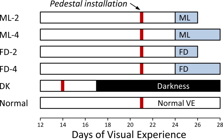
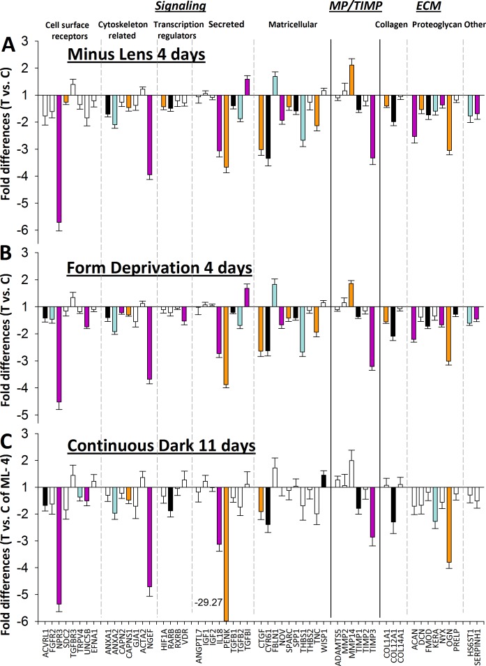
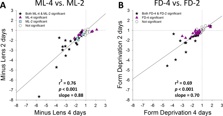
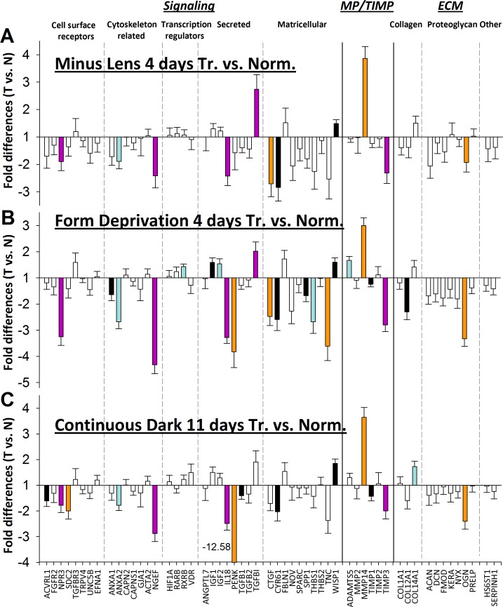
Methods: Six groups of tree shrews (n = 7 per group) were used. Starting 24 days after normal eye-opening (days of visual experience [DVE]), two minus-lens groups wore a monocular -5 diopter (D) lens for 2 days (ML-2) or 4 days (ML-4); two form-deprivation groups wore a monocular translucent diffuser for 2 days (FD-2) or 4 days (FD-4). A dark-treatment (DK) group was placed in continuous darkness for 11 days after experiencing a light/dark environment until 17 DVE. A normal colony-reared group was examined at 28 DVE. Quantitative PCR was used to measure the relative differences in mRNA levels for 55 candidate genes in the sclera that were selected, either because they showed differential expression changes in previous ML studies or because a whole-transcriptome analysis suggested they would change during myopia development.

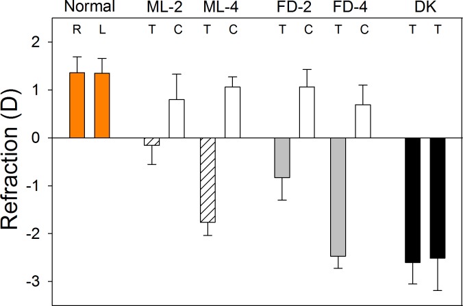
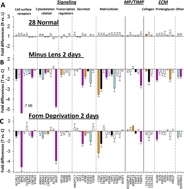
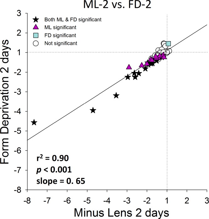
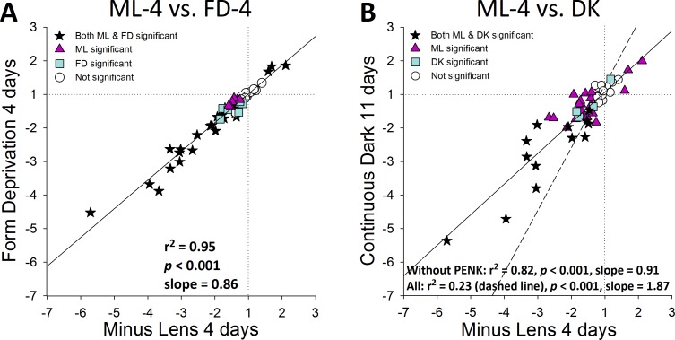
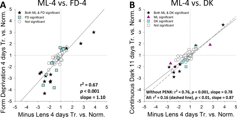
Results: The treated eyes in all groups responded with a significant myopic shift, indicating that the myopia was actively progressing. In the ML-2 group, 27 genes were significantly downregulated in the treated eyes, relative to control eyes. In the treated eyes of the FD-2 group, 16 of the same genes also were significantly downregulated and one was upregulated. The two gene expression patterns were significantly correlated (r(2) = 0.90, P < 0.001). After 4 days of treatment, 31 genes were significantly downregulated in the treated eyes of the ML-4 group and three were upregulated. Twenty-nine of the same genes (26 down- and 3 up-regulated) and six additional genes (all downregulated) were significantly affected in the FD-4 group. The response patterns were highly correlated (r(2) = 0.95, P < 0.001). When the DK group (mean of right and left eyes) was compared to the control eyes of the ML-4 group, the direction and magnitude of the gene expression patterns were similar to those of the ML-4 (r(2) = 0.82, P < 0.001, excluding PENK). Similar patterns also were found when the treated eyes of the ML-4, FD-4, and DK groups were compared to the age-matched normal eyes.

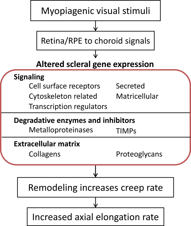
Conclusions: The very similar gene expression signatures produced in the sclera by the three different myopiagenic visual conditions at different time points suggests that there is a "scleral remodeling signature" in this mammal, closely related to primates. The scleral genes examined did not distinguish between the specific visual stimuli that initiate the signaling cascade that results in axial elongation and myopia.

Keywords: animal models; axial elongation; emmetropization; gene expression; myopia; refractive error; sclera.


Figures

Figure 1
Figure 1
Information flow produced by three myopiagenic conditions (minus-lens wear, form deprivation, and dark treatment). Retinal neurons detect these stimuli, and generate signals that cascade through the RPE and choroid to produce remodeling in the sclera. The remodeled sclera has increased viscoelasticity that produces an increase in the axial elongation rate. The focus of this paper (central box) is the gene expression changes that occur in the sclera in response to these three visual conditions, including altered gene expression related to signaling, degradative enzymes and inhibitors, and extracellular matrix.
Figure 2
Figure 2
Experimental groups and duration of treatments. The red vertical bar indicates the point when a dental acrylic pedestal was installed under anesthesia. Filled regions indicate the type and duration of visual treatment. The right end of each bar indicates the time point when mRNA levels were measured.
Figure 3
Figure 3
End-of-treatment refractive measures for the normal, ML, FD, and DK groups. Values are the mean refraction ± SEM for the right (R) and left (L) eyes of the normal and DK groups, and for the treated (T) and control (C) eyes of the ML and FD groups. Treated eyes in all groups were significantly myopic relative to control (or normal) eyes.
Figure 4
Figure 4
Gene expression fold differences. (A) Normal eyes (right eyes versus left eyes). (B) 2 days of minus-lens wear (treated eyes versus control eyes). (C) 2 days of form deprivation (treated eyes versus control eyes). Filled bars represent statistically significant differences between the treated and control eyes (P < 0.05). Bar color is arbitrary and intended to help in comparing the same gene in the three different conditions. Error bars indicate SEM. In (B), the off-scale fold difference for NPR3 (−7.66) is indicated next to the bar.
Figure 5
Figure 5
Comparison of the gene expression differences (treated eye versus control eye) in Figure 4B (ML-2) with the differences in Figure 4C (FD-2). The amount of differential expression in both conditions was very similar. Stars: significant fold differences for both ML and FD. Triangles: significant fold differences only for ML. Squares: significant fold differences only for FD. Circles: fold differences not significant in either treatment.
Figure 6
Figure 6
Gene expression fold differences. (A) 4 days of minus-lens wear (treated eyes versus control eyes). (B) 4 days of form deprivation (treated eyes versus control eyes). (C) 11 days of dark treatment (mean right and left versus ML-4 control eyes).
Figure 7
Figure 7
Comparison of the treated versus control gene expression differences in Figure 6. (A) ML-4 (Fig. 6A) versus FD-4 (Fig. 6B). (B) ML-4 (Fig. 6A) versus 11 days of continuous darkness (Fig. 6C). Stars: significant fold differences for both treatments. Triangles: significant fold differences only for ML-4. Squares: significant fold differences only for FD-4 (A) or DK (B). Circles: fold differences not significant for either treatment.
Figure 8
Figure 8
Comparison of the 4-day (Fig. 6) versus 2-day (Fig. 4) treated versus control gene expression differences produced by (A) minus-lens wear (Figs. 6A versus 4B) and (B) form deprivation (Figs. 6B versus 4C). Stars: significant fold differences for both 2- and 4-day treatments. Triangles: significant fold differences only for 4-day. Squares: significant fold differences only for 2-day. Circles: fold differences not significant at either treatment duration.
Figure 9
Figure 9
Gene expression fold differences. (A) 4 days of minus-lens wear (treated eyes versus normal eyes). (B) 4 days of form deprivation (treated eyes versus normal eyes). (C) 11 days of dark treatment (mean right and left versus normal eyes).
Figure 10
Figure 10
Comparison of gene expression patterns in the treated eyes of ML, FD, and DK with normal eyes as a common reference (Fig. 9). (A) minus-lens wear compared to form deprivation (Figs. 9A versus 9B). (B) minus-lens wear compared to continuous darkness (Figs. 9A versus 9C). Because the DK group treatment was binocular, the fold difference for DK eyes is the mean of values of the right and left eyes.

References

    1. Sperduto RD, Seigel D, Roberts J, Rowland M. Prevalence of myopia in the United States. Arch Ophthalmol. 1983; 101: 405–407 - PubMed
    1. Fledelius HC. Myopia prevalence in Scandinavia. A survey, with emphasis on factors of relevance for epidemiological refraction studies in general. Acta Ophthalmol Suppl. 1988; 185: 44–50 - PubMed
    1. Wang Q, Klein BE, Klein R, Moss SE. Refractive status in the Beaver Dam Eye Study. Invest Ophthalmol Vis Sci. 1994; 35: 4344–4347 - PubMed
    1. Attebo K, Ivers RQ, Mitchell P. Refractive errors in an older population: the Blue Mountains Eye Study. Ophthalmology. 1999; 106: 1066–1072 - PubMed
    1. Wensor M, McCarty CA, Taylor HR. Prevalence and risk factors of myopia in Victoria, Australia. Arch Ophthalmol. 1999; 117: 658–663 - PubMed

Publication types