

Case Reports
doi: 10.1007/s12105-013-0491-7.
Epub 2013 Sep 18.
Extramedullary plasmacytoma of the trachea
Affiliations
- PMID: 24046059
- PMCID: PMC4022935
- DOI: 10.1007/s12105-013-0491-7
Item in Clipboard
Case Reports
Extramedullary plasmacytoma of the trachea
Head Neck Pathol.
2014 Jun.
Abstract
Extramedullary plasmacytomas are plasma cell tumors that occur outside the bone marrow. They constitute around 4 % of all plasma cell neoplasms. The most common site of extramedullary plasmacytoma is the upper aerodigestive tract-nasal cavity, paranasal sinuses and oronasopharynx. We are presenting a case of extramedullary plasmacytoma of the trachea. Trachea is an extremely rare site of plasmacytoma. When extraosseous plasmacytoma occur in uncommon sites, the distinction from B cell lymphomas showing extensive plasmacytic differentiation can be difficult and diagnostically challenging.
Figures
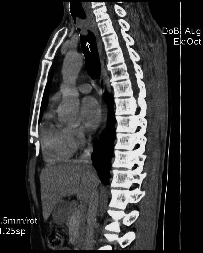
Computerized tomography (sagittal section) of the thorax showing a lobulated soft tissue attenuation mass lesion in the trachea, arising from the posterior wall and protruding intra luminally
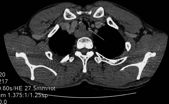
Post contrast axial section showing mass lesion in the trachea, causing partial luminal compromise
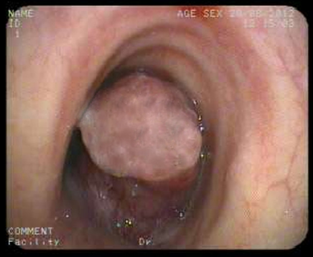
Bronchoscopy showing broad based pedunculated intraluminal tracheal mass which markedly narrow the lumen
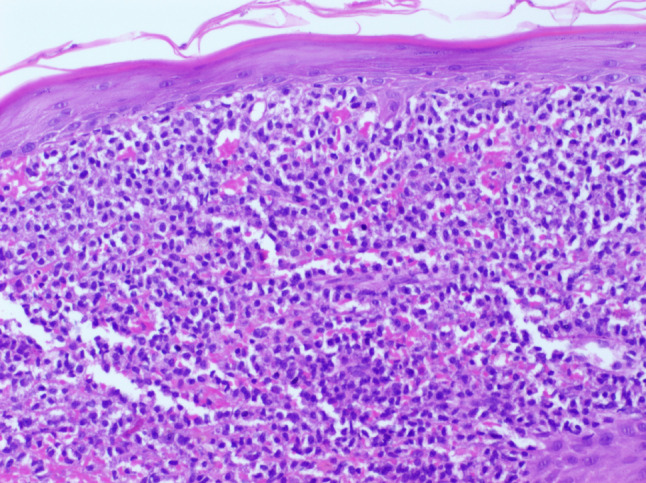
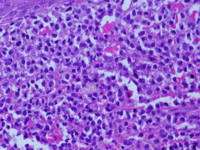
Microscopy showing neoplasm beneath the intact squamous epithelium (H and E, ×100)
Higher power showing sheets of plasmacytoid cells. (H and E, ×400)
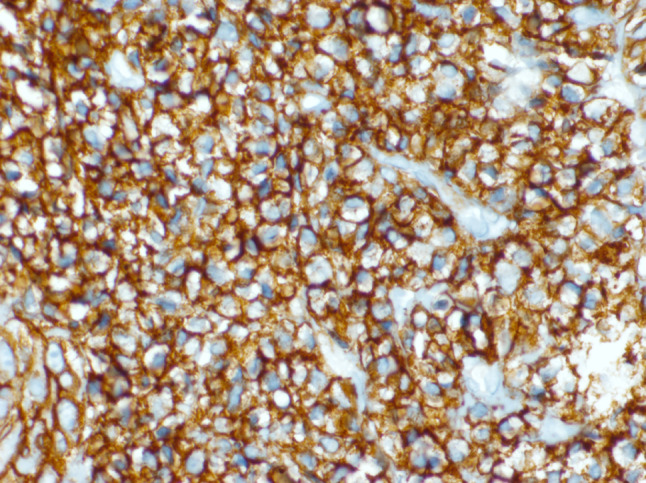
Tumour cells showing intense positivity for CD138. (IHC, ×400)
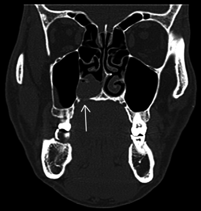
Coronal CT sections through the paranasal sinuses (bone window) reveal a soft tissue mass in the right nasal cavity, region of inferior turbinate, with irregular destruction of adjacent hard palate
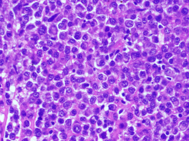
Microscopy of paranasal sinus mass showing sheets of plasmacytoid cells including immature and binucleate forms. (H and E, ×400)
References
-
- Swerdlow SH, Campo E, Harris NL, Jaffe ES, Pileri SA, Stein H, Thiele J, Vardiman JW, editors. WHO classification of tumors of haematopoetic and lymphoid tissues. IARC: Lyon, France; 2008.
-
- Reyhan M, Rercan F, Ergin M, Sukan A, Aydin M, Yapar F. Sonographic diagnosis of a tracheal extramedullary plasmacytoma. J Ultrasound Med. 2005;24:1031–1034. - PubMed
-
- Rai SP, Kumar R, Bharadwaj R, Panda BN. Solitary tracheal plasmacytoma. Indian J Chest Dis Allied Sci. 2003;45:269–272. - PubMed
Publication types
MeSH terms
Substances
LinkOut - more resources
Full Text Sources
Other Literature Sources
