Review
. 2013 Oct;140(19):3939-49.
doi: 10.1242/dev.080549.

The developmental origins of adipose tissue

Affiliations
Review

The developmental origins of adipose tissue

Daniel C Berry et al. Development. 2013 Oct.

Abstract

Adipose tissue is formed at stereotypic times and locations in a diverse array of organisms. Once formed, the tissue is dynamic, responding to homeostatic and external cues and capable of a 15-fold expansion. The formation and maintenance of adipose tissue is essential to many biological processes and when perturbed leads to significant diseases. Despite this basic and clinical significance, understanding of the developmental biology of adipose tissue has languished. In this Review, we highlight recent efforts to unveil adipose developmental cues, adipose stem cell biology and the regulators of adipose tissue homeostasis and dynamism.

Keywords: Adipocyte; Adipose; Development; Niche; Stem cells; Vascular niche.


Figures
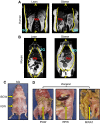
Fig. 1.
Fig. 1.
The anatomical distributions of fat. (A,B) Coronal MRI of a lean (left) and an obese (right) human (A) and mouse (B); yellow lines delineate subcutaneous and visceral depots. A, anterior; P, posterior; SQ, subcutaneous; Vis, visceral. (C,D) Physical orientation, distribution and location of subcutaneous and visceral depots. Subcutaneous fat (C) is located in the inguinal (IGW) and interscapular (ISCW) regions. Visceral fat (D) is positioned at perigonadal (PGW), retroperitoneal (RPW) and mesenteric (MWAT) regions of the body. Yellow arrows indicate the denoted depot location within the body of the mouse.
Fig. 2.
Fig. 2.
Heterogeneity of fat. (A-C) Adipose depots are positioned at stereotypic times and locations with defined sizes; shown are subcutaneous (SQ) fat depots (A), perigonadal (visceral) fat depots (B) and intrascapular brown adipose tissue (C) from three unrelated mice. Histologically, white adipose depots differ in adipocyte size; subcutaneous depots are more heterogeneous, containing both uni- and multilocular adipocytes and interstitial tissue, whereas visceral depots are more uniform and contain mostly unilocular adipocytes. Morphologically, brown adipose tissue is very distinct from white adipose tissue with uniformity of multilocular lipid droplets.
Fig. 3.
Fig. 3.
From the vascular niche to adipocyte. (A) Illustration conceptualizing the adipose vascular niche depicting blood vessels (red), adipose stem cells (green with green nuclei), mural cells (green with black nuclei) and mature adipocytes (yellow with green nuclei). (B) Mouse subcutaneous adipose tissue stained with Hematoxylin and Eosin (H&E) stain. (C) Immunofluorescence image of mouse subcutaneous adipose tissue section showing the expression of GFP (marking adipose stem cells, green), PECAM (an endothelial marker, red) and smooth muscle actin (SMA) (a mural cell marker, blue). (D) Stromal vascular particulates (SVPs) were isolated from mouse subcutaneous fat depots and immunostained for GFP (adipose stem cell, green), PECAM (endothelial marker, red) and SMA (mural cell marker, blue). Yellow arrows indicate adipose stem cells (green) residing at the vascular interface.
Fig. 4.
Fig. 4.
Fat expansion. (A) Isolated postnatal day (P)2 and P30 subcutaneous (inguinal) fat depots, demonstrating the extensive expansion that occurs within this short window of time. (B) Adipo-trak mice (Tang et al., 2008) were treated with placebo (-Dox) or doxycycline (+Dox) at denoted times. In the absence of doxycycline the GFP marker is expressed and the adipose depots are green. In the presence of doxycycline, all GFP signal is lost, indicating that there was a massive recruitment of new stem cells and adipocyte replacement. (C) White adipose depots enlarge with age; subcutaneous and visceral fat depots were isolated from 30-, 90- and 180-day-old mice. Depot weight nearly quintupled between 30 and 180 days. (D) Subcutaneous inguinal (SQ) and visceral perigonadal depots obtained from mice on a standard diet (chow) or a high fat diet (HFD), demonstrating the dramatic expansion in response to HFD.
Fig. 5.
Fig. 5.
Manipulating the adipose stem cell: kill the stem, kill the tree. (A) Illustration conceptualizing the importance of adipose stem cells to the formation of adipose tissue. This illustration demonstrates the extensive branching of the vascular network and shows that adipose stem cells (green nuclei) and mural cells (black nuclei) reside on the vasculature. Adipose stem cells then transition to a multilocular progenitor giving rise to the unilocular adipocyte. Inset: Histological image of subcutaneous fat from a wild-type mouse. (B) Disrupting adipose stem cells by activating WNT signaling results in a fate change that promotes the formation of fibrotic tissue (purple). Inset: Histological image of subcutaneous fat from a mouse with activated WNT signaling.

References

    1. Akazawa S., Sun F., Ito M., Kawasaki E., Eguchi K. (2000). Efficacy of troglitazone on body fat distribution in type 2 diabetes. Diabetes Care 23, 1067–1071 - PubMed
    1. Atit R., Sgaier S. K., Mohamed O. A., Taketo M. M., Dufort D., Joyner A. L., Niswander L., Conlon R. A. (2006). Beta-catenin activation is necessary and sufficient to specify the dorsal dermal fate in the mouse. Dev. Biol. 296, 164–176 - PubMed
    1. Baglioni S., Cantini G., Poli G., Francalanci M., Squecco R., Di Franco A., Borgogni E., Frontera S., Nesi G., Liotta F., et al. (2012). Functional differences in visceral and subcutaneous fat pads originate from differences in the adipose stem cell. PLoS ONE 7, e36569 - PMC - PubMed
    1. Barak Y., Nelson M. C., Ong E. S., Jones Y. Z., Ruiz-Lozano P., Chien K. R., Koder A., Evans R. M. (1999). PPAR gamma is required for placental, cardiac, and adipose tissue development. Mol. Cell 4, 585–595 - PubMed
    1. Berry R., Rodeheffer M. S. (2013). Characterization of the adipocyte cellular lineage in vivo. Nat. Cell Biol. 15, 302–308 - PMC - PubMed

Publication types