. 2013 Dec 15;126(Pt 24):5541-52.
doi: 10.1242/jcs.115972. Epub 2013 Sep 17.

eNOS-derived nitric oxide regulates endothelial barrier function through VE-cadherin and Rho GTPases

Affiliations

eNOS-derived nitric oxide regulates endothelial barrier function through VE-cadherin and Rho GTPases

Annarita Di Lorenzo et al. J Cell Sci. .

Erratum in

  • J Cell Sci. 2014 May 1;127(Pt 9):2120

Abstract

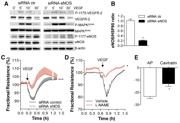
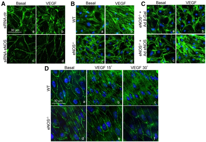
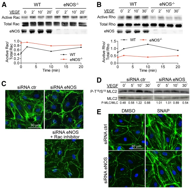
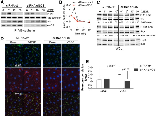
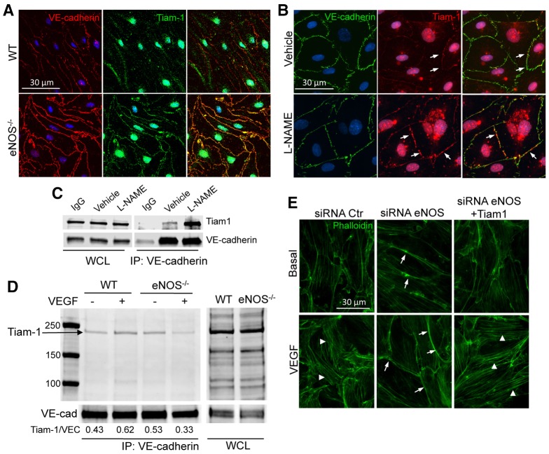
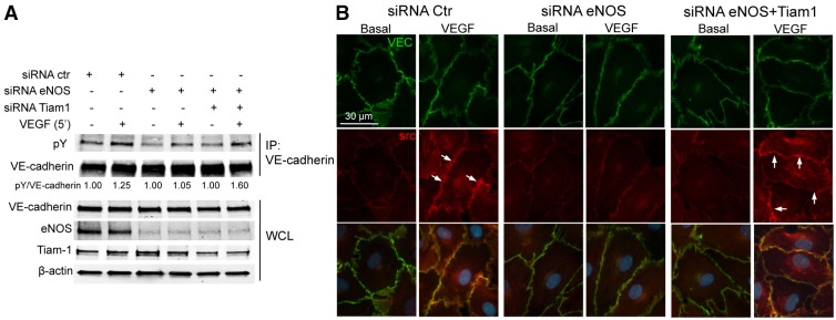
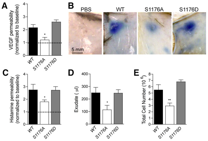
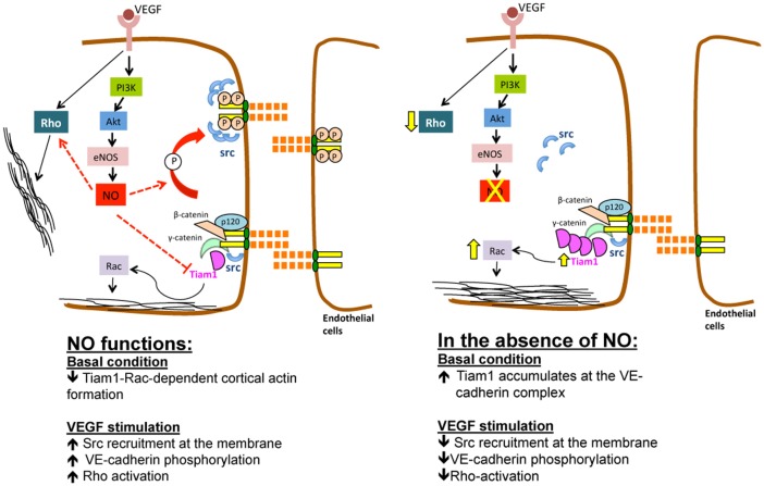
Transient disruption of endothelial adherens junctions and cytoskeletal remodeling are responsible for increases in vascular permeability induced by inflammatory stimuli and vascular endothelial growth factor (VEGF). Nitric oxide (NO) produced by endothelial NO synthase (eNOS) is crucial for VEGF-induced changes in permeability in vivo; however, the molecular mechanism by which endogenous NO modulates endothelial permeability is not clear. Here, we show that the lack of eNOS reduces VEGF-induced permeability, an effect mediated by enhanced activation of the Rac GTPase and stabilization of cortical actin. The loss of NO increased the recruitment of the Rac guanine-nucleotide-exchange factor (GEF) TIAM1 to adherens junctions and VE-cadherin (also known as cadherin 5), and reduced Rho activation and stress fiber formation. In addition, NO deficiency reduced VEGF-induced VE-cadherin phosphorylation and impaired the localization, but not the activation, of c-Src to cell junctions. The physiological role of eNOS activation is clear given that VEGF-, histamine- and inflammation-induced vascular permeability is reduced in mice bearing a non-phosphorylatable knock-in mutation of the key eNOS phosphorylation site S1176. Thus, NO is crucial for Rho GTPase-dependent regulation of cytoskeletal architecture leading to reversible changes in vascular permeability.

Keywords: Cadherin 5; Cytoskeleton; Nitric oxide; Src; VE-cadherin; VEGF; eNOS.


Figures

Fig. 1.
Fig. 1.
eNOS activity is crucial for VEGF-induced permeability in HDEMCs. (A) HDMECs with control or eNOS siRNA were stimulated with vehicle (control, 0 minutes) or VEGF (100 ng/ml) for 5, 10 and 30 minutes and the cell lysates were analyzed by western blotting for eNOS, P-eNOS1177, P-1175-VEGFR-2, VEGFR-2, P-MAPK42/44, MAPK42/44 (where P indicates the phosphorylated form). (B) Quantification of eNOS levels in HDEMCs by densitometry from four independent experiments and expressed relative to the amount of β-actin. The data shown represent the mean±s.e.m. from five individual experiments. *P<0.05 compared to HDEMCs with siRNA control. (C) TEER measurements of post-confluent HDEMCs transfected with control (black line) or eNOS (red line) siRNA expressed as fractional resistance (%) of the TEER basal values. HDEMCs were stimulated with VEGF (100 ng/ml; addition indicated by the arrow) and TEER measured overtime. *P<0.05 compared to HDEMCs with control siRNA. (D) TEER measurements of HDEMCs treated with L-NAME (1 mM) or vehicle followed by VEGF (100 ng/ml) stimulation. *P<0.05 compared to vehicle-treated HDEMCs. (E) TEER measurements of HDEMCs treated with the eNOS inhibitor cavtratin peptide (10 µM) or the peptide control (antennapedia internalization peptide, AP) for 1 hour, measured after VEGF stimulation for 5 minutes. *P<0.05 compared to AP-treated cells (n = 3). The data shown represent the mean±s.e.m. from three individual experiments.
Fig. 2.
Fig. 2.
The loss of eNOS prevents VEGF-induced stress fiber formation in ECs in vitro and ECs of intrapulmonary arteries in situ. (A) HDEMCs with control or eNOS siRNA were stimulated with vehicle (basal) or VEGF (100 ng/ml) for 30 minutes, then fixed and stained with phalloidin to label F-actin (green) and nuclei (blue). (B) WT and eNOS−/− MLECs were stimulated with vehicle or VEGF (100 ng/ml) for 30 minutes and labeled as in A. (C) eNOS−/− MLECs were transduced with adenovirus encoding either control lac Z (Ad β-gal) or eNOS (Ad eNOS) at 100 MOI for 3 days before stimulation with vehicle or VEGF (100 ng/ml) and immunolabeled similarly to in A. These figures are representative of three separate experiments. (D) Intrapulmonary arteries isolated from WT or eNOS−/− mice were stimulated with vehicle or VEGF (100 ng/ml) for 15 or 30 minutes. Blood vessels were then fixed, cut open longitudinally and immunolabeled with phalloidin as described in A. These figures are representative of five animals from each group.
Fig. 3.
Fig. 3.
eNOS deficiency enhances Rac activity while reducing Rho activity. (A) WT and eNOS−/− MLECs were stimulated with VEGF (100 ng/ml) for the indicated times and Rac activity was measured as described in the Materials and Methods. Total Rac and eNOS levels were determined in the whole cell lysates before GST–PAK incubation. The graph shows the densitometric ratio of active Rac to total Rac. The data are representative of three independent experiments. (B) WT and eNOS−/− MLEC were stimulated with VEGF (100 ng/ml) for the indicated times and cell lysates processed for Rho activity. Total Rho and eNOS levels were determined in the whole cell lysates before GST–Rhotekin incubation. The graph shows the densitometric of active Rho to total Rho. The data are representative of three independent experiments. (C) HDEMCs with control or eNOS siRNA were pre-incubated with Rac inhibitor (25 µM, 6 hours) and stimulated with VEGF (100 ng/ml) following immunolabeling of F-actin (green) and nuclei (blue). (D) Western blotting for P-T18/19 MLC2 and MLC2 in whole cell lysates from HDEMCs with eNOS or control siRNA stimulated with VEGF (100 ng/ml) for the indicated times. The data represent three independent experiments. (E) HDEMCs with eNOS or control siRNA were stimulated with vehicle or SNAP, an NO donor (100 µM, 1 hour) and immunolabeled for F-actin (green) and nuclei (blue). Images are representative of three experiments.
Fig. 4.
Fig. 4.
eNOS is required for VEGF-driven c-Src-translocation to cellular junctions and VE-cadherin phosphorylation. (A) HDEMCs transfected with eNOS or control siRNA were serum starved and stimulated with VEGF for the indicated times. VE-cadherin immunoprecipitates were analyzed by immunoblotting against phosphorylated tyrosine (4G10, PY20 clone), VE-cadherin and also for co-immunoprecipitation of c-Src and eNOS proteins. (B) The amount of tyrosine-phosphorylated VE-cadherin was quantified by densitometry from four independent experiments and expressed as fold increase compared with untreated controls. (C) Western blot analysis on whole cell lysates (WCL) from HDEMCs treated as described above. The activation of additional pathways (P-419-Src, Src, P-861-FAK, FAK, P-p38, p38; where P indicates the phosphorylated form) was examined after VEGF (100 ng/ml) stimulation. Densitometric quantification of phosphorylated:total for each protein is shown below the blot. (D) Immunofluorescent co-staining of VE-cadherin (green) and c-Src (red) in HDEMCs transfected with eNOS or control siRNA, then serum starved and stimulated with VEGF (100 ng/ml) for 15 minutes. Scale bar: 20 µm. The images have been captured at a 0.3 µm slice thickness (z-stack) by using a Zeiss Axiovert epifluorescence microscope and a 63× oil immersion objective, following deconvolution by Openlab software (Improvision, Lexington, MA). Data are representative of at least three experiments. Scale bar: 30 µm. (E) Pearson's correlation analysis of VE-cadherin and Src colocalization to the plasma membrane.
Fig. 5.
Fig. 5.
NO regulates VEGF-induced F-actin formation through TIAM1 localization to cellular junctions. (A) WT and eNOS−/− MLECs were plated onto glass coverslips, and were treated with 0.5% Triton X-100-containing buffer for 5 minutes on ice. The soluble fraction was removed and the ECs were gently washed with PBS before fixation with paraformaldehyde 4% and immunofluorescent staining of VE-cadherin (red) and TIAM1 (green). (B) Immunofluorescent co-staining of VE-cadherin (green) and TIAM1 (red) in HDEMCs plated on glass-bottomed dishes (MatTek) transfected with eNOS siRNA or control siRNA and treated with 0.5% Triton X-100-containing buffer for 5 minutes on ice. The arrows show TIAM1 at the junctions. (C) HDEMCs were treated with L-NAME or vehicle, and VE-cadherin was immunoprecipitated from the cell lysates. Western blotting for TIAM1 and VE-cadherin was carried out on the whole cell lysates (WCL) and on the immunoprecipitates (IP). (D) Basal and VEGF-stimulated primary WT and eNOS−/− MLEC lysates were subjected to immunoprecipitation of VE-cadherin and immunoprecipitates were analyzed by immunoblotting against TIAM1. The amount of TIAM1 co-immunoprecipitated was quantified and is reported as a ratio to VE-cadherin (VEC). (E) Immunofluorescent staining of F-actin (green) in HDEMCs transfected with eNOS siRNA or control siRNA, or co-transfected with eNOS siRNA and TIAM1 siRNA, serum starved and stimulated with VEGF (100 ng/ml) for 30 minutes. Arrows indicate cortical actin; arrowheads indicate stress fibers formed upon VEGF stimulation. The images have been captured with Zeiss Axiovert epifluorescence microscope and a 63× oil immersion objective, using Openlab software (Improvision, Lexington, MA). Data are representative of three independent experiments.
Fig. 6.
Fig. 6.
Partial knockdown of TIAM1, in eNOS-deficient cells, restores the phosphorylation of VE-cadherin and the recruitment of c-Src to cellular junctions upon VEGF stimulation. (A) HDEMCs transfected with siRNA control, or siRNA against eNOS or eNOS and TIAM1 were serum starved and stimulated with VEGF (100 ng/ml) for 10 minutes. VE-cadherin was immunoprecipitated and the associated proteins were analyzed by immunoblotting against phosphorylated tyrosine (pY; 4G10, PY20 clone) and VE-cadherin. The whole cell lysates (WCL) were analyzed by western blotting for VE-cadherin, eNOS, TIAM1 and β-actin expression before the incubation with anti-VE-cadherin antibody. (B) HDEMCs transfected with eNOS siRNA or control siRNA, or with eNOS siRNA and TIAM1 siRNA, were co-labeled for VE-cadherin (green) and c-Src (red) after before and incubation with VEGF (100 ng/ml) for 15 minutes. The images have been captured with a Zeiss Axiovert epifluorescence microscope and a 63× oil immersion objective, using Openlab software (Improvision, Lexington, MA). Arrows depict c-Src recruitment to the plasma membrane, which colocalizes with VE-cadherin. Data are representative of three independent experiments.
Fig. 7.
Fig. 7.
The phosphorylation of eNOS on S1176 is crucial for vascular permeability and inflammation in vivo. (A) VEGF-induced dermal vascular permeability was reduced in S1176A eNOS (loss of function) mice compared to WT and S1176D eNOS (gain of function) mice. The data are plotted as the VEGF:PBS ratio of individual mice. *P<0.05; n = 5 mice per group. (B) Representative images of Evan's blue leakage upon PBS or VEGF injection of the mice described in A. (C) Histamine-induced dermal vascular permeability was reduced in S1176A eNOS mice compared to WT and S1176D eNOS mice. (D,E) S1176A eNOS mice displayed a significant reduction of carrageenan-induced (D) exudate formation and (E) neutrophil recruitment into the air pouches 4 hours post-instillation compared to WT and S1176D eNOS mice (n = 5, per group). The data shown represent the mean±s.e.m. *P<0.05, **P<0.01 compared to WT group.
Fig. 8.
Fig. 8.
Schematic view of VEGF-induced NO regulation of the VE-cadherin complex. In basal conditions, the accumulation of TIAM1 at the VE-cadherin complex in basal conditions is limited by eNOS-derived NO (left panel), while VEGF-induced Rho activation and Src-dependent VE-cadherin phosphorylation are promoted by the NO. The lack of NO increases the amount of VE-cadherin-bound TIAM1, leading to the formation of stable cortical actin in basal and VEGF-stimulated conditions (right panel), reduces stress fiber formation and VE-cadherin phosphorylation, with consequent improvement of endothelial barrier function.

References

    1. Ackah E., Yu J., Zoellner S., Iwakiri Y., Skurk C., Shibata R., Ouchi N., Easton R. M., Galasso G., Birnbaum M. J. et al.(2005). Akt1/protein kinase Balpha is critical for ischemic and VEGF-mediated angiogenesis. J. Clin. Invest. 115, 2119–2127 10.1172/JCI24726 - DOI - PMC - PubMed
    1. Amano M., Ito M., Kimura K., Fukata Y., Chihara K., Nakano T., Matsuura Y., Kaibuchi K. (1996). Phosphorylation and activation of myosin by Rho-associated kinase (Rho-kinase). J. Biol. Chem. 271, 20246–20249 10.1074/jbc.271.34.20246 - DOI - PubMed
    1. Andriopoulou P., Navarro P., Zanetti A., Lampugnani M. G., Dejana E. (1999). Histamine induces tyrosine phosphorylation of endothelial cell-to-cell adherens junctions. Arterioscler. Thromb. Vasc. Biol. 19, 2286–2297 10.1161/01.ATV.19.10.2286 - DOI - PubMed
    1. Angelini D. J., Hyun S. W., Grigoryev D. N., Garg P., Gong P., Singh I. S., Passaniti A., Hasday J. D., Goldblum S. E. (2006). TNF-alpha increases tyrosine phosphorylation of vascular endothelial cadherin and opens the paracellular pathway through fyn activation in human lung endothelia. Am. J. Physiol. 291, L1232–L1245 10.1152/ajplung.00109.2006 - DOI - PubMed
    1. Aramoto H., Breslin J. W., Pappas P. J., Hobson R. W., 2nd, Durán W. N. (2004). Vascular endothelial growth factor stimulates differential signaling pathways in in vivo microcirculation. Am. J. Physiol. 287, H1590–H1598 10.1152/ajpheart.00767.2003 - DOI - PubMed

Publication types

MeSH terms