. 2013 Sep 6:4:350.
doi: 10.3389/fpls.2013.00350. eCollection 2013.

New insights into Fe localization in plant tissues

Affiliations

New insights into Fe localization in plant tissues

Hannetz Roschzttardtz et al. Front Plant Sci. .

Abstract

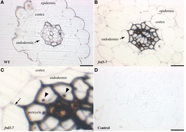
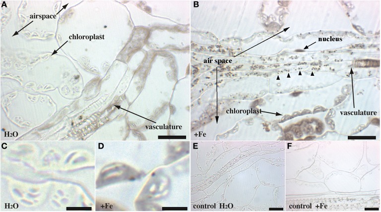
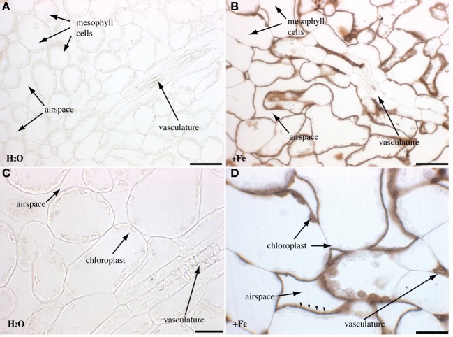
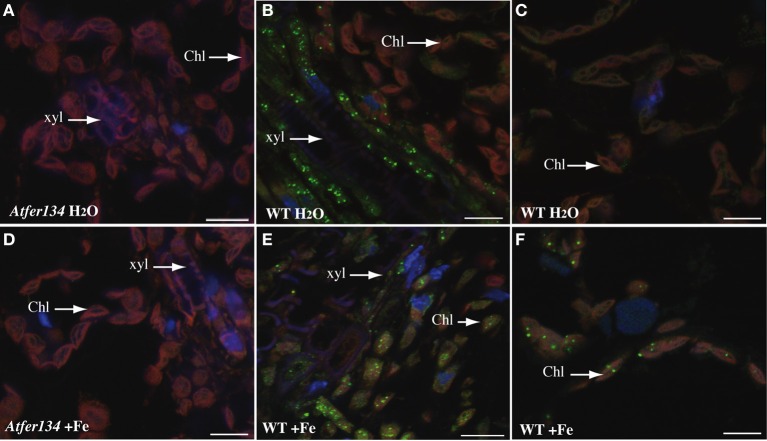
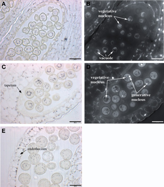
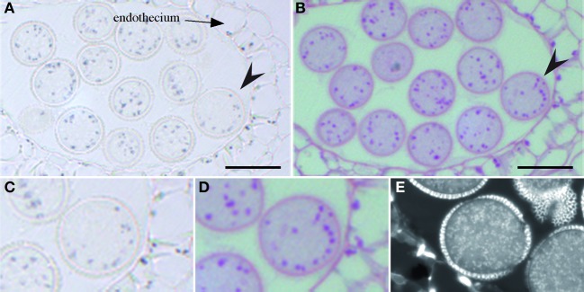
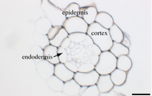
Deciphering cellular iron (Fe) homeostasis requires having access to both quantitative and qualitative information on the subcellular pools of Fe in tissues and their dynamics within the cells. We have taken advantage of the Perls/DAB Fe staining procedure to perform a systematic analysis of Fe distribution in roots, leaves and reproductive organs of the model plant Arabidopsis thaliana, using wild-type and mutant genotypes affected in iron transport and storage. Roots of soil-grown plants accumulate iron in the apoplast of the central cylinder, a pattern that is strongly intensified when the citrate effluxer FRD3 is not functional, thus stressing the importance of citrate in the apoplastic movement of Fe. In leaves, Fe level is low and only detected in and around vascular tissues. In contrast, Fe staining in leaves of iron-treated plants extends in the surrounding mesophyll cells where Fe deposits, likely corresponding to Fe-ferritin complexes, accumulate in the chloroplasts. The loss of ferritins in the fer1,3,4 triple mutant provoked a massive accumulation of Fe in the apoplastic space, suggesting that in the absence of iron buffering in the chloroplast, cells activate iron efflux and/or repress iron influx to limit the amount of iron in the cell. In flowers, Perls/DAB staining has revealed a major sink for Fe in the anthers. In particular, developing pollen grains accumulate detectable amounts of Fe in small-size intracellular bodies that aggregate around the vegetative nucleus at the binuclear stage and that were identified as amyloplasts. In conclusion, using the Perls/DAB procedure combined to selected mutant genotypes, this study has established a reliable atlas of Fe distribution in the main Arabidopsis organs, proving and refining long-assumed intracellular locations and uncovering new ones. This "iron map" of Arabidopsis will serve as a basis for future studies of possible actors of iron movement in plant tissues and cell compartments.

Keywords: Arabidopsis; amyloplast; chloroplast; ferritin; iron; mitochondria; pollen; root.


Figures

Figure 1
Figure 1
Iron detection in Arabidopsis roots. Sections of 4-week old WT (A) or frd3-7 (B–D) mutant plants stained with Perls/DAB. Panel (C) corresponds to a magnified image of panel (B), both showing iron restriction within the boundary of the Casparian strip (arrow), nucleoli are indicated by arrowheads. (D), control section stained with DAB without previous Perls reaction. Scale bars: 50 μm (A,B,D) or 25 μm (C).
Figure 2
Figure 2
Iron excess in Arabidopsis rosette leaves. Sections of 4-week old WT plants irrigated with either water (A,C,E) or Fe-EDDHA 2 mM during 48 h (B,D,F) were stained with either Perls/DAB (A–D) or DAB alone as a negative control (E,F). The panels (C,D) correspond to a magnified image of regions of panels (A,B), respectively. Arrowheads in panel (B) indicate Fe-rich structures in the vascular tissues. Scale bars: 20 μm (A,B,E,F) or 5 μm (C,D).
Figure 3
Figure 3
Iron excess in rosette leaves of the Atfer134 triple mutant. Sections of 4-week old Atfer134 plants irrigated with either water (A,C) or Fe-EDDHA 2 mM during 48 h (B,D) were stained with either Perls/DAB (A,B,D) or DAB alone as a negative control. (C) Fe accumulation in the extracellular space is indicated by arrowheads in panel (D). Scale bars: 50 μm (A,B) or 20 μm (C,D).
Figure 4
Figure 4
Immunolocalization of Ferritin in Arabidopsis leaves. Sections of rosette leaves from 3 week-old WT (B,C,E,F) or triple mutant Atfer1,3,4 (A,D) plants irrigated with either water (A–C) or 2 mM Fe-EDDHA (D–F) were probed with an anti-ferritin antibody and revealed with a secondary anti-rabbit antibody coupled to the Alexa Fluor® 488 fluorophore. Sections were stained with DAPI to reveal cell nuclei. Ferritin localization appears in green, DAPI fluorescence in blue and chlorophyll auto-fluorescence in red. Scale bars: 10 μm.
Figure 5
Figure 5
Arabidopsis flowers stained with Perls/DAB. (A) Open mature flowers, (B) stigmatic papillae with sticking pollen grains, (C) anther with mature pollen grains.
Figure 6
Figure 6
Iron distribution during Arabidopsis pollen development. Sections of anthers at three different stages of pollen development were stained with Perls/DAB and DAPI. (A,B) mononuclear pollen grain, (C,D) binuclear pollen grain, (E) mature pollen grain. (A,C,E) Bright field images; (B,D) Epifluorescence images from slides (A,C), respectively, showing DAPI-stained vegetative and generative nuclei. Scale bars: 20 μm.
Figure 7
Figure 7
Iron co-localizes with starch in pollen. Serial sections of the same anther were stained with either Perls/DAB (A,C), periodic acid-Schiff (B,D) or DiOC6 (E). The arrow heads in (A,B) indicate the pollen grain that is magnified in (C,D), respectively. Scale bars: 20 μm.
Figure A1
Figure A1
Iron detection in Arabidopsis roots from plants grown in plate. Three-week old wild-type plants grown in MS/2 medium supplemented with 50 μM Fe-EDTA were embedded in Technovit resin and the sections were stained with Perls/DAB. The scale bar represents 50 μm.

References

    1. Bienfait H. F., Briel W. V. D., Mesland-Mul N. T. (1985). Free space iron pools in roots: generation and mobilization. Plant Physiol. 78, 596–600 10.1104/pp.78.3.596 - DOI - PMC - PubMed
    1. Branton D., Jacobson L. (1962). Iron transport in pea plants. Plant Physiol. 37, 539–545 10.1104/pp.37.4.539 - DOI - PMC - PubMed
    1. Briat J. F., Lebrun M. (1999). Plant responses to metal toxicity. C. R. Acad. Sci. III, Sci. Vie 322, 43–54 10.1016/S0764-4469(99)80016-X - DOI - PubMed
    1. Briat J. F., Lobreaux S. (1998). Iron storage and ferritin in plants. Met. Ions Biol. Syst. 35, 563–584 - PubMed
    1. Connolly E. L., Campbell N. H., Grotz N., Prichard C. L., Guerinot M. L. (2003). Overexpression of the FRO2 ferric chelate reductase confers tolerance to growth on low iron and uncovers posttranscriptional control. Plant Physiol. 133, 1102–1110 10.1104/pp.103.025122 - DOI - PMC - PubMed