

Branchial cleft cyst
- PMID: 24046795
- PMCID: PMC3774901
- DOI: 10.1007/s40477-013-0004-2
Branchial cleft cyst
Abstract
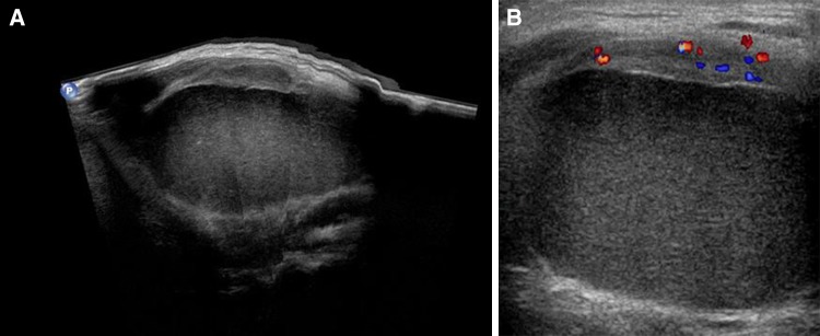
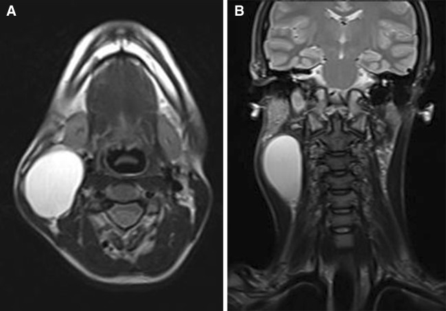
Branchial cleft cysts are benign lesions caused by anomalous development of the brachial cleft. This report describes a 20-year-old girl with swelling on the right lateral aspect of the neck, which expanded slowly but progressively. The clinical suspicion was that of a branchial cleft cyst. Sonography revealed a homogeneously hypo- to anechoic mass with well-defined margins and no intralesional septa. Color Doppler reviewed no internal vascularization. The ultrasound examination confirmed the clinical diagnosis of a second branchial cleft cyst, demonstrating the cystic nature of the mass and excluding the presence of complications. For superficial lesions like these, ultrasound is the first-level imaging study of choice because it is non-invasive, rapid, low-cost, and does not involve exposure to ionizing radiation.
Le cisti branchiali sono lesioni benigne dovute ad anomalie di sviluppo degli archi branchiali. Viene presentato il caso di una Paziente, 20 anni, con tumefazione laterocervicale a lenta e progressiva crescita, sospetta clinicamente per cisti branchiale. Ecograficamente la lesione mostrava struttura omogenea ipo- anecogena, con margini ben definiti, priva di setti intralesionali e di patologica vascolarizzazione alla valutazione color-Doppler. L’ecografia confermava quindi il sospetto clinico di cisti del II arco branchiale, dimostrando la sua natura cistica ed escludendo possibili complicanze. Trattandosi di lesioni superficiali l’ecografia rappresenta la metodica di imaging di primo livello essendo non invasiva, di rapida esecuzione, poco costosa, non esponendo il paziente a radiazioni ionizzanti.
Keywords: Neck mass; Second branchial cleft cyst; Ultrasonography.
Figures

References
-
- Thomaidis V, Seretis K, Tamiolakis D, Papadopoulos N, Tsamis I. Branchial cysts. A report of 4 cases. Acta Dermatovenerol Alp Panonica Adriat. 2006;15(2):85–89. - PubMed
-
- Mitroi M, Dumitrescu D, Simionescu C, Popescu C, Mogoanta C, Cioroianu L, et al. Management of second branchial cleft anomalies. Rom J Morphol Embryol. 2008;49(1):69–74. - PubMed
LinkOut - more resources
Full Text Sources
Other Literature Sources
