

Uterine arteriovenous malformation
- PMID: 24046800
- PMCID: PMC3774899
- DOI: 10.1007/s40477-013-0007-z
Uterine arteriovenous malformation
Abstract
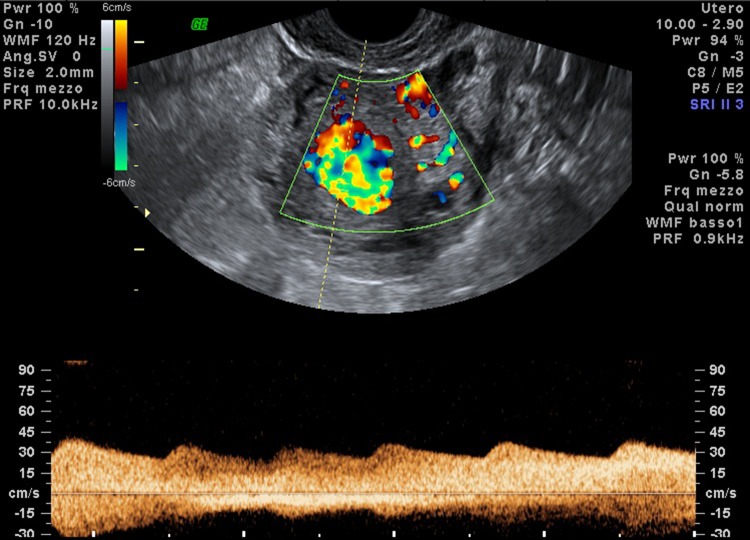
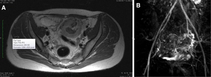
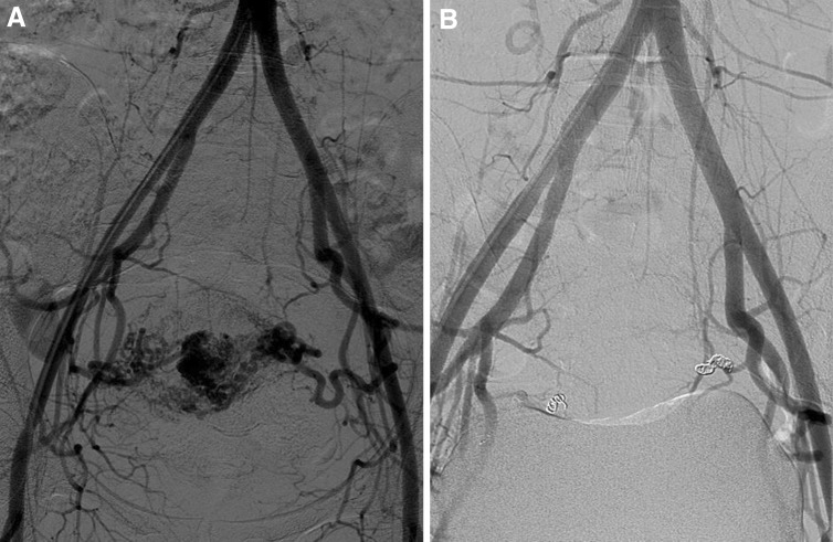
Uterine arteriovenous vascular malformations (UAVM) are uncommon vascular diseases, occurring during reproductive age. Patients affected by UAMVs usually present with recurrent pregnancy loss or menorrhagia. Initial evaluation of UAVMs is made with ultrasonography (US) and US-Doppler. Magnetic resonance is used when a UAMV is suspected at US. Treatment can be surgical (hysterectomy or surgical removal of AVM), or with selective uterine arterial embolization. We report a case of UAMV, from its clinical signs to diagnostic confirmation and subsequent treatment.
Le malformazioni arterovenose dell’utero (MAVU) sono disordini vascolari poco comuni, che spesso si presentano in donne in età fertile; si manifestano come aborti ricorrenti o con menorragia. La valutazione iniziale viene eseguita con l’ecografia e eco colour-Doppler. La risonanza magnetica è utilizzata in caso di sospetto ecografico di MAVU. Il trattamento può essere chirurgico o mediante embolizzazione delle arterie uterine. In questo articolo è riportato un caso di MAVU, dal sospetto clinico alla conferma diagnostica e al trattamento effettivo.
Keywords: Embolization; MRI; US-Doppler; Uterine arteriovenous malformation; Uterine artery.
Figures
References
-
- Polat P, Suma S, Kantarcý M, Alper F, Levent A. Color Doppler US in the evaluation of uterine vascular abnormalities. Radiographics. 2002;22(1):47–53. - PubMed
-
- Dubreuil G, Loubat E. Aneurysme cirsoide de l’uterus. Ann Anat Pathol. 1926;3:697–718.
LinkOut - more resources
Full Text Sources
Other Literature Sources
