. 2013 Sep 18:11:67.
doi: 10.1186/1478-811X-11-67.

Direct transdifferentiation of spermatogonial stem cells to morphological, phenotypic and functional hepatocyte-like cells via the ERK1/2 and Smad2/3 signaling pathways and the inactivation of cyclin A, cyclin B and cyclin E

Affiliations

Direct transdifferentiation of spermatogonial stem cells to morphological, phenotypic and functional hepatocyte-like cells via the ERK1/2 and Smad2/3 signaling pathways and the inactivation of cyclin A, cyclin B and cyclin E

Zhenzhen Zhang et al. Cell Commun Signal. .

Abstract

Background: Severe shortage of liver donors and hepatocytes highlights urgent requirement of extra-liver and stem cell source of hepatocytes for treating liver-related diseases. Here we hypothesized that spermatogonial stem cells (SSCs) can directly transdifferentiate to hepatic stem-like cells capable of differentiating into mature hepatocyte-like cells in vitro without an intervening pluripotent state.

Results: SSCs first changed into hepatic stem-like cells since they resembled hepatic oval cells in morphology and expressed Ck8, Ck18, Ck7, Ck19, OV6, and albumin. Importantly, they co-expressed CK8 and CK19 but not ES cell markers. Hepatic stem-like cells derived from SSCs could differentiate into small hepatocytes based upon their morphological features and expression of numerous hepatic cell markers but lacking of bile epithelial cell hallmarks. Small hepatocytes were further coaxed to differentiate into mature hepatocyte-like cells, as identified by their morphological traits and strong expression of Ck8, Ck18, Cyp7a1, Hnf3b, Alb, Tat, Ttr, albumin, and CYP1A2 but not Ck7 or CK19. Notably, these differentiated cells acquired functional attributes of hepatocyte-like cells because they secreted albumin, synthesized urea, and uptake and released indocyanine green. Moreover, phosphorylation of ERK1/2 and Smad2/3 rather than Akt was activated in hepatic stem cells and mature hepatocytes. Additionally, cyclin A, cyclin B and cyclin E transcripts and proteins but not cyclin D1 or CDK1 and CDK2 transcripts or proteins were reduced in mature hepatocyte-like cells or hepatic stem-like cells derived from SSCs compared to SSCs.

Conclusions: SSCs can transdifferentiate to hepatic stem-like cells capable of differentiating into cells with morphological, phenotypic and functional characteristics of mature hepatocytes via the activation of ERK1/2 and Smad2/3 signaling pathways and the inactivation of cyclin A, cyclin B and cyclin E. This study thus provides an invaluable source of mature hepatocytes for treating liver-related diseases and drug toxicity screening and offers novel insights into mechanisms of liver development and cell reprogramming.


Figures

Figure 1
Figure 1
A novel protocol for transdifferentiation of SSCs to mature hepatocyte-like cells and their morphological features. (A) Schematic diagram showed the procedures for inducing SSCs to transdifferentiate to mature hepatocyte-like cells. (B-E) Morphology was characterized for SSCs (B), hepatic stem-like cells (C), small hepatocytes (D), and mature hepatocyte-like cells (E). Scale bars in B, C, D, and E = 50 μm. (F-G) Ultrastructure of hepatic stem cells (F) and mature hepatocyte-like cells (G) from SSCs. Note: mitochondria (Mi), endoplasmic reticulum (Er), lysosome (Ly), Golgi apparatus (Go), and chromatin (Ch), nucleus (Nu). Scale bars in F and G= 2 μm.
Figure 2
Figure 2
Transcriptional characteristics of transdifferentiation of SSCs into hepatic stem-like cells and hepatocyte-like cells. (A) RT-PCR revealed mRNA expression of Ck8, Ck18, Ck7, and Ck19 in hepatic stem-like cells derived from SSCs. (B) RT-PCR showed the transcripts of Ck8, Ck18, Cyp1a2, Cyp7a1, Hnf3b, Hnf4a, Alb, Tat, and Ttr in SSCs (lane 1), SSC induction for 7 days (lane 2), SSC induction for 10 days (lane 3), small hepatocytes (lane 4), and mature hepatocyte-like cells (lane 5) derived from SSCs. The expression of these genes in liver tissues of adult mice (lane 6) was used as positive controls. (C) The transcription of Ck8, Ck18, and Ck7 in mature hepatocyte-like cells derived from SSCs. Gapdh was used as loading controls of total RNA. (D) The transcription of Ck18, Ck7 and Ck19 in the cells derived from primary mouse SSCs. Gapdh was used as loading controls of total RNA. (E) RT-PCR showed the transcripts of Ck8, Ck18, Cyp1a2, Cyp7a1, Hnf3b, Hnf4a, Alb, Tat, and Ttr in mature hepatocyte-like cells derived from primary SSCs. Gapdh was used as loading controls of total RNA.
Figure 3
Figure 3
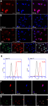
The expression of OV6, CYP1A2, ALB, CK8, and CK19 of hepatic stem-like cells derived from transdifferentiation of SSCs. (A-D) Immunocytochemistry showed protein expression of OV6 (A), CYP1A2 (B), ALB (C), as well as co-expression of CK8 and CK19 (D) in transdifferentiated cells derived from C18-4 cells. Scale bars in A, B, C, and D = 20 μm. (E) Flow cytometry showed expression of CK8 in the transdifferentiated cells derived from C18-4 cells in the conditioned medium without PD98059 (left panel) or with PD98059 (right panel). (F-G) Immunocytochemistry showed protein expression of ALB (F) as well as co-expression of CK8 and CK19 (G) in transdifferentiated cells derived from mouse primary SSCs. Scale bars in F and G = 20 μm.
Figure 4
Figure 4
Translational characterization of small hepatocytes derived from SSCs. (A-C) Immunocytochemistry displayed expression of ALB (A), CYP1A2 (B), as well as co-expression of CK8 and CK19 (C) in small hepatocytes derived from SSCs. Scale bars in A, B, and C = 20 μm.
Figure 5
Figure 5
Characterization of mature hepatocyte-like cells derived from SSCs. (A-C) Immunocytochemistry showed expression of ALB (A), CYP1A2 (B), as well as co-expression of CK8 and CK19 (C) in mature hepatocyte-like cells derived from SSCs. Scale bars in A, B, and C = 50 μm.
Figure 6
Figure 6
Albumin synthesis, urea productions, and cellular uptake and release of ICG of mature hepatocyte-like cells derived from SSCs. (A) ELISA showed albumin synthesis of SSCs (lane 1), mature hepatocyte-like cells derived from SSCs (lane 2), and primary hepatocytes (lane 3). “*” indicated statistically significant differences (p< 0.05) between SSCs and mature hepatocyte-like cells derived from SSCs or primary hepatocytes. (B) Urea assay displayed urea production of SSCs (lane 1), mature hepatocyte-like cells derived from SSCs (lane 2), and primary hepatocytes (lane 3). “*” indicated statistically significant differences (p< 0.05) between SSCs and mature hepatocyte-like cells derived from SSCs or primary hepatocytes. (C-G) Cellular uptake (C) and release (D) of ICG in mature hepatocyte-like cells derived from SSCs, uptake of ICG in SSCs (E), as well as uptake (F) and release (G) of ICG in mouse primary hepatocytes. Scale bars in C-G = 50 μm.
Figure 7
Figure 7
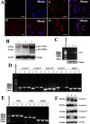
Expression of ERK1/2 phosphorylation, c-fos transcription and cell cycle proteins in SSCs as well as in hepatic stem-like cells and mature hepatocyte-like cells derived from SSCs. (A) Immunocytochemistry revealed the expression of phosph-ERK1/2 in SSCs (Panel i), hepatic stem-like cells derived from SSCs (Panel ii), small hepatocytes derived from SSCs (Panel iii), and mature hepatocyte-like cells derived from SSCs (Panel iv). Scale bars in A = 50 μm. (B) Western blots showed the expression of ERK1/2 phosphorylation in SSCs (lane 1), hepatic stem-like cells derived from SSCs (lane 2), and mature hepatocyte-like cells derived from SSCs (lane 3). The expression of ERK2 was used as a loading control of total proteins. (C) RT-PCR revealed the transcription of c-fos mRNA in SSCs (lane 1), hepatic stem-like cells derived from SSCs (lane 2), and mature hepatocyte-like cells derived from SSCs (lane 3). Housekeeping gene Gapdh served as a loading control of total RNA. (D) RT-PCR displayed mRNA expression of cyclin A, cyclin B, cyclin D1, and cyclin E in SSCs (lane 1), hepatic stem-like cells derived from SSCs (lane 2), and mature hepatocyte-like cells derived from SSCs (lane 3). Gapdh was used as loading control of total RNA. (E) RT-PCR showed transcripts of CDK1 and CDK2 in SSCs (lane 1), hepatic stem-like cells derived from SSCs (lane 2), and mature hepatocyte-like cells derived from SSCs (lane 3). Gapdh served as loading control of total RNA. (F) Western blots revealed the expression of cyclin A, cyclin B, cyclin D1, and cyclin E in SSCs (lane 1), hepatic stem-like cells derived from SSCs (lane 2), and mature hepatocyte-like cells derived from SSCs (lane 3). The expression of ACTB was used as a loading control of total proteins.
Figure 8
Figure 8
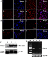
Expression of Smad2-, and Stat3- phosphorylation and Oct-4 in SSCs, hepatic stem-like cells, small hepatocytes, and mature hepatocyte-like cells. (A) Immunocytochemistry revealed expression of Expression of phosph-Smad2 in SSCs (Panel i), hepatic stem-like cells (Panel ii), small hepatocytes (Panel iii), and mature hepatocyte-like cells (Panel iv). (B) Expression of phosph-Stat3 in SSCs (Panel i), hepatic stem-like cells (Panel ii), small hepatocytes (Panel iii), and mature hepatocyte-like cells (Panel iv). Scale bars in A-B = 50 μm. (C) Western blots showed the expression of phosph-Smad2 in SSCs (lane 1), hepatic stem-like cells (lane 2), and mature hepatocyte-like cells (lane 3). (D) RT-PCR revealed Oct-4 expression in SSCs (lane 1), hepatic stem-like cells (lane 2), and mature hepatocyte-like cells (lane 3).
Figure 9
Figure 9
Schematic diagram demonstrated the transdifferentiation of SSCs into mature hepatocyte-like cells and ERK1/2 and Smad2/3 signaling pathways. “P” indicated “phosphorylate”; solid arrow denoted “promote”; dotted arrow indicated “inhibit”; dotted line showed “no change”.

References

    1. Ferlay J, Shin HR, Bray F. et al.Estimates of worldwide burden of cancer in 2008: GLOBOCAN 2008. Int J Cancer. 2010;11:2893–2917. doi: 10.1002/ijc.25516. - DOI - PubMed
    1. Williams R. Global challenges in liver disease. Hepatology. 2006;11:521–526. doi: 10.1002/hep.21347. - DOI - PubMed
    1. Fuster J, Charco R, Llovet JM. et al.Liver transplantation in hepatocellular carcinoma. Transpl Int. 2005;11:278–282. doi: 10.1111/j.1432-2277.2004.00046.x. - DOI - PubMed
    1. Michalopoulos GK, DeFrances MC. Liver regeneration. Science. 1997;11:60–66. doi: 10.1126/science.276.5309.60. - DOI - PubMed
    1. Muraca M, Gerunda G, Neri D. et al.Hepatocyte transplantation as a treatment for glycogen storage disease type 1a. Lancet. 2002;11:317–318. doi: 10.1016/S0140-6736(02)07529-3. - DOI - PubMed

Publication types