. 2013 Nov 1;188(9):1137-46.
doi: 10.1164/rccm.201304-0803OC.

S100A8/A9 proteins mediate neutrophilic inflammation and lung pathology during tuberculosis

Affiliations

S100A8/A9 proteins mediate neutrophilic inflammation and lung pathology during tuberculosis

Radha Gopal et al. Am J Respir Crit Care Med. .

Abstract

Rationale: A hallmark of pulmonary tuberculosis (TB) is the formation of granulomas. However, the immune factors that drive the formation of a protective granuloma during latent TB, and the factors that drive the formation of inflammatory granulomas during active TB, are not well defined.

Objectives: The objective of this study was to identify the underlying immune mechanisms involved in formation of inflammatory granulomas seen during active TB.

Methods: The immune mediators involved in inflammatory granuloma formation during TB were assessed using human samples and experimental models of Mycobacterium tuberculosis infection, using molecular and immunologic techniques.

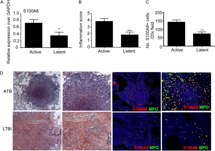
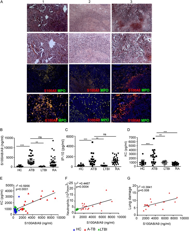
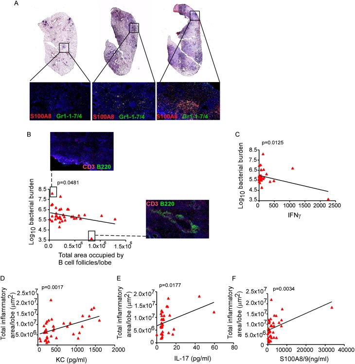
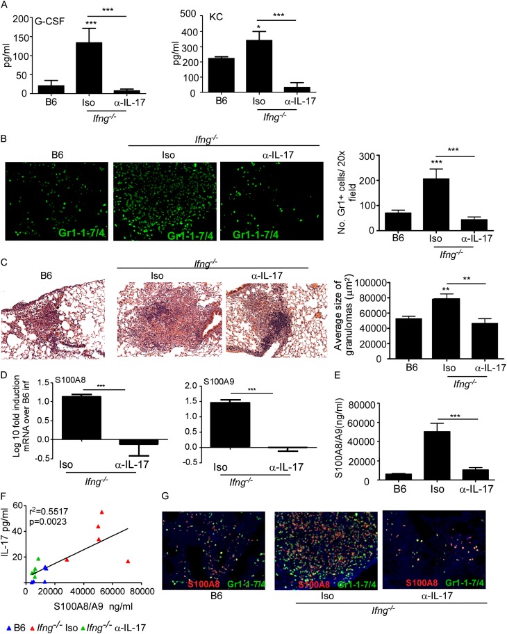
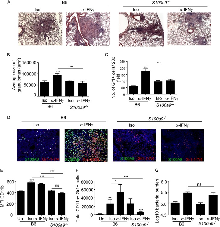
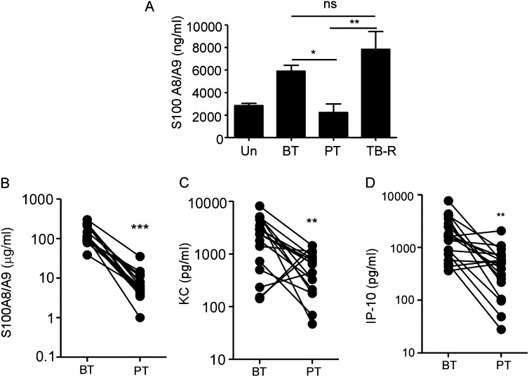
Measurements and main results: We demonstrate that in human patients with active TB and in nonhuman primate models of M. tuberculosis infection, neutrophils producing S100 proteins are dominant within the inflammatory lung granulomas seen during active TB. Using the mouse model of TB, we demonstrate that the exacerbated lung inflammation seen as a result of neutrophilic accumulation is dependent on S100A8/A9 proteins. S100A8/A9 proteins promote neutrophil accumulation by inducing production of proinflammatory chemokines and cytokines, and influencing leukocyte trafficking. Importantly, serum levels of S100A8/A9 proteins along with neutrophil-associated chemokines, such as keratinocyte chemoattractant, can be used as potential surrogate biomarkers to assess lung inflammation and disease severity in human TB.

Conclusions: Our results thus show a major pathologic role for S100A8/A9 proteins in mediating neutrophil accumulation and inflammation associated with TB. Thus, targeting specific molecules, such as S100A8/A9 proteins, has the potential to decrease lung tissue damage without impacting protective immunity against TB.


Figures

<i>Figure 1.</i>
Figure 1.
S100A8/A9 expression coincides with severity of inflammation in nonhuman primates (NHP) with active tuberculosis (ATB). NHPs aerosol infected with Mycobacterium tuberculosis CDC1551 exhibited either latent TB infection (LTBI) or ATB as described under Methods. (A) mRNA expression levels of S100A8 from lung granulomatous lesions obtained from ATB and LTBI NHPs was determined by reverse transcriptase polymerase chain reaction and expression relative to glyceraldehyde phosphate dehydrogenase (GAPDH) expression shown. (B) The inflammation score was determined by a pathologist as mild, moderate, or severe and degree of inflammation annotated as inflammatory score was scored from 1 to 4 (with 4 reflecting maximum pathology) for each NHP sample described. (C) Formalin-fixed paraffin embedded lungs from ATB and LTBI were stained with hematoxylin and eosin or with antibodies specific for S100A8 (red) and MPO (green) and the number of S100A8+ neutrophils counted. (D) A representative image from ATB and LTBI sample is shown (left, ×100 original magnification; right, ×200 original magnification). Staining from an individual NHP shown. The data points represent the mean (±SD) of values from 4–7 NHPs (A–C). *P ≤ 0.05, ***P ≤ 0.0005.
<i>Figure 2.</i>
Figure 2.
S100A8/A9 expression correlates with active disease and lung inflammation in humans with active tuberculosis (ATB). Formalin-fixed paraffin embedded lung sections from patients with ATB (A) were stained with hematoxylin and eosin or with antibodies specific for S100A8 (red) and MPO (green). Three representative human ATB samples (A) are shown (top, hematoxylin and eosin, original magnification ×100; bottom, ×200). Immunohistochemistry, 4 × 4 mosaic (top, ×200 original magnification; bottom, ×200) (A). S100A8/A9 (B), induced protein-10 (IP-10) (C), and keratinocyte chemoattractant (KC) (D) protein levels were measured in serum collected from healthy control subjects (HC), ATB, latent TB infection (LTBI), and patients with rheumatoid arthritis (RA) using ELISA or Luminex assays. Linear correlation analysis of S100A8/9 protein levels and KC (E), S100A8/A9 protein levels and neutrophils in peripheral blood (F), and S100A8/A9 levels and lung damage score (G) in HCs (blue triangles), patients with ATB (red triangles), and patients with LTBI (green triangles) was performed using GraphPad Prism (GraphPad Software, Inc., La Jolla, CA). The data points represent the mean (±SD) of values from 15 HCs, 17 ATB, 8 LTBI, and 33 RA patients (B–G). **P ≤ 0.005, ***P ≤ 0.0005. ns = not significant.
<i>Figure 3.</i>
Figure 3.
Inflammation in Mycobacterium tuberculosis (Mtb) infected genetically diverse Diversity Outbred (DO) mouse population is associated with increased IL-17 production and S100A8/A9 proteins. (A) Genetically diverse mice from DO strain were aerosol infected with approximately 100 CFU of Mtb H37Rv and on Day 60 postinfection, formalin-fixed paraffin embedded (FFPE) lung sections were stained with hematoxylin and eosin or analyzed by immunofluorescence using antibodies specific for S100A8 (red) and Gr1-1-7/4 (green). FFPE lung sections were also analyzed by immunofluorescence using antibodies specific for B220 (green) and CD3 (red), and the total area occupied by B-cell follicles per lobe quantified using the morphometric tool of the Zeiss Axioplan (Carl Zeiss, Oberkochen, Germany) (4 × 4 mosaic, ×200 original magnification). Lung CFU was determined by plating. Linear correlation analysis between total area occupied by B-cell follicles per lung lobe and bacterial burden (B) and lung IFN-γ levels and bacterial burden (C) was performed using GraphPad Prism. A representative image demonstrating absence of B-cell follicles in a mouse exhibiting the highest Mtb bacterial burden and a representative image showing well-formed B-cell follicles in a mouse exhibiting the lowest Mtb burden is also shown (×100 original magnification). Total area occupied by inflammatory lesions per lobe was quantified in the Mtb-infected hematoxylin and eosin–stained FFPE lungs using the morphometric tool of the Zeiss Axioplan microscope. Lung protein levels of keratinocyte chemoattractant (KC) (D), IL-17 (E), and S100A8/A9 proteins (F) were measured and linear regression analysis was determined using GraphPad Prism. The data points represent values from 37 mice (A–F).
<i>Figure 4.</i>
Figure 4.
IL-17–dependent induction of S100A8/A9 proteins in Ifng−/− Mycobacterium tuberculosis (Mtb)-infected mice mediates lung inflammation. B6, Ifng−/− mice were aerosol infected with approximately 100 CFU Mtb H37Rv. Starting at Day 9 postinfection, groups of Ifng−/− infected mice received either isotype control antibody (Iso) or IL-17 neutralizing antibody (α−IL-17) (300 μg/mouse every 48 h) and samples for the below described analyses were harvested on Day 30 postinfection. Induction of granulocyte colony–stimulating factor (G-CSF) and keratinocyte chemoattractant (KC) (A) levels were measured in lung homogenates from infected mice. Formalin-fixed paraffin embedded lung sections were stained with rat antimouse Ly6G/Ly6C and rat antimouse Ly6-B.2 antigen to detect pulmonary neutrophils in mice (Gr1-1-7/4, green) (B) or underwent hematoxylin and eosin staining (C). The number of Gr1+ cells per ×20 field were counted (B) or average size of inflammatory lesions were quantified using the morphometric tool of the Zeiss Axioplan microscope (C) (×100 original magnification for hematoxylin and eosin images; ×200 original magnification for fluorescent images). Log10 fold induction of S100A8 or S100A9 mRNA in B6 and different groups of Ifng−/− Mtb-infected lungs was measured by reverse transcriptase polymerase chain reaction and fold induction in Ifng−/− infected lungs over B6 Mtb-infected lungs is shown (D). Protein levels of S100A8/A9 (E) or IL-17 (F) were measured from lung homogenates by ELISA and linear regression analysis was determined using GraphPad Prism. Red triangles, Ifn−/− Mtb infected isotype control treated mice; green triangles, Ifng−/− Mtb-infected mice treated with IL-17 neutralizing antibody; blue triangles, B6 Mtb-infected mice (F). Formalin-fixed paraffin embedded lung sections from Mtb-infected mice were assayed by immunofluorescence staining for Gr1-1-7/4 (green) and S100A8 (red) (G). The data points represent the mean (±SD) of values from 4–6 mice (A–G). *P ≤ 0.05, **P ≤ 0.005, ***P ≤ 0.0005. One experiment representative of two is shown.
<i>Figure 5.</i>
Figure 5.
S100A8/A9 proteins mediate exacerbated inflammation during Mycobacterium tuberculosis (Mtb) infection. B6 and S100a9−/− mice were infected with approximately 100 CFU Mtb H37Rv. From Day 9 postinfection, B6 and S100a9−/− Mtb-infected mice received either isotype (Iso) or IFN-γ neutralizing antibody (α−IFN-γ) (300 μg/mouse every 48 h) and samples for the analyses below were collected on Day 30 postinfection. Formalin-fixed paraffin embedded lung sections were stained with hematoxylin and eosin (A and B) or analyzed by immunofluorescence using antibodies specific for Gr1-1-7/4 (red) and S100A8 (green) (C and D). Average size of inflammatory lesions (B) was quantified in the Mtb-infected lungs using the morphometric tool of the Zeiss Axioplan microscope or number of Gr1+ cells per ×20 field counted (C) (×100 original magnification for hematoxylin and eosin images; ×200 original magnification for fluorescent images). Single cell suspensions from lungs of Mtb-infected mice were stained with antibodies specific for CD11b and Gr1 and the mean fluorescent intensity (MFI) of CD11b expression (E) and number of lung neutrophils (Gr1+ CD11b+) determined by flow cytometry (F). Lung bacterial burden in Mtb-infected mice was determined by plating (G). The data points represent the mean (±SD) of values from 4–6 mice (A–G). *P ≤ 0.05, **P ≤ 0.005, ***P ≤ 0.0005. ns = not significant. One experiment representative of two is shown.
<i>Figure 6.</i>
Figure 6.
S100A8/A9 protein levels decrease with Mycobacterium tuberculosis clearance and are surrogate indicators of tuberculosis (TB) reactivation. (A) S100A8/A9 lung protein levels in mice left untreated (Un), M. tuberculosis infected before antibiotic therapy (BT) or treated with antibiotic therapy (PT), or allowed to reactivate (TB-R) was determined by ELISA (n = 4–5). Serum S100A8/A9 proteins (B), keratinocyte chemoattractant (KC) (C), and induced protein-10 (IP-10) (D) levels in patients with ATB before treatment (BT) with antibiotics, or post-treatment (PT) was determined in the samples from India. The data points represent the mean (±SD) of values from 20 patients with ATB (B–D). *P ≤ 0.05, **P ≤ 0.005, ***P ≤ 0.0005. ns = not significant.

Similar articles

Cited by

References

    1. Saunders BM, Cooper AM. Restraining mycobacteria: role of granulomas in mycobacterial infections. Immunol Cell Biol. 2000;78:334–341. - PubMed
    1. Slight SR, Rangel-Moreno J, Gopal R, Lin Y, Fallert Junecko BA, Mehra S, Selman M, Becerril-Villanueva E, Baquera-Heredia J, Pavon L, et al. CXCR5+ T helper cells mediate protective immunity against tuberculosis. J Clin Invest. 2013;123:712–726. - PMC - PubMed
    1. Sacks LV, Pendle S. Factors related to in-hospital deaths in patients with tuberculosis. Arch Intern Med. 1998;158:1916–1922. - PubMed
    1. Eruslanov EB, Lyadova IV, Kondratieva TK, Majorov KB, Scheglov IV, Orlova MO, Apt AS. Neutrophil responses to Mycobacterium tuberculosis infection in genetically susceptible and resistant mice. Infect Immun. 2005;73:1744–1753. - PMC - PubMed
    1. Pichugin AV, Yan BS, Sloutsky A, Kobzik L, Kramnik I. Dominant role of the sst1 locus in pathogenesis of necrotizing lung granulomas during chronic tuberculosis infection and reactivation in genetically resistant hosts. Am J Pathol. 2009;174:2190–2201. - PMC - PubMed

Publication types