. 2013 Dec;49(6):885-91.
doi: 10.1165/rcmb.2013-0191OC.

Sirtuin 3 deficiency does not augment hypoxia-induced pulmonary hypertension

Affiliations

Sirtuin 3 deficiency does not augment hypoxia-induced pulmonary hypertension

Gregory B Waypa et al. Am J Respir Cell Mol Biol. 2013 Dec.

Abstract

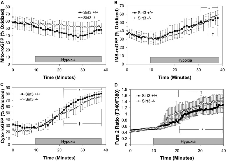
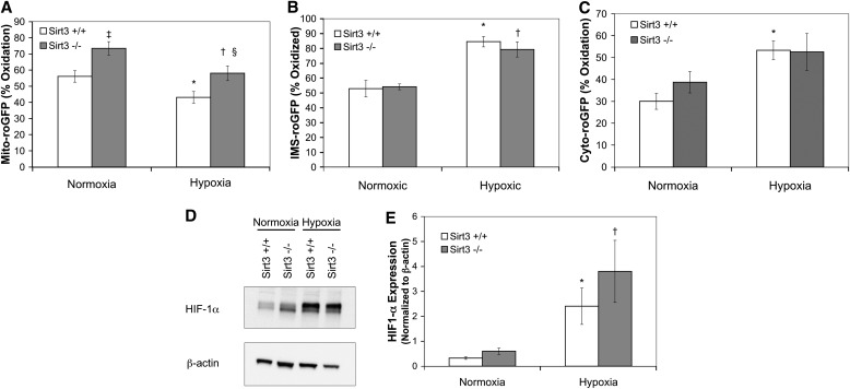
Alveolar hypoxia elicits increases in mitochondrial reactive oxygen species (ROS) signaling in pulmonary arterial (PA) smooth muscle cells (PASMCs), triggering hypoxic pulmonary vasoconstriction. Mice deficient in sirtuin (Sirt) 3, a nicotinamide adenine dinucleotide-dependent mitochondrial deacetylase, demonstrate enhanced left ventricular hypertrophy after aortic banding, whereas cells from these mice reportedly exhibit augmented hypoxia-induced ROS signaling and hypoxia-inducible factor (HIF)-1 activation. We therefore tested whether deletion of Sirt3 would augment hypoxia-induced ROS signaling in PASMCs, thereby exacerbating the development of pulmonary hypertension (PH) and right ventricular hypertrophy. In PASMCs from Sirt3 knockout (Sirt3(-/-)) mice in the C57BL/6 background, we observed that acute hypoxia (1.5% O2; 30 min)-induced changes in ROS signaling, detected using targeted redox-sensitive, ratiometric fluorescent protein sensors (roGFP) in the mitochondrial matrix, intermembrane space, and the cytosol, were indistinguishable from Sirt3(+/+) cells. Acute hypoxia-induced cytosolic calcium signaling in Sirt3(-/-) PASMCs was also indistinguishable from Sirt3(+/+) cells. During sustained hypoxia (1.5% O2; 16 h), Sirt3 deletion augmented mitochondrial matrix oxidant stress, but this did not correspond to an augmentation of intermembrane space or cytosolic oxidant signaling. Sirt3 deletion did not affect HIF-1α stabilization under normoxia, nor did it augment HIF-1α stabilization during sustained hypoxia (1.5% O2; 4 h). Sirt3(-/-) mice housed in chronic hypoxia (10% O2; 30 d) developed PH, PA wall remodeling, and right ventricular hypertrophy that was indistinguishable from Sirt3(+/+) littermates. Thus, Sirt3 deletion does not augment hypoxia-induced ROS signaling or its consequences in the cytosol of PASMCs, or the development of PH. These findings suggest that Sirt3 responses may be cell type specific, or restricted to certain genetic backgrounds.


Figures

<i>Figure 1.</i>
Figure 1.
Genotyping sirtuin (Sirt) 3 mice. (A) Sirt3 RT-PCR genotyping, a roughly 560-bp product indicates a wild-type (Sirt3+/+) mouse, a roughly 150-bp product indicates a knockout (Sirt3−/−) mouse, and both products indicate a heterozygous (Sirt3+/−) mouse. (B) Western blot of lysates of pulmonary arterial (PA) smooth muscle cells (PASMCs) isolated from Sirt3+/+ or Sirt3−/− mice probed with antibodies against Sirt3 or β-actin.
<i>Figure 2.</i>
Figure 2.
Effect of Sirt3 deletion on acute hypoxia–induced changes in reactive oxygen species (ROS) and calcium signaling in PASMCs. (AD) PASMCs isolated from Sirt3+/+ or Sirt3−/− mice. PASMCs expressing the redox-sensitive, ratiometric fluorescent protein sensor (roGFP) in the mitochondrial matrix (Mito-roGFP) (A), intermembrane space (IMS)–roGFP (B), cytosolic roGFP (Cyto-roGFP) (C), or loaded with Fura-2 (D) were superfused with normoxic (21% O2) or hypoxic (1.5% O2) media for 30 minutes. Values are means (± SEM); n = 6 cover slips, 4–10 cells/cover slip. *P < 0.05 compared with normoxic baseline of Sirt3+/+ PASMCs; P < 0.05 compared with normoxic baseline of Sirt3−/− PASMCs.
<i>Figure 3.</i>
Figure 3.
Effect of Sirt3 deletion on sustained hypoxia–induced changes in ROS signaling and hypoxia-inducible factor (HIF)-1α expression in PASMCs. (AE) PASMCs isolated from Sirt3+/+ or Sirt3−/− mice. PASMCs expressing Mito-roGFP (A), IMS-roGFP (B), or Cyto-roGFP (C) were incubated under normoxic (21% O2; Normoxia) or hypoxic (1.5% O2; Hypoxia) conditions for 16 hours. (D) Representative Western blot of lysates of PASMCs isolated from Sirt3+/+ or Sirt3−/− mice incubated under normoxic (21% O2; Normoxia) or hypoxic (1.5% O2; Hypoxia) conditions for 4 hours, probed with antibodies against HIF-1α or β-actin. (E) Quantitative analysis of multiple Western blots from (D). Values are means (± SEM); n = 6 cover slips, 4–10 cells/cover slip (AC). Values are means (± SEM); n = 6 dishes of PASMCs (E). *P < 0.05 compared with Normoxia Sirt3+/+ PASMCs; P < 0.05 compared with Normoxia Sirt3−/− PASMCs; P < 0.05 compared with Normoxia Sirt3+/+ PASMCs; §P < 0.05 compared with Hypoxia Sirt3+/+ PASMCs.
<i>Figure 4.</i>
Figure 4.
Effect of Sirt3 deletion on chronic hypoxia–induced pulmonary hypertension. (AF) Sirt3+/+ or Sirt3−/− mice housed under normoxic (room air; Normoxia) or hypoxic (10% O2; Hypoxia) conditions for 30 days. (A) Chronic hypoxia decreased pulmonary acceleration time (PAT)/ejection time (ET) in Sirt3−/− mice, but this response was not different from that in Sirt3+/+ animals. (B) Chronic hypoxia increased right ventricular (RV) systolic pressure (RVSP) in Sirt3−/− mice, but this response was not different from that in Sirt3+/+ animals. (C) Chronic hypoxia increased the acute hypoxia (5% O2; 1-min ventilation)–induced change in RVSP in Sirt3−/− mice, but this response was not different from that in Sirt3+/+ animals. (D) Inflation-fixed, hematoxylin and eosin–stained lung sections demonstrate chronic hypoxia–induced vascular remodeling. “PA” denotes small PAs. Left image denotes a lung section from a Normoxia Sirt3+/+ mouse; right image is from a Hypoxia Sirt3+/+ mouse. (E) Chronic hypoxia–induced vascular remodeling as assessed by PA wall thickness in both Sirt3+/+ and Sirt3−/− mice compared with their normoxic counterparts. (F) Chronic hypoxia–induced RV hypertrophy as assessed by the Fulton index in both Sirt3+/+ and Sirt3−/− mice compared with their normoxic counterparts. Values are means (± SEM); n = 6–10 mice per experimental group. *P < 0.05 compared with Normoxia Sirt3+/+ mice; P < 0.05 compared with Normoxia Sirt3−/− mice. LV, left ventricular; S, septum.
<i>Figure 5.</i>
Figure 5.
Effect of Sirt3 deletion on the acetylation status of manganese superoxide dismutase (MnSOD). MnSOD was immunoprecipitated from lung tissue lysates from Sirt3+/+ and Sirt3−/− mice using a rabbit antibody against MnSOD, and the resulting product was assessed by immunoblotting for protein lysine acetylation. (A) Representative Western blot of the lung lysates probed with antibodies against acetylated lysine (acetylated MnSOD) or MnSOD. (B) Quantitative analysis of Western blots from (A). Values are means (± SEM); n = 3 mice per experimental condition.

References

    1. Waypa GB, Guzy R, Mungai PT, Mack MM, Marks JD, Roe MW, Schumacker PT. Increases in mitochondrial reactive oxygen species trigger hypoxia-induced calcium responses in pulmonary artery smooth muscle cells. Circ Res. 2006;99:970–978. - PubMed
    1. Waypa GB, Marks JD, Mack MM, Boriboun C, Mungai PT, Schumacker PT. Mitochondrial reactive oxygen species trigger calcium increases during hypoxia in pulmonary arterial myocytes. Circ Res. 2002;91:719–726. - PubMed
    1. Waypa GB, Chandel NS, Schumacker PT. Model for hypoxic pulmonary vasoconstriction involving mitochondrial oxygen sensing. Circ Res. 2001;88:1259–1266. - PubMed
    1. Guzy RD, Sharma B, Bell E, Chandel NS, Schumacker PT. Loss of the SdhB, but not the SdhA, subunit of complex II triggers reactive oxygen species–dependent hypoxia-inducible factor activation and tumorigenesis. Mol Cell Biol. 2008;28:718–731. - PMC - PubMed
    1. Mansfield KD, Guzy RD, Pan Y, Young RM, Cash TP, Schumacker PT, Simon MC. Mitochondrial dysfunction resulting from loss of cytochrome c impairs cellular oxygen sensing and hypoxic HIF-alpha activation. Cell Metab. 2005;1:393–399. - PMC - PubMed

Publication types

MeSH terms