. 2013 Oct 1;12(19):3228-36.
doi: 10.4161/cc.26462. Epub 2013 Sep 12.

Pom1 and cell size homeostasis in fission yeast

Affiliations

Pom1 and cell size homeostasis in fission yeast

Elizabeth Wood et al. Cell Cycle. .

Abstract

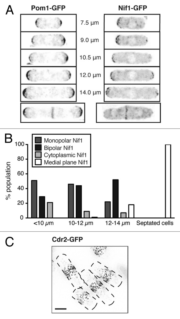
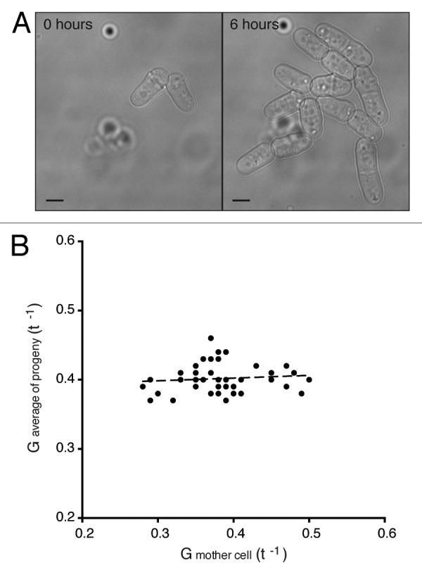
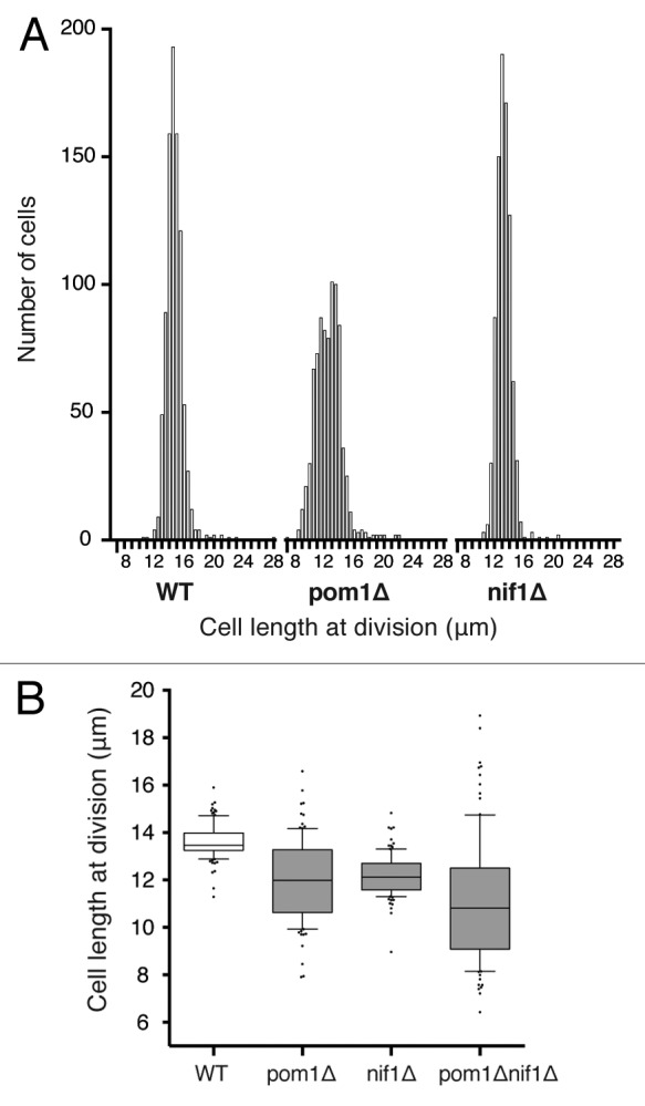
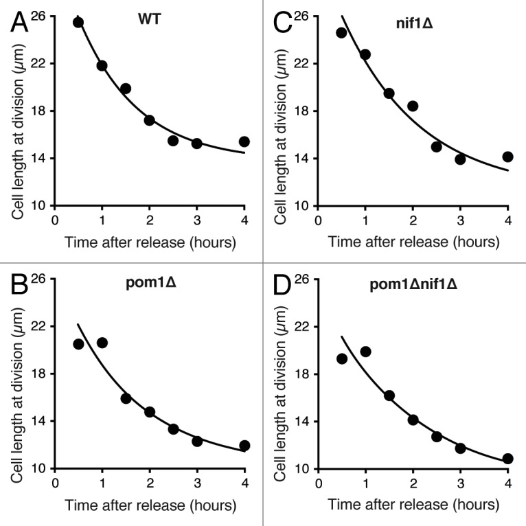
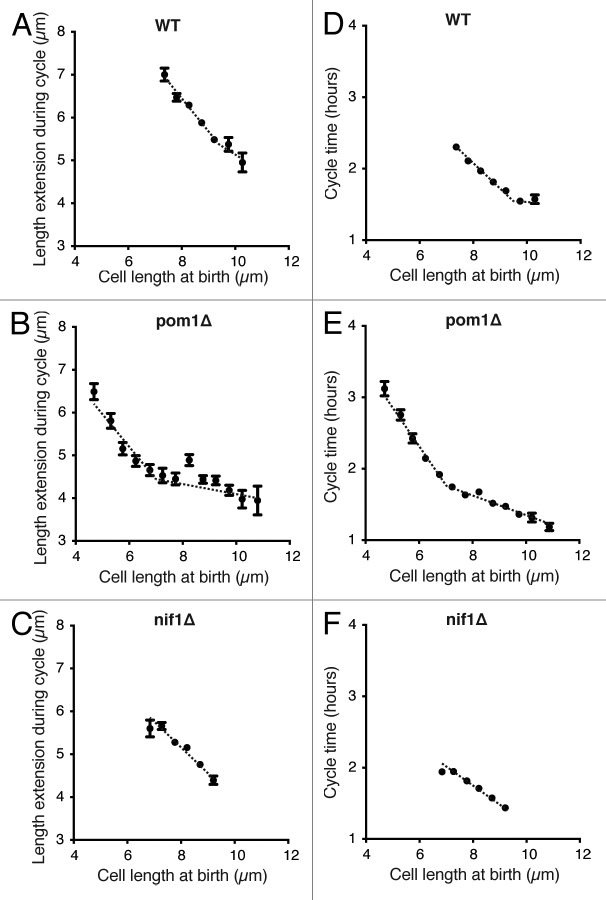
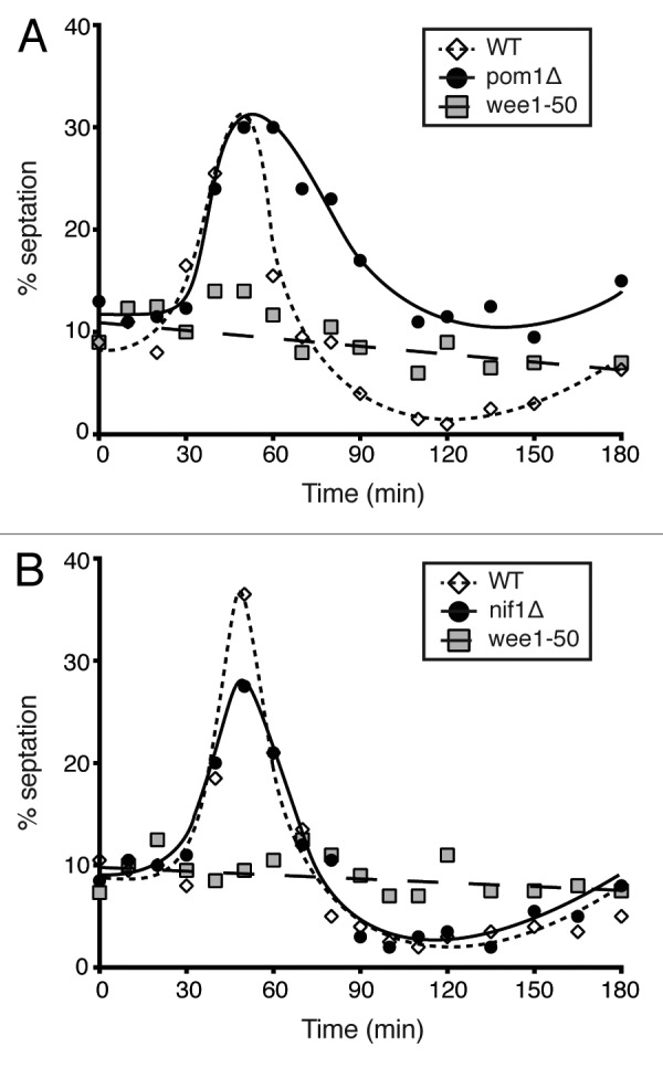
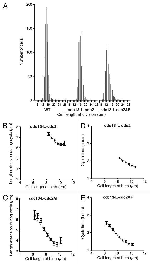
Cells sense their size and use this information to coordinate cell division with cell growth to maintain a constant cell size within a given population. A model has been proposed for cell size control in the rod-shaped cells of the fission yeast, Schizosaccharomyces pombe. This involves a protein localized to the cell ends, which inhibits mitotic activators in the middle of the cell in a cell size-dependent manner. This protein, Pom1, along with another tip-localized protein, Nif1, have been implicated as direct sensors of cell size controlling the onset of mitosis. Here we have investigated cell size variability and size homeostasis at the G 2/M transition, focusing on the role of pom1 and nif1. Cells deleted for either of these 2 genes show wild-type size homeostasis both in size variability analyses and size homeostasis experiments. This indicates that these genes do not have a critical role as direct cell size sensors in the control mechanism. Cell size homeostasis also seems to be independent of Cdc2-Tyr15 phosphorylation, suggesting that the size sensing mechanism in fission yeast may act through an unidentified pathway regulating CDK activity by an unknown mechanism.

Keywords: Pom1; cell cycle; cell size control; cell size variability; fission yeast; growth rate.


Figures

Figure 1.
Figure 1.
(A) Localization of GFP-tagged protein in cells of indicated size. Inverted maximum intensity projections of deconvolved images. (B) Quantification of Nif1 localization in cells of indicated size. Cytoplasmic Nif1 refers to cells with no visible protein enrichment. All septated cells were 12–14 μm in length. (C) Cdr2-GFP tagged cells showing protein localization to medial cortical nodes. Inverted maximum intensity projection of deconvolved image, cell outlines shown by black dashed lines. Scale bar, 4 μm.
Figure 2.
Figure 2.
(A) Brightfield images of wild-type cells in a microfluidic chamber after loading (0 h) and then after 6 h of growth in the chamber. Scale bar, 4 μm. (B) Growth rate values of mother cells plotted against the average values calculated for the 14 progeny. G = E/(BL ∙ CT). Regression slope 0.04.
Figure 3.
Figure 3.
(A) Histogram distribution of cell length at division of wild-type, pom1Δ and nif1Δ cells. For each strain, 800–900 cells were measured in a microfluidic chamber using brightfield images. Bin size 0.5 μm. (B) Cell length at division of WT, pom1Δ, nif1Δ, and pom1Δnif1Δ calcofluor-stained cells. Boxes represent 25–75% of the data and the central lines indicate the median. Whiskers show 10–90% of the data and individual dots represent the outer 20%. One hundred cells of each strain were measured in exponential growth phase.
Figure 4.
Figure 4.
(A–D) Cell length recovery of the indicated strains after elongation with the S phase inhibitor hydroxyurea. HU added for 4 h then washed out at time zero. Average cell length at division was measured for each time point and exponential curves fitted to the data.
Figure 5.
Figure 5.
(A–C) Cell length at birth plotted against length extension from time-lapse images of around 800 cells for each strain indicated. Data shown excludes the outer 2% of birth length and length extension values. Points show mean value and error bars represent SEM. Regression lines best fitted to the data are shown. (D–F) Same cells as in (A–C) with cycle time plotted against birth length. Data shown excludes the outer 2% of birth length and cycle time values. Mean with error bars showing SEM. Regression lines best fitted to the data are shown.
Figure 6.
Figure 6.
(A) Septation index of a pom1Δ population compared with wild-type and wee150 cells after nutritional shift-down from glutamate to proline as a nitrogen source. (B) Experiment as in (A) with nif1Δ cells compared with wild-type and wee150 controls. All cells grown at 25 °C in EMM-N + 20 mM glutamic acid and shifted to 36 °C for 2 cycles then transferred at time zero to EMM-N + 10 mM proline. Number of septated cells counted and represented as a percentage of the whole population.
Figure 7.
Figure 7.
(A) Histogram distribution of cell length at division of wild-type, cdc13-L-cdc2, and cdc13-L-cdc2AF cells. For each strain, 700–900 cells were measured in a microfluidic chamber using brightfield images. Bin size 0.5 μm. (B and C) Cell length at birth plotted against length extension from time-lapse images of around 800 cells for each strain indicated. Data shown excludes the outer 2% of birth length and length extension values. Points show mean value and error bars represent SEM. Regression lines best fitted to the data are shown. (D and E) Same cells as in (BB and CC) with cycle time plotted against birth length. Data shown excludes the outer 2% of birth length and cycle time values. Mean with error bars showing SEM. Regression lines best fitted to the data are shown.

Comment in

  • Pom1 is not the size ruler.
    Novák B. Novák B. Cell Cycle. 2013 Nov 15;12(22):3463-4. doi: 10.4161/cc.26818. Epub 2013 Oct 15. Cell Cycle. 2013. PMID: 24131919 Free PMC article. No abstract available.

References

    1. Schaechter M, Williamson JP, Hood JR, Jr., Koch AL. Growth, cell and nuclear divisions in some bacteria. J Gen Microbiol. 1962;29:421–34. doi: 10.1099/00221287-29-3-421. - DOI - PubMed
    1. Killander D, Zetterberg A. Quantitative Cytochemical Studies on Interphase Growth. I. Determination of DNA, Rna and Mass Content of Age Determined Mouse Fibroblasts in Vitro and of Intercellular Variation in Generation Time. Exp Cell Res. 1965;38:272–84. doi: 10.1016/0014-4827(65)90403-9. - DOI - PubMed
    1. Killander D, Zetterberg A. A quantitative cytochemical investigation of the relationship between cell mass and initiation of DNA synthesis in mouse fibroblasts in vitro. Exp Cell Res. 1965;40:12–20. doi: 10.1016/0014-4827(65)90285-5. - DOI - PubMed
    1. Donachie WD. Relationship between cell size and time of initiation of DNA replication. Nature. 1968;219:1077–9. doi: 10.1038/2191077a0. - DOI - PubMed
    1. Nurse P. Genetic control of cell size at cell division in yeast. Nature. 1975;256:547–51. doi: 10.1038/256547a0. - DOI - PubMed

Publication types

MeSH terms

LinkOut - more resources