. 2013 Nov 1;12(21):3405-20.
doi: 10.4161/cc.26421. Epub 2013 Sep 17.

The estrogen receptor α is the key regulator of the bifunctional role of FoxO3a transcription factor in breast cancer motility and invasiveness

Affiliations

The estrogen receptor α is the key regulator of the bifunctional role of FoxO3a transcription factor in breast cancer motility and invasiveness

Diego Sisci et al. Cell Cycle. .

Abstract

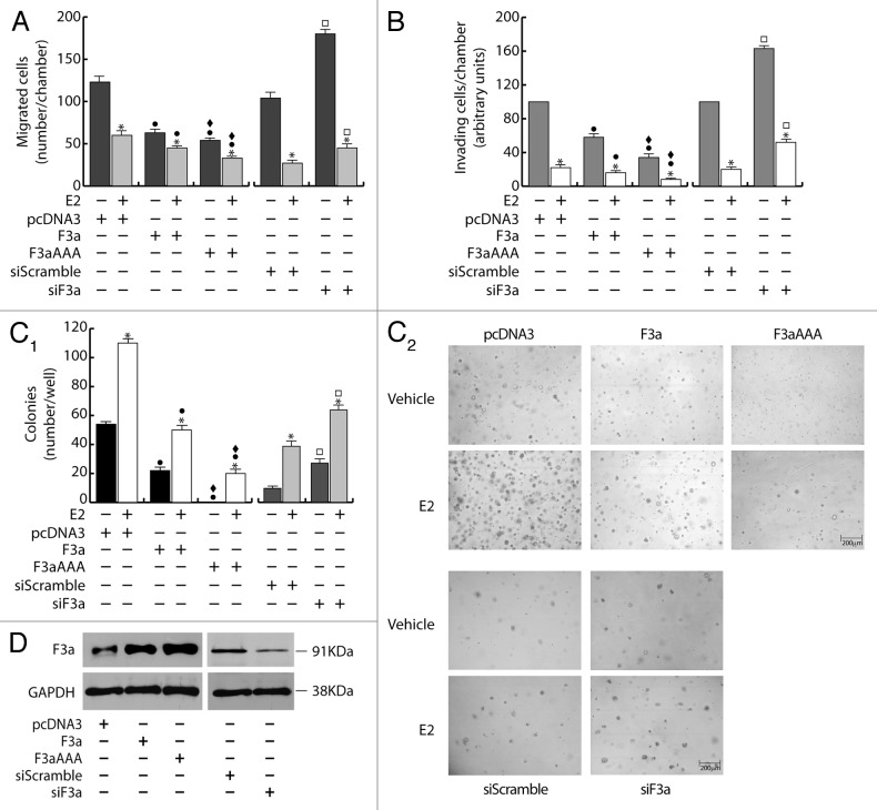
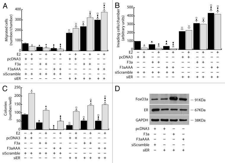
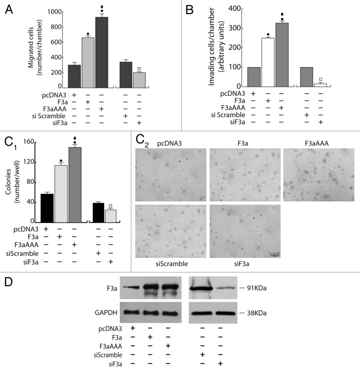
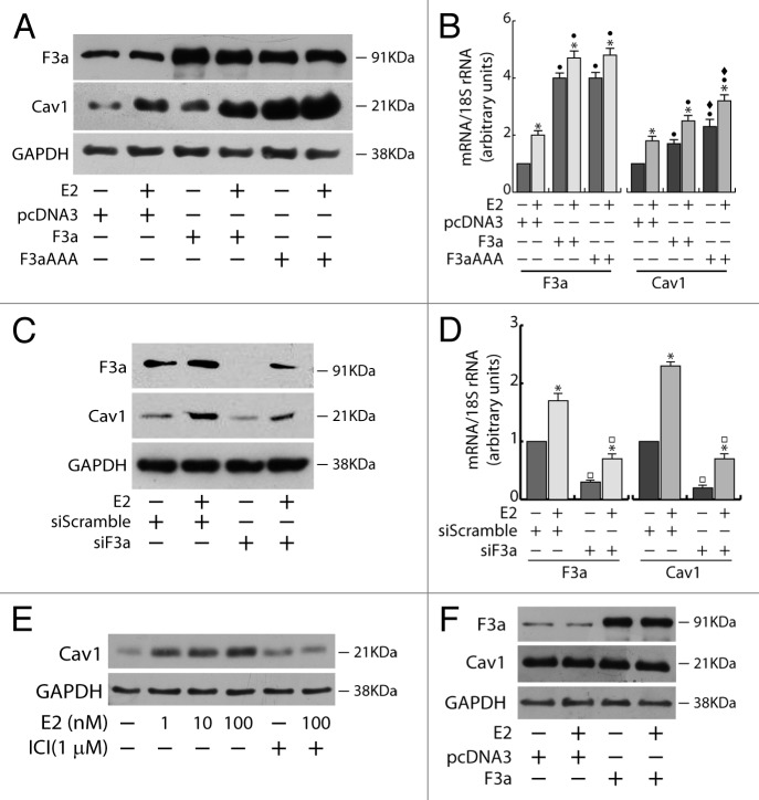
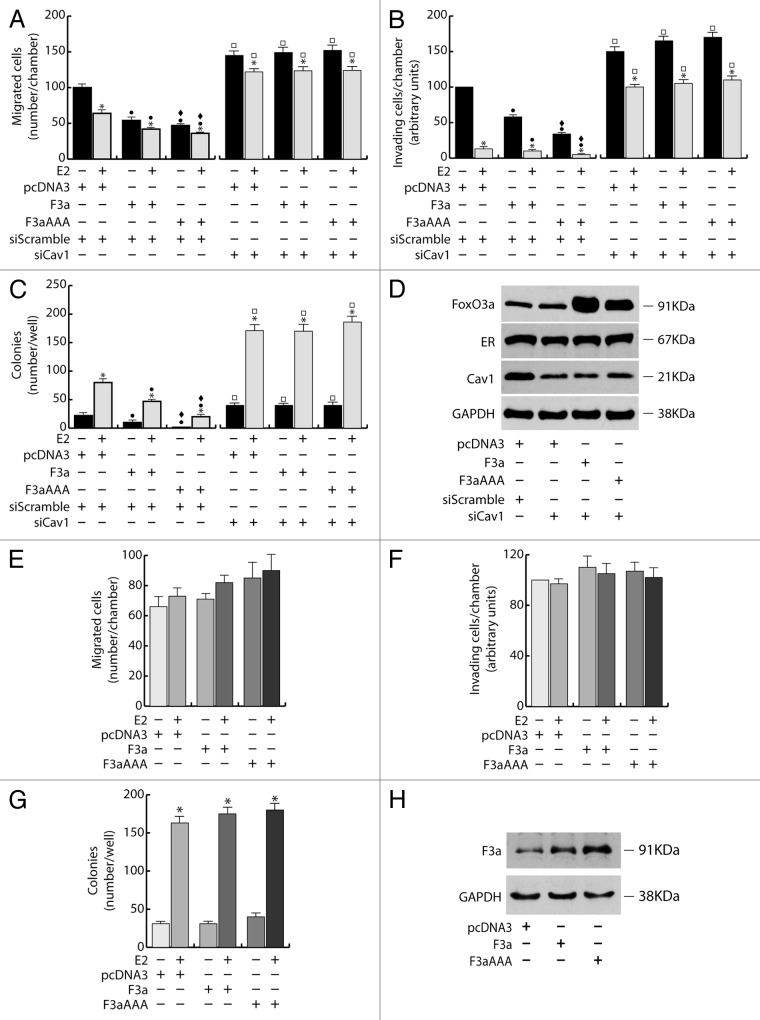
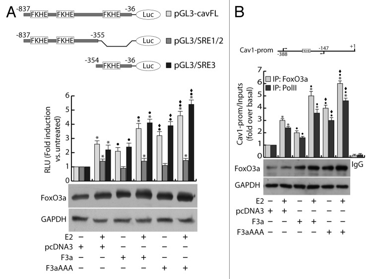
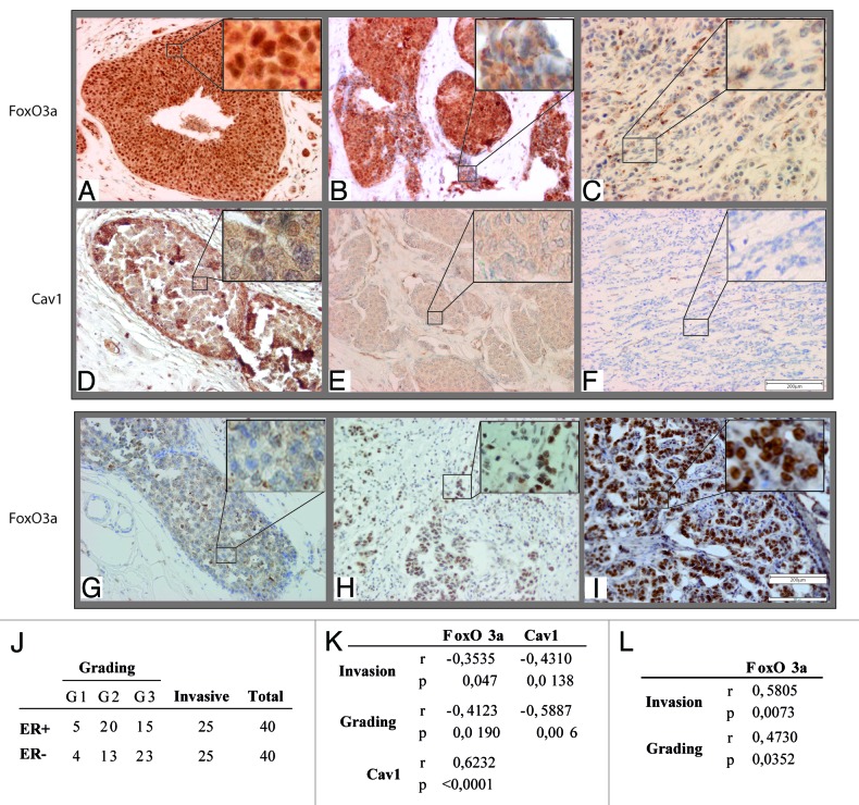
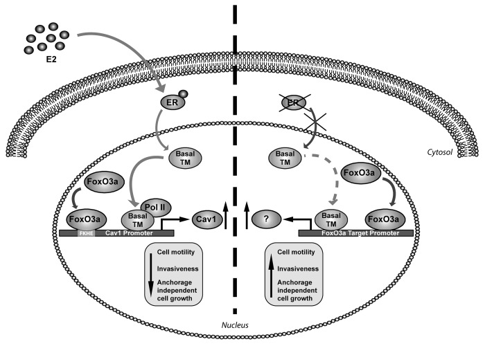
The role of the Forkhead box class O (FoxO)3a transcription factor in breast cancer migration and invasion is controversial. Here we show that FoxO3a overexpression decreases motility, invasiveness, and anchorage-independent growth in estrogen receptor α-positive (ERα+) cancer cells while eliciting opposite effects in ERα-silenced cells and in ERα-negative (ERα-) cell lines, demonstrating that the nuclear receptor represents a crucial switch in FoxO3a control of breast cancer cell aggressiveness. In ERα+ cells, FoxO3a-mediated events were paralleled by a significant induction of Caveolin-1 (Cav1), an essential constituent of caveolae negatively associated to tumor invasion and metastasis. Cav1 induction occurs at the transcriptional level through FoxO3a binding to a Forkhead responsive core sequence located at position -305/-299 of the Cav1 promoter. 17β-estradiol (E2) strongly emphasized FoxO3a effects on cell migration and invasion, while ERα and Cav1 silencing were able to reverse them, demonstrating that both proteins are pivotal mediators of these FoxO3a controlled processes. In vivo, an immunohistochemical analysis on tissue sections from patients with ERα+ or ERα- invasive breast cancers or in situ ductal carcinoma showed that nuclear FoxO3a inversely (ERα+) or directly (ERα-) correlated with the invasive phenotype of breast tumors. In conclusion, FoxO3a role in breast cancer motility and invasion depends on ERα status, disclosing a novel aspect of the well-established FoxO3a/ERα interplay. Therefore FoxO3a might become a pursuable target to be suitably exploited in combination therapies either in ERα+ or ERα- breast tumors.

Keywords: breast cancer; estrogen receptor; forkhead transcription factors; invasion; motility.


Figures

None
Figure 1. FoxO3a inhibits migration, invasion and anchorage independent growth in ERα+ MCF-7 breast cancer cells. A double set of MCF-7 cells was transiently transfected with 1 μg/35 mm dish of F3a, F3aAAA, or pcDNA3 as control. Another double set was silenced for FoxO3a expression (siF3a), using a siScramble as control (60 pmol siRNAs/35 mm dish). After 5 h cells were switched to PRF-SFM, and the next day one of each set of cells was harvested and subjected to migration (A), invasion (B), and soft agar assay (C1 and C2). Migration and invasion assays were conducted as described in “Materials and Methods”, adding 100 nM E2 in the bottom of the wells where indicated. Migrated and invading cells were evaluated after 24 h and 72 h of incubation, respectively. In soft agar assay, colonies >50 μm diameter formed after 14 d from plating were photographed at 4× magnification (C2) and counted under the microscope (C1). The second set of either transfected or silenced MCF-7 cells was used for total protein extractions and WB analysis to assess transfections efficiency; GAPDH was evaluated as a loading control (D). Results are reported as the mean ± s.d. of at least 3 independent experiments. In all experiments, significance values were as follows: *, P < 0.01 vs. untreated; ●, P < 0.01 vs. corresponding pcDNA3; ♦, P < 0.05 vs. corresponding F3a; □, P < 0.01 vs. corresponding siScramble.
None
Figure 2. FoxO3a mediated inhibition of breast cancer cell migration, invasion and growth in suspension depends on ERα Two double sets of MCF-7 cells were silenced either for ERα (siER), using siScramble as control. After 5 h cells were switched to PRF-SFM and transiently transfected with F3a, F3aAAA, or pcDNA3. Next day cells were harvested and one set of each experiment was subjected to migration, invasion, and soft agar assay in the presence or in the absence of E2. Migrated (A) and invading (B) cells were evaluated after 24 h and 72 h of incubation, respectively. In soft agar assay, colonies ≥50 μm diameter formed after 14 d from plating were counted under the microscope (C). The second set of each experiment was used for total protein extraction to evaluate transfections efficiency by WB analysis; GAPDH was used as loading control (D). Results are the mean ± s.d. of at least three independent experiments. *, P < 0.05 vs. untreated; ●, P < 0.01 vs. corresponding pcDNA3; ♦, P < 0.01 vs. corresponding F3a; □, P < 0.01 vs. corresponding siScramble.
None
Figure 3. FoxO3a promotes migration, invasion, and anchorage-independent growth in ERα− MDA-MB-231 breast cancer cells. A double set of MDA-MB-231 cells were transiently transfected with 1 μg/35 mm dish of F3a, F3aAAA, or pcDNA3 or silenced for FoxO3a expression (siF3a) using a siScramble as control (60 pmol siRNAs/35 mm dish). Both transfection and silencing were made on cells in suspended PRF-GM. After 5 h cells were serum starved and, 24 h later, harvested. One set was subjected to migration (A), invasion (B), or soft agar assay (C1 and C2). Migrated and invading cells were evaluated after 16 h and 48 h of incubation, respectively. In soft agar assay, colonies > 50 μm diameter formed after 14 d from plating were photographed at 4× magnification (C2) and counted under the microscope (C1). The second set of either transfected or silenced MCF-7 cells was used to assess transfections efficiency by WB analysis on total protein extracts; GAPDH was evaluated as a loading control (D). Results are reported as the mean ± s.d. of at least 3 independent experiments. ●, P < 0.01 vs. pcDNA3; ♦, P < 0.01 vs. F3a; □, P < 0.05 vs. siScramble.
None
Figure 4. Cav1 expression depends on E2 and FoxO3a in ERα+ MCF-7 breast cancer cells. A double set of MCF-7 cells were either transiently transfected with F3a, F3aAAA, or pcDNA3 or silenced for FoxO3a, serum starved after 5 h and treated the next day with 100 nM E2 for 24 h. Cells were then harvested and total proteins and RNA were extracted, and subjected to WB (A and C) and RT-PCR analysis (B and D), respectively, for F3a and Cav1 expression assessment. (E) MCF-7 cells were seeded in growing medium, serum starved the next day for 24 h, pre-treated or not for 1 h with the pure antiestrogen ICI 182.780 and then treated with increasing concentrations of E2 (0, 1, 10, and 100 nM). (F) MDA-MB-231 cells were transiently transfected with F3a or pcDNA3 as control, serum starved for 24 h and then treated or not with 100 nM E2. After 24 h of E2 treatment, total proteins were extracted and subjected to WB analysis. GAPDH was analyzed as loading control in WB assays. For RT-PCR assays, each sample was normalized to its 18S rRNA content. Results are reported as the mean ± s.d. of at least 3 independent experiment. *, P < 0.01 vs. untreated; ●, P < 0.01 vs. pcDNA3; ♦, P < 0.01 vs. F3a; □, P < 0.05 vs. siScramble.
None
Figure 5. Cav1 is a mediator of FoxO3a dependent inhibition of migration, invasion and growth in suspension of ERα+ breast cancer cells. (A–D) Two double sets of MCF-7 cells were silenced for Caveolin-1 (siCav1), using siScramble as control. After 5 h cells were switched to PRF-SFM and transiently transfected with F3a, F3aAAA, or pcDNA3. Next day cells were harvested and one set of each experiment was subjected to migration, invasion, and soft agar assay, in the presence or in the absence of E2. Migrated (A) and invading (B) cells were evaluated after 24 h and 72 h of incubation, respectively. In soft agar assay, colonies ≥50 μm diameter formed after 14 d from plating were counted under the microscope (C). Transfection efficiency was evaluated by WB analysis on total protein extracted by the second set of cells; GAPDH was used as loading control (D). Results are the mean ± s.d. of at least 3 independent experiments. *, P < 0.05 vs. untreated; ●, P < 0.01 vs. corresponding pcDNA3; ♦, P < 0.01 vs. corresponding F3a; □, P < 0.01 vs. corresponding siScramble. (E–H) A double set of T47D cells were transiently transfected with F3a, F3aAAA or pcDNA3. After 5h cells were switched to PRF-SFM and the next day one set of cells was harvested and subjected to migration (E), invasion (F), or soft agar assay (G), with or without 100 nM E2. Migrated and invading cells were counted after 24 h and 72 h of incubation, respectively. In soft agar assay, colonies formed after 14 d from plating were exposed to MTT and counted under the microscope. The second set of cells was lysed, and total protein was used for WB analysis to assess transfections efficiency; GAPDH was used as loading control (H). Results are the mean ± s.d. of at least 3 independent experiments. *, P < 0.01 vs. untreated.
None
Figure 6. FoxO3a binds to and transactivates the Cav1 promoter. (A) MCF-7 were seeded in culture medium on 24-well plates, serum starved for 24 h, co-transfected in PRF-CT with pGL3-cavFL, or pGL3/SRE1/2, or pGL3/SRE3 and pRL-Tk, in presence of either pcDNA3 or F3a or F3aAAA vectors. After 6 h, E2 (100 nM) was added to the medium, where opportune, and the next day cells were harvested, and luciferase activity was evaluated. Cell extracts were also processed by WB analysis to assess F3a and F3aAAA transfection efficiency; GAPDH was used as loading control. (B) ChIP analysis was performed on the nuclear extracts from subconfluent MCF-7 cells seeded in 15 cm dish diameter, switched to PRF-SFM, and transfected with pcDNA3, F3a, or F3aAAA vectors. Twenty-four hours after transfection, the cells were treated with 100 nM E2 for 30 min or left untreated. The FKHE-containing Cav1 promoter region, precipitated with either anti-FoxO3a or anti-PolII pAbs were amplified using a specific pair of primers reported in “Materials and Methods”. E2-treated samples were also precipitated with normal rabbit IgG and used as negative control. FoxO3a expression in transfected samples was analyzed by WB on Cytosolic lysates from the same set of cells. Data represents the mean ± s.d. of 3 independent experiments. *, P < 0.05 vs. untreated; ●, P < 0.05 vs. corresponding pcDNA3; ♦, P < 0.05 vs. corresponding F3a.
None
Figure 7. Nuclear FoxO3a is highly expressed in non-invasive ERα+, and in invasive ERα− breast tumors. FoxO3a (A–C) and Cav1 (D–F) expression in ERα+ breast tumors and FoxO3a (G–I) in ERα− breast tumor samples. IHC was conducted on tissue sections deriving from biopsies diagnosed as DCIS (A and D), microinvasive DCIS (B and E), DCIS with contiguous IDC areas (G and H) and highly aggressive IDC (C, F, and I). Representative fields were photographed at 20× magnification. Insets, showing details of proteins subcellular localization, were taken at 100× magnification. (J) Samples descriptions and classification; (K) correlation between nuclear FoxO3a or Cav1 content and the tumor grading and invasive potential in ERα+ breast cancer samples; (L) correlation between nuclear FoxO3a content and the tumor grading and invasive potential in ERα− breast cancer samples. The correlation coefficient (r) and the statistical significance (P) are reported.
None
Figure 8. Proposed model for FoxO3a-mediated control of cell motility and invasiveness in presence or absence of ERα. F3a and ERα synergistically induce the expression of Cav1, which, in turn, reduces cell motility and invasiveness of ERα+ breast cancer cells. Transcriptionally active F3a binds to a FKHE located on the Cav1 proximal promoter and increases the recruitment of RNA Polymerase II, which is enhanced upon E2 stimulation. The lack of the hormone receptor enables active F3a to behave in an opposite fashion, thus increasing cell motility and invasion. Basal TM, basal transcriptional machinery.

References

    1. Greer EL, Brunet A. FOXO transcription factors at the interface between longevity and tumor suppression. Oncogene. 2005;24:7410–25. doi: 10.1038/sj.onc.1209086. - DOI - PubMed
    1. Yang JY, Hung MC. Deciphering the role of forkhead transcription factors in cancer therapy. Curr Drug Targets. 2011;12:1284–90. doi: 10.2174/138945011796150299. - DOI - PMC - PubMed
    1. Hu MC, Lee DF, Xia W, Golfman LS, Ou-Yang F, Yang JY, Zou Y, Bao S, Hanada N, Saso H, et al. IkappaB kinase promotes tumorigenesis through inhibition of forkhead FOXO3a. Cell. 2004;117:225–37. doi: 10.1016/S0092-8674(04)00302-2. - DOI - PubMed
    1. Yang JY, Zong CS, Xia W, Yamaguchi H, Ding Q, Xie X, Lang JY, Lai CC, Chang CJ, Huang WC, et al. ERK promotes tumorigenesis by inhibiting FOXO3a via MDM2-mediated degradation. Nat Cell Biol. 2008;10:138–48. doi: 10.1038/ncb1676. - DOI - PMC - PubMed
    1. Zou Y, Tsai WB, Cheng CJ, Hsu C, Chung YM, Li PC, Lin SH, Hu MC. Forkhead box transcription factor FOXO3a suppresses estrogen-dependent breast cancer cell proliferation and tumorigenesis. Breast Cancer Res. 2008;10:R21. doi: 10.1186/bcr1872. - DOI - PMC - PubMed

Publication types

MeSH terms