. 2014 Mar-Apr;20(2):155-9.
doi: 10.5152/dir.2013.13139.

Applications of the Amplatzer Vascular Plug to various vascular lesions

Affiliations

Applications of the Amplatzer Vascular Plug to various vascular lesions

Serkan Güneyli et al. Diagn Interv Radiol. 2014 Mar-Apr.

Abstract

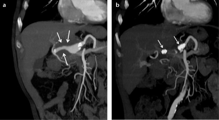
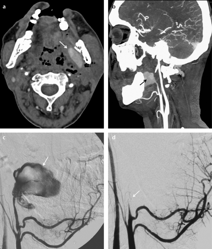
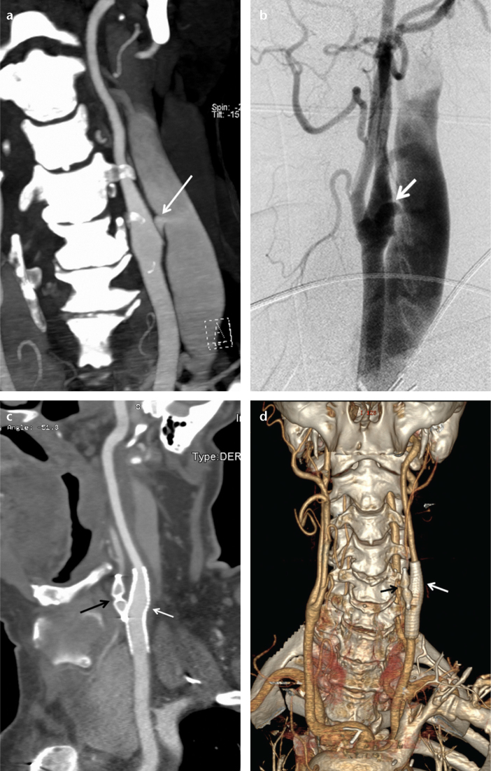
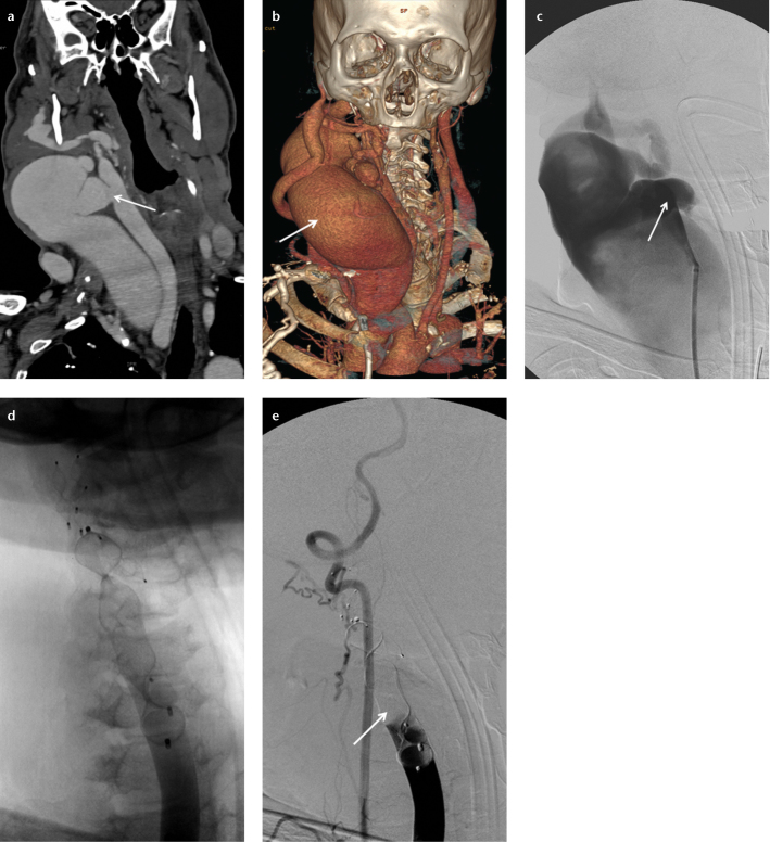
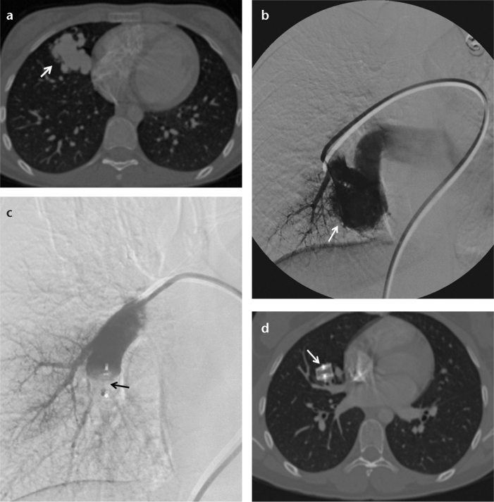
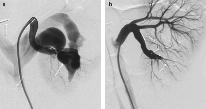
The Amplatzer® Vascular Plug (AVP) can be used to embolize medium-to-large high-flow vessels in various locations. Between 2009 and 2012, 41 AVPs (device size, 6-22 mm in diameter) were used to achieve occlusion in 31 patients (24 males, seven females) aged 9-92 years (mean age, 54.5 years). The locations and indications for embolotherapy were as follows: internal iliac artery embolization before stent-graft repair for aorto-iliac (n=6) and common iliac artery (n=3) aneurysms, subclavian artery embolization before stent-graft repair for thoracic aorta (n=3) and arcus aorta (n=1) aneurysms, brachiocephalic trunk embolization before stent-graft repair for a thoracic aorta aneurysm (n=1), embolization of aneurysms and pseudoaneurysms (n=5), embolization for carotid blow-out syndrome (n=3), closure of arteriovenous fistula (n=8), and closure of a portosystemic fistula (n=1). Of the 41 AVPs, 30 were AVP 2 and 11 were AVP 4. The mean follow-up duration was 4.7 months (range, 1-24 months). During follow-up, there was one migration, one insufficient embolization, and one recanalization. The remaining vascular lesions were successfully excluded from the circulation. The AVP, which can be used in a wide spectrum of pathologies, is easy to use and causes few complications. This essay presents our experience with the AVP.


Figures

Figure 1. a, b.
Figure 1. a, b.
Coronal CT angiography reconstruction images of a 63-year-old male show a partially thrombosed common hepatic artery aneurysm (a, arrows) and two AVPs placed in the proximal and distal parts of the aneurysm (b, arrows) one month after the occlusion.
Figure 2. a–d.
Figure 2. a–d.
Axial CT angiography image (a) of a 47-year-old male shows a pseudoaneurysm (arrow) in the left parapharyngeal space. A sagittal CT angiography reconstruction image (b) shows a left internal maxillary artery pseudoaneurysm (arrow). Selective left external carotid angiography (c) shows active contrast extravasation from the pseudoaneurysm (arrow). Selective left external carotid angiography after the deployment of an 8 mm AVP 2 (d) shows total embolization of the left internal maxillary artery (arrow).
Figure 3. a–d.
Figure 3. a–d.
Coronal CT angiography reconstruction image (a) of a 62-year-old male shows a caroticojugular fistula (arrow) between the internal carotid artery and internal jugular vein. Carotid angiography (b) shows the caroticojugular fistula (arrow). One month following the embolization, a sagittal CT angiography reconstruction image (c) shows the occluded left external carotid artery with an 8 mm AVP 4 (black arrow) and the occluded fistula with a stent-graft (white arrow). One month following the embolization, a coronal CT angiography reconstruction image (d) shows the occluded left external carotid artery with the AVP 4 (black arrow) and the occluded fistula with the stent-graft (white arrow).
Figure 4. a–e.
Figure 4. a–e.
Coronal CT angiography reconstruction image (a) of a 57-year-old male shows a caroticojugular fistula (arrow) between the common carotid artery and internal jugular vein. A coronal CT angiography reconstruction image (b) shows the dilated internal jugular vein (arrow) secondary to the caroticojugular fistula. Carotid angiography (c) shows the caroticojugular fistula (arrow). Carotid angiography after deployment of the AVPs (d) shows multiple AVPs. Carotid angiography after deployment of the AVPs (e) shows total embolization of the common carotid artery (arrow).
Figure 5. a–d.
Figure 5. a–d.
Axial CT angiography (a) and pulmonary angiography (b) images of a 17-year-old female show a pulmonary arteriovenous fistula (arrow) at the right middle lobe. Pulmonary angiography after deployment of a 16 mm AVP 2 (c) shows total occlusion of the feeding artery and the AVP 2 (arrow) in the right middle lobe segment pulmonary artery. Six months later, an axial CT angiography image (d) shows the AVP 2 (arrow) in the right middle lobe segmental pulmonary artery.
Figure 6. a, b.
Figure 6. a, b.
Renal angiography image (a) of an 11-year-old male shows a renal arteriovenous fistula (arrow) at the mid to lower third of the kidney. Renal angiography after deploying an 8 mm AVP 4 (b) shows total occlusion (arrow) of the fistula.

References

    1. Ong CK, Lam DV, Ong MT, Power MA, Parkinson RJ, Wenderoth JD. Neuroapplication of Amplatzer Vascular Plug for therapeutic sacrifice of major craniocerebral arteries: an initial clinical experience. Ann Acad Med Singap. 2009;38:763–768. - PubMed
    1. Hill SL, Hijazi ZM, Hellenbrand WE, Cheatham JP. Evaluation of the Amplatzer Vascular Plug for embolization of peripheral vascular malformations associated with congenital heart disease. Catheter Cardiovasc Interv. 2006;67:113–119. - PubMed
    1. Mangini M, Lagana D, Fontana F, et al. Use of Amplatzer Vascular Plug (AVP) in emergency embolisation: preliminary experience and review of literature. Emerg Radiol. 2008;15:153–160. - PubMed
    1. Schwartz M, Glatz AC, Rome JJ, Gillespie MJ. The Amplatzer vascular plug and Amplatzer Vascular Plug II for vascular occlusion procedures in 50 patients with congenital cardiovascular disease. Catheter Cardiovasc Interv. 2010;76:411–417. - PubMed
    1. Tabori NE, Love BA. Transcatheter occlusion of pulmonary arteriovenous malformations using the Amplatzer Vascular Plug II. Catheter Cardiovasc Interv. 2008;71:940–943. - PubMed

Publication types