. 2013 Nov;163(3):1376-86.
doi: 10.1104/pp.113.226803. Epub 2013 Sep 18.

The importance of size and disorder in the cryoprotective effects of dehydrins

Affiliations

The importance of size and disorder in the cryoprotective effects of dehydrins

Stephanie L Hughes et al. Plant Physiol. 2013 Nov.

Abstract

Dehydrins protect plant proteins and membranes from damage during drought and cold. Vitis riparia K2 is a 48-residue protein that can protect lactate dehydrogenase from freeze-thaw damage by preventing the aggregation and denaturation of the enzyme. To further elucidate its mechanism, we used a series of V. riparia K2 concatemers (K4, K6, K8, and K10) and natural dehydrins (V. riparia YSK2, 60 kilodalton peach dehydrin [PCA60], barley dehydrin5 [Dhn5], Thellungiella salsuginea dehydrin2 [TsDHN-2], and Opuntia streptacantha dehydrin1 [OpsDHN-1]) to test the effect of the number of K-segments and dehydrin size on their ability to protect lactate dehydrogenase from freeze-thaw damage. The results show that the larger the hydrodynamic radius of the dehydrin, the more effective the cryoprotection. A similar trend is observed with polyethylene glycol, which would suggest that the protection is simply a nonspecific volume exclusion effect that can be manifested by any protein. However, structured proteins of a similar range of sizes did not show the same pattern and level of cryoprotection. Our results suggest that with respect to enzyme protection, dehydrins function primarily as molecular shields and that their intrinsic disorder is required for them to be an effective cryoprotectant. Lastly, we show that the cryoprotection by a dehydrin is not due to any antifreeze protein-like activity, as has been reported previously.


Figures

Figure 1.
Figure 1.
Sequences of the dehydrin constructs. The sequences and architectures of K2 and variants are shown using the single-letter amino acid code. The complete sequences are shown for K2, K-peptide, and KK-peptide. The insert used to generate the Kn constructs is shown as the base repeating unit that is added to the C terminus of K2, where n = 1 attached to the C terminus of the K2 sequence would be the sequence of the K4 construct.
Figure 2.
Figure 2.
CD spectra of K2 and the various constructs. Far UV spectra were collected as described in “Materials and Methods.” A, K-peptide, black squares; KK-peptide, white squares; K2, black circles. B, K2, black circles; K4, white triangles; K6, gray triangles; K8, black triangles; K10, white diamonds. C, Relationship between the length of a dehydrin construct and the hydrodynamic radius. Symbols are as in A and B. The line is a linear fit through all of the data points using Equation 1. MW, Molecular weight.
Figure 3.
Figure 3.
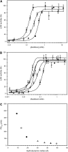
Cryoprotection of LDH by K2 and the various constructs. The ability of the dehydrins to protect LDH from freeze-thaw damage was plotted as percentage recovery of LDH activity versus additive concentration. The data were fitted to Equation 3. The error bars represent sd of four measurements. A, K-peptide, black squares; KK-peptide, white squares; K2, black circles. B, K2, black circles, K4, white triangles; K6, gray triangles; K8, black triangles; K10, white diamonds. C, Comparison of PD50 versus hydrodynamic radius. The plot shows the relationship between the size of the protein and the cryoprotective efficiency, which is expressed as the PD50. K-peptide, black squares; KK-peptide, white squares; K2, black circles, K4, white triangles; K6, gray triangles; K8, black triangles; K10, white diamonds.
Figure 4.
Figure 4.
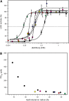
A, Cryoprotection of LDH by natural dehydrins. The ability of the larger dehydrins to protect LDH from freeze-thaw damage was plotted as percentage recovery of LDH activity versus protein concentration. The error bars represent the sd of four measurements. The dotted line indicates 50% recovery of LDH activity. K2, black circles; YSK2, yellow circles; TsDHN-2, purple circles; OpsDHN-1, blue circles; PCA60, red circles; Dhn5, green circles. B, Plot of PD50 versus hydrodynamic radius. Symbol colors are as in A, with the data from the Kn constructs overlaid as black circles. [See online article for color version of this figure.]
Figure 5.
Figure 5.
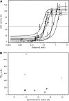
Cryoprotection of LDH by structured proteins. A, The ability of the ordered proteins to protect LDH from freeze-thaw damage was plotted as percentage recovery of LDH activity versus protein concentration. The error bars represent the sd of four measurements. The dotted line indicates 50% recovery of LDH activity. B, Comparison of PD50 with the hydrodynamic radius. Lysozyme, gray squares; carbonic anhydrase, white squares; β-casein, white upside-down triangles; ovalbumin, white circles; BSA, black upside-down triangles; phosphorylase, gray upside-down triangles. For comparison purposes, the PD50 of K2 is included as black circles.
Figure 6.
Figure 6.
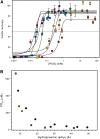
Cryoprotection of LDH by PEG. A, The ability of this polymer to protect LDH from freeze-thaw damage was plotted as percentage recovery of LDH activity versus polymer concentration. The error bars represent the sd of four measurements. The dotted line indicates 50% recovery of LDH activity. PEG-1000, red squares; PEG-1500, orange squares; PEG-2000, yellow squares; PEG-4000, green squares; PEG-8000, light blue squares; PEG-10000, dark blue squares; PEG-20000, purple squares. B, Plot of PD50 versus hydrodynamic radius. PEG, colored squares; Kn concatemers, black circles. [See online article for color version of this figure.]
Figure 7.
Figure 7.
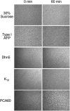
Ice recrystallization inhibition assay. The ability of the various dehydrins to inhibit the recrystallization of ice was qualitatively examined, with the proteins tested indicated on the left. The first column of images represents the ice crystals at time zero, whereas the second column shows the recrystallization state of ice after 60 min at −6°C. The 30% Suc sample is the negative control, while the winter flounder type I AFP (HPLC6 isoform) is the positive control.

References

    1. Allagulova CR, Gimalov FR, Shakirova FM, Vakhitov VA. (2003) The plant dehydrins: structure and putative functions. Biochemistry (Mosc) 68: 945–951 - PubMed
    1. Alsheikh MK, Heyen BJ, Randall SK. (2003) Ion binding properties of the dehydrin ERD14 are dependent upon phosphorylation. J Biol Chem 278: 40882–40889 - PubMed
    1. Anchordoquy TJ, Carpenter JF. (1996) Polymers protect lactate dehydrogenase during freeze-drying by inhibiting dissociation in the frozen state. Arch Biochem Biophys 332: 231–238 - PubMed
    1. Andrews P. (1970) Estimation of molecular size and molecular weights of biological compounds by gel filtration. Methods Biochem Anal 18: 1–53 - PubMed
    1. Bravo LA, Close TJ, Corcuera LJ, Guy CL. (1999) Characterization of an 80-kDa dehydrin-like protein in barley responsive to cold acclimation. Physiol Plant 106: 177–183

Publication types

MeSH terms

LinkOut - more resources