

Working memory performance and neural activity in prefrontal cortex of peripubertal monkeys
- PMID: 24047904
- PMCID: PMC3882774
- DOI: 10.1152/jn.00370.2013
Working memory performance and neural activity in prefrontal cortex of peripubertal monkeys
Abstract
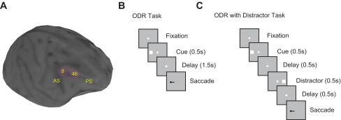
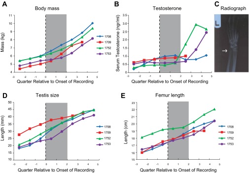
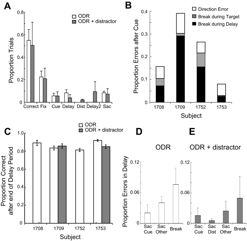
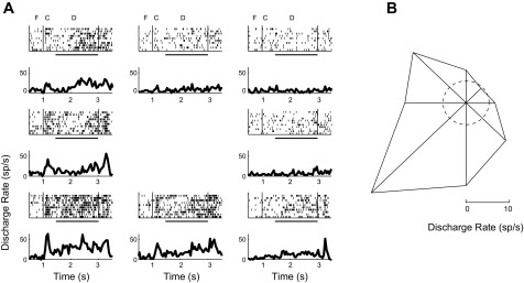
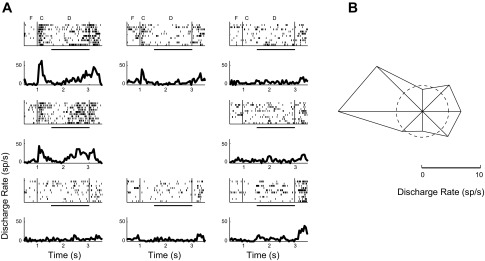
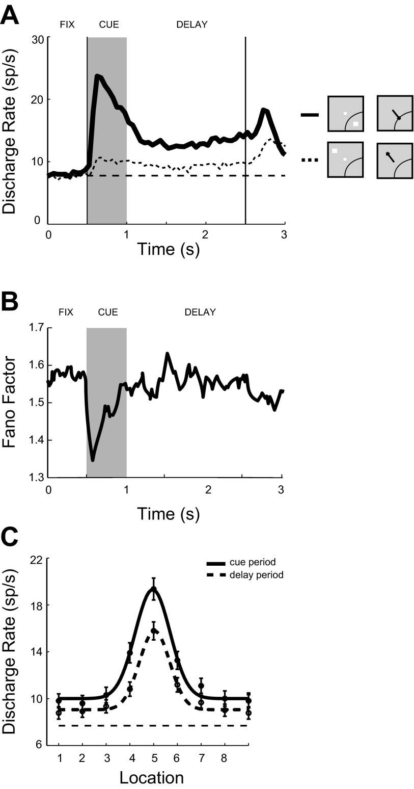
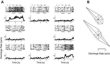
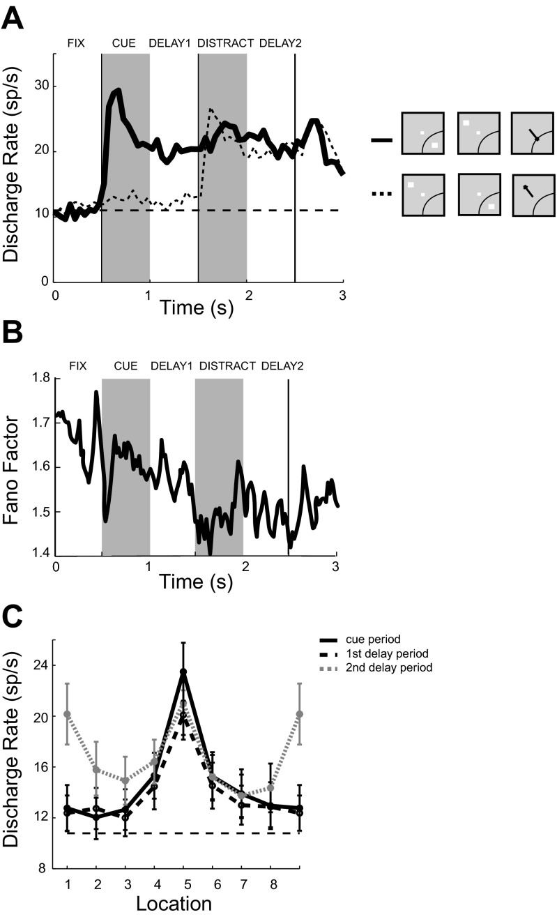
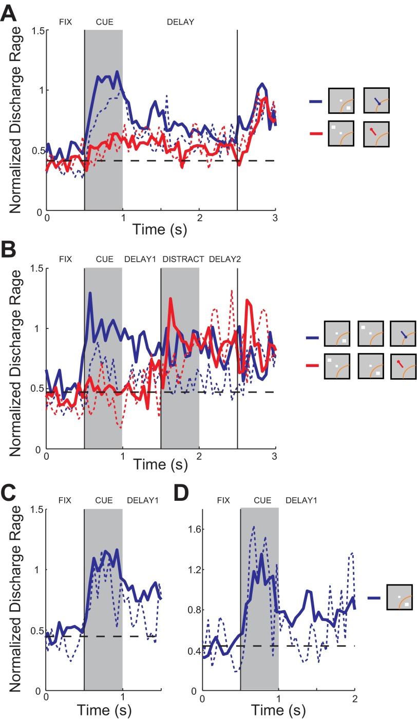
The dorsolateral prefrontal cortex matures late into adolescence or early adulthood. This pattern of maturation mirrors working memory abilities, which continue to improve into adulthood. However, the nature of the changes that prefrontal neuronal activity undergoes during this process is poorly understood. We investigated behavioral performance and neural activity in working memory tasks around the time of puberty, a developmental event associated with the release of sex hormones and significant neurological change. The developmental stages of male rhesus monkeys were evaluated with a series of morphometric, hormonal, and radiographic measures. Peripubertal monkeys were trained to perform an oculomotor delayed response task and a variation of this task involving a distractor stimulus. We found that the peripubertal monkeys tended to abort a relatively large fraction of trials, and these were associated with low levels of task-related neuronal activity. However, for completed trials, accuracy in the delayed saccade task was high and the appearance of a distractor stimulus did not impact performance significantly. In correct trials delay period activity was robust and was not eliminated by the presentation of a distracting stimulus, whereas in trials that resulted in errors the sustained cue-related activity was significantly weaker. Our results show that in peripubertal monkeys the prefrontal cortex is capable of generating robust persistent activity in the delay periods of working memory tasks, although in general it may be more prone to stochastic failure than in adults.
Keywords: development; eye movement; neurophysiology; persistent activity; principal sulcus.
Figures
Similar articles
-
Neural correlates of working memory development in adolescent primates.Nat Commun. 2016 Nov 9;7:13423. doi: 10.1038/ncomms13423. Nat Commun. 2016. PMID: 27827365 Free PMC article.
-
Matching patterns of activity in primate prefrontal area 8a and parietal area 7ip neurons during a spatial working memory task.J Neurophysiol. 1998 Jun;79(6):2919-40. doi: 10.1152/jn.1998.79.6.2919. J Neurophysiol. 1998. PMID: 9636098
-
Prefrontal task-related activity representing visual cue location or saccade direction in spatial working memory tasks.J Neurophysiol. 2002 Jan;87(1):567-88. doi: 10.1152/jn.00249.2001. J Neurophysiol. 2002. PMID: 11784772
-
Prefrontal cortex and working memory processes.Neuroscience. 2006 Apr 28;139(1):251-61. doi: 10.1016/j.neuroscience.2005.07.003. Epub 2005 Dec 1. Neuroscience. 2006. PMID: 16325345 Review.
-
Information processes in the primate prefrontal cortex in relation to working memory processes.Rev Neurosci. 2002;13(4):313-45. doi: 10.1515/revneuro.2002.13.4.313. Rev Neurosci. 2002. PMID: 12542260 Review.
Cited by
-
Drifts in Prefrontal and Parietal Neuronal Activity Influence Working Memory Judgments.Cereb Cortex. 2021 Jul 5;31(8):3650-3664. doi: 10.1093/cercor/bhab038. Cereb Cortex. 2021. PMID: 33822919 Free PMC article.
-
The meso-connectomes of mouse, marmoset, and macaque: network organization and the emergence of higher cognition.Cereb Cortex. 2024 May 2;34(5):bhae174. doi: 10.1093/cercor/bhae174. Cereb Cortex. 2024. PMID: 38771244 Free PMC article. Review.
-
Selective Loss of Thin Spines in Area 7a of the Primate Intraparietal Sulcus Predicts Age-Related Working Memory Impairment.J Neurosci. 2018 Dec 5;38(49):10467-10478. doi: 10.1523/JNEUROSCI.1234-18.2018. Epub 2018 Oct 24. J Neurosci. 2018. PMID: 30355632 Free PMC article.
-
Strong Gamma Frequency Oscillations in the Adolescent Prefrontal Cortex.J Neurosci. 2022 Apr 6;42(14):2917-2929. doi: 10.1523/JNEUROSCI.1604-21.2022. Epub 2022 Feb 23. J Neurosci. 2022. PMID: 35197317 Free PMC article.
-
Laminar pattern of adolescent development changes in working memory neuronal activity.bioRxiv [Preprint]. 2023 Jul 29:2023.07.28.550982. doi: 10.1101/2023.07.28.550982. bioRxiv. 2023. Update in: J Neurophysiol. 2023 Oct 1;130(4):980-989. doi: 10.1152/jn.00294.2023. PMID: 37546979 Free PMC article. Updated. Preprint.
References
-
- Alexander GE. Functional development of frontal association cortex in monkeys: behavioural and electrophysiological studies. Neurosci Res Prog Bull 20: 471–479, 1982 - PubMed
-
- Alexander GE, Goldman PS. Functional development of the dorsolateral prefrontal cortex: an analysis utilizing reversible cryogenic depression. Brain Res 143: 233–249, 1978 - PubMed
-
- Bercovitch FB. Dominance rank and reproductive maturation in male rhesus macaques (Macaca mulatta). J Reprod Fertil 99: 113–120, 1993 - PubMed
-
- Bourgeois JP, Goldman-Rakic PS, Rakic P. Synaptogenesis in the prefrontal cortex of rhesus monkeys. Cereb Cortex 4: 78–96, 1994 - PubMed
Publication types
MeSH terms
Grants and funding
LinkOut - more resources
Full Text Sources
Other Literature Sources