Review
. 2013 Oct;14(10):1014-22.
doi: 10.1038/ni.2703.

Innate and adaptive immune cells in the tumor microenvironment

Affiliations
Review

Innate and adaptive immune cells in the tumor microenvironment

Thomas F Gajewski et al. Nat Immunol. 2013 Oct.

Abstract

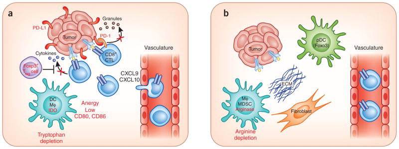
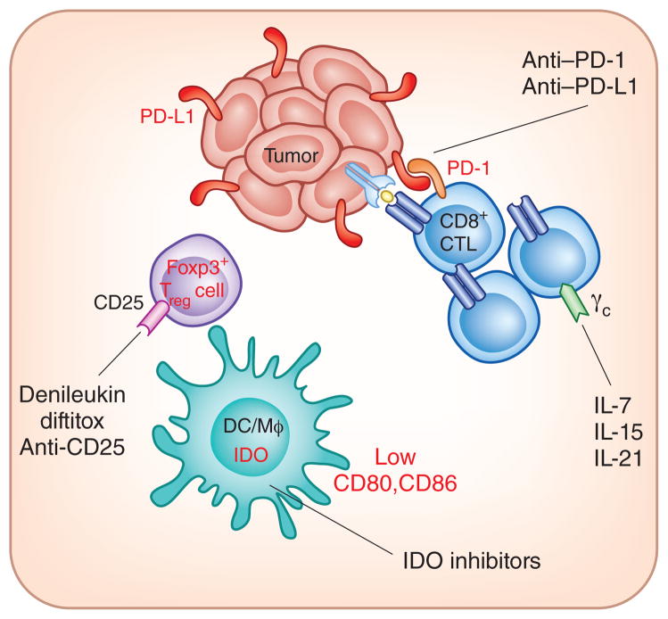
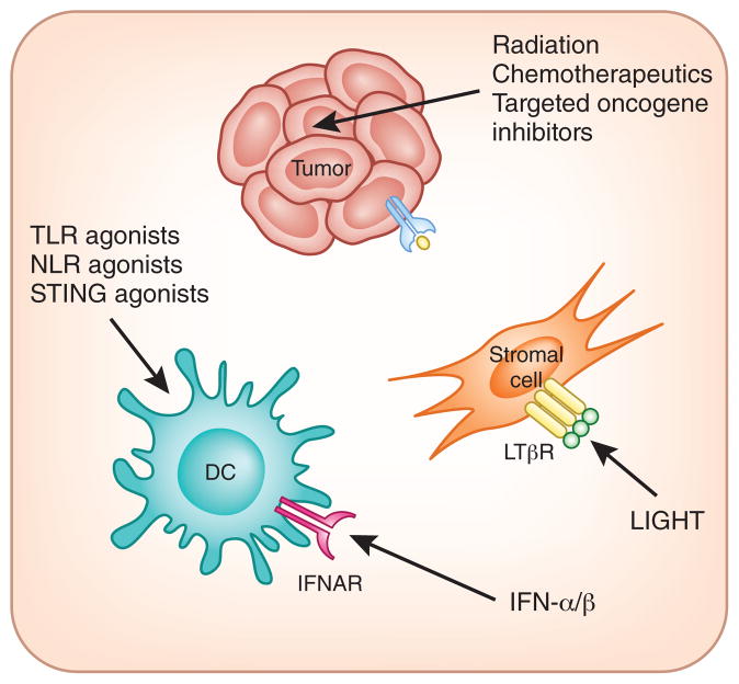
Most tumor cells express antigens that can mediate recognition by host CD8(+) T cells. Cancers that are detected clinically must have evaded antitumor immune responses to grow progressively. Recent work has suggested two broad categories of tumor escape based on cellular and molecular characteristics of the tumor microenvironment. One major subset shows a T cell-inflamed phenotype consisting of infiltrating T cells, a broad chemokine profile and a type I interferon signature indicative of innate immune activation. These tumors appear to resist immune attack through the dominant inhibitory effects of immune system-suppressive pathways. The other major phenotype lacks this T cell-inflamed phenotype and appears to resist immune attack through immune system exclusion or ignorance. These two major phenotypes of tumor microenvironment may require distinct immunotherapeutic interventions for maximal therapeutic effect.


Conflict of interest statement

COMPETING FINANCIAL INTERESTS

The authors declare no competing financial interests.

Figures

Figure 1
Figure 1
Working model for the segregation of tumors based on immune system regulatory pathways in the tumor microenvironment. (a) In T cell–infiltrated tumors, chemokines support influx of CD8+ effector T cells, but these subsequently become functionally inhibited by the effects of PD-L1, IDO, Treg cells and anergy. The development of this phenotype appears, in part, to be promoted by type I interferon signaling and the CD8 α+ DC lineage. (b) In non–T cell–infiltrated tumors, there is poor chemokine expression and lack of T cell infiltration but also minimal presence of defined immune inhibitory pathways. It is speculated that these tumors also have denser stroma and alternative myeloid or macrophage (MΦ) populations. Although this distinction has been best characterized in patients with melanoma, similar immune phenotypes may be operational in other solid tumors in which a subset of tumors shows T cell infiltration.
Figure 2
Figure 2
Therapeutic interventions being investigated that target immune inhibitory pathways in the tumor microenvironment. In melanoma, T cell–infiltrated tumors show the highest expression of PD-L1, IDO and Treg cells, and indirect evidence suggests T cell–intrinsic anergy as well. Monoclonal antibodies blocking PD-1–PD-L1 interactions, small-molecule inhibitors of IDO, CD25-targeting agents to deplete Treg cells and γc-binding cytokines to promote homeostatic proliferation of T cells and anergy reversal are all being investigated clinically.
Figure 3
Figure 3
Therapeutic strategies being considered that may promote appropriate inflammation and/or innate immune activation in the tumor microenvironment. Non–T cell–infiltrated tumors may require novel interventions to trigger innate immune activation and to facilitate signals for effector T cell trafficking. Radiation therapy, some chemotherapy drugs and targeted oncogene pathway inhibitors may achieve this effect through immunogenic cell death and altered tumor cell biology. The TNF superfamily member LIGHT induces chemokine production by stromal cells via binding to the LTβR. Agonists of innate immune pathways including TLRs, NLRs and STING are being considered to activate DCs and initiate productive priming of T cells. Introduction of type I interferon into tumor sites also may activate dendritic cells through the IFN-αR and/or IFN-βR and has additional effects on the tumor vasculature. IFNAR

References

    1. van der Bruggen P, et al. A gene encoding an antigen recognized by cytolytic T lymphocytes on a human melanoma. Science. 1991;254:1643–1647. - PubMed
    1. Topalian SL, et al. Recognition of shared melanoma antigens by human tumor-infiltrating lymphocytes. J Immunother. 1992;12:203–206. - PubMed
    1. Monach PA, Meredith SC, Siegel CT, Schreiber H. A unique tumor antigen produced by a single amino acid substitution. Immunity. 1995;2:45–59. - PubMed
    1. Hodi FS, et al. Improved survival with ipilimumab in patients with metastatic melanoma. N Engl J Med. 2010;363:711–723. - PMC - PubMed
    1. Brichard VG, Lejeune D. GSK’s antigen-specific cancer immunotherapy programme: pilot results leading to Phase III clinical development. Vaccine. 2007;25 (suppl. 2):B61–B71. - PubMed