. 2013 Nov;24(22):3496-510.
doi: 10.1091/mbc.E12-07-0539. Epub 2013 Sep 18.

Bves and NDRG4 regulate directional epicardial cell migration through autocrine extracellular matrix deposition

Affiliations

Bves and NDRG4 regulate directional epicardial cell migration through autocrine extracellular matrix deposition

Emily C Benesh et al. Mol Biol Cell. 2013 Nov.

Abstract

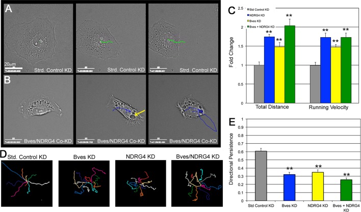
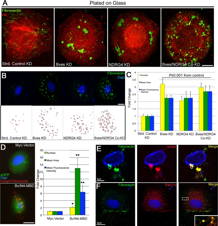
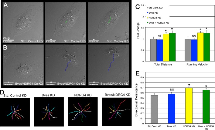
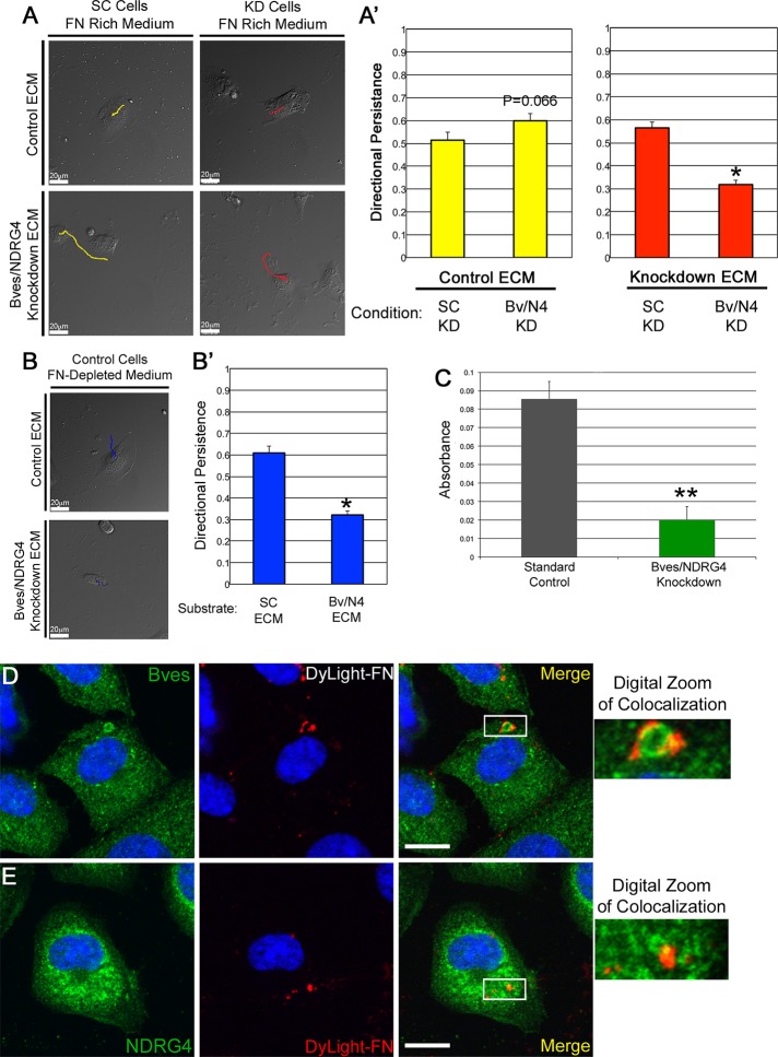
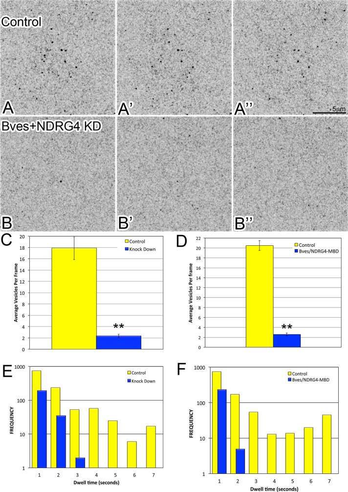
Directional cell movement is universally required for tissue morphogenesis. Although it is known that cell/matrix interactions are essential for directional movement in heart development, the mechanisms governing these interactions require elucidation. Here we demonstrate that a novel protein/protein interaction between blood vessel epicardial substance (Bves) and N-myc downstream regulated gene 4 (NDRG4) is critical for regulation of epicardial cell directional movement, as disruption of this interaction randomizes migratory patterns. Our studies show that Bves/NDRG4 interaction is required for trafficking of internalized fibronectin through the "autocrine extracellular matrix (ECM) deposition" fibronectin recycling pathway. Of importance, we demonstrate that Bves/NDRG4-mediated fibronectin recycling is indeed essential for epicardial cell directional movement, thus linking these two cell processes. Finally, total internal reflectance fluorescence microscopy shows that Bves/NDRG4 interaction is required for fusion of recycling endosomes with the basal cell surface, providing a molecular mechanism of motility substrate delivery that regulates cell directional movement. This is the first evidence of a molecular function for Bves and NDRG4 proteins within broader subcellular trafficking paradigms. These data identify novel regulators of a critical vesicle-docking step required for autocrine ECM deposition and explain how Bves facilitates cell-microenvironment interactions in the regulation of epicardial cell-directed movement.


Figures

FIGURE 1:
FIGURE 1:
NDRG4 protein interacts with Bves residues 307–316. (A) Exogenously expressed Bves-Myc protein pulled down NDRG4-GFP. NDRG4-GFP did not pull down in the absence of Bves-Myc. (B) NDRG4-GFP was pulled down by GST-Bves357 (amino acids 115–357), whereas other truncations of GST-Bves357 (amino acids 115 to 218, 275, or 300, respectively) or GST-only control failed to pull down NDRG4-GFP. (B′) GFP-vector lysates fail to be pulled down by GST-Bves357 or other truncations. (C) In the SPOTs assay, 13-mer synthesized peptides from amino acid 115 to the end of the Bves protein were fused to a cellulose membrane, with 10–amino acid overlap between adjacent peptides. GFP-NDRG4 lysate binds to three consecutive Bves peptides on the cellulose membrane, peptides 63–65 (depicted in grid overlay), corresponding to Bves residues 307–316: HHFLRGSSST. (D) A domain from Bves residues 292–331, containing the Bv/N4-MBD identified in C, is cloned into the myc-3tag vector. n ≥ 4 assays/experiment.
FIGURE 2:
FIGURE 2:
Bves and NDRG4 colocalize and interact in mammalian epicardial cells. (A) Bves and NDRG4 colocalize in trabecular cardiomyocytes (merge) and epicardial cells (box denotes digitally zoom inset) from E14.5 murine heart sections. Scale, 75 μm. (B) A column cross-linked to Bves antibody pulls down NDRG4 protein from epicardial cell lysate, whereas another antibody, MF-20, does not; n ≥ 4 pull downs. (C) Epicardial cells at low confluence exhibit Bves and NDRG4 colocalization in plasma membrane protrusions (box denotes digitally zoomed inset); graph depicts colocalization in confocal images (D). Punctate, vesicle-like colocalization of Bves and NDRG4 are apparent in high-magnification confocal images of epicardial cells with motile morphology (box denotes digitally zoomed inset; white arrows demarcate vesicle-like structure). (E) Bves/NDRG4 fail to colocalize in confocal images of epicardial cell sheets (box denotes inset of cell borders). Epi, epicardium; Myo, myocardium; optical slices, 1 AU. Colocalizations quantified by ImageJ intensity profiling, Bar, 10 μm or as marked.
FIGURE 3:
FIGURE 3:
The Bves and NDRG4 interaction regulates epicardial cell movement. 3′UTR targeting NDRG4 and Bves oligos specifically reduce protein levels of each gene, respectively (A, B). Cyclophilin blots are protein loading controls (α-cyclophilin). (A′) Densitometry analysis of three blots demonstrates significant depletion of NDRG4 from NDRG4 and Bves + NDRG4 siRNA–treated epicardial cells (graphs relative to SC levels; p < 0.05; NS, not significant). (B′) Significant depletion of Bves is demonstrated from three blots in Bves and Bves + NDRG4 siRNA–treated epicardial cells (*p < 0.05; NS, not significant). (C) Overexpressed Bv/N4-MBD localizes in cytoplasmic puncta in epicardial cells. Bar, 10 μm. (D) Single-pixel colocalization of the Bv/N4-MBD construct (green) with native NDRG4 protein (red) in multiple Z-planes is (E) demarcated by white pixels (0.5-μm optical slices; bar, 10 μm). (F) Random epicardial transwell migration is elevated with Bves/NDRG4 siRNA treatment and rescued by stable overexpression of each gene. (G) Expression of Bves-full length plasmid (Myc-BvFL) rescues transwell migration of epicardial cells expressing Bves siRNA, whereas expression of a Bves plasmid truncated at residue 275 (Myc-Bv275) does not. (H) Overexpression of two clones of the Bv/N4-MBD construct (Bv/N4-MBD1 and 2, respectively) induces an increase in random cell motility due to disruption of Bves–NDRG4 interaction. For F–H, n ≥ 3 assays/experiment, *p ≤ 0.05, **p ≤ 0.0003, from SCs.
FIGURE 4:
FIGURE 4:
Bves and NDRG4 coregulate epicardial cell directional persistence and fibronectin trafficking. Lines overlaid on time-lapse movies using manual tracking analysis (ImageJ) demarcate cell movements per minute. (A) The migration pattern of epicardial cells treated with SC siRNAs is directional. (B) The migration pattern of Bves/NDRG4 co-knockdown epicardial cells shows increased distance traveled and lacks directionality compared with controls. Cells also retain large cytoplasmic vacuoles (yellow arrow). (C) Bves and/or NDRG4 knockdown causes significantly accelerated cell movement speed and increased distance traveled. (D) An overlay of 10 representative tracks of cells illustrates the randomized cell movements due to Bves and/or NDRG4depletion. Scale bars, 20 μm; n ≥ 40 cells/condition. (E) Directional persistence is highly impaired in Bves- and/or NDRG4-depleted epicardial cells (1 = highly directional, 0 = random); **p ≤ 0.001.
FIGURE 5:
FIGURE 5:
Bves/NDRG4-depleted cells contain large fibronectin-positive objects in late endosomal/lysosomal and recycling endosome compartments. (A) Endogenous fibronectin-positive cytoplasmic objects are apparent in Bves- and/or NDRG4-depleted epicardial cells (DiI, red; fibronectin antibody, green). (B, C) Objects were measured using ImageJ particle analysis (top, original image; bottom, included particles). Bves- and/or NDRG4-depleted cells have significantly more fibronectin-positive objects that are larger with stronger fluorescence intensity compared with SC. (D) Bv/N4-MBD epicardial cells demonstrate increased number of internalized cytoplasmic DyLight-fibronectin 550 granules that are larger, with stronger fluorescence intensity compared with controls, *p ≤ 0.001, **p ≤ 0.0001, from respective control cells. (E) Bves/NDRG4-depleted epicardial cells exhibit fibronectin-positive objects (green) traffic in late endosomal/lysosomal compartments labeled with Lamp2 (red) in. Bar, 5 μm. (F) Fibronectin-positive objects (green) are also apparent in recycling endosomes labeled with Rab11a (red; box denotes inset) in these cells. Scale bars, 10 μm unless otherwise indicated, n ≥ 50 cells/condition; optical slices, 1 AU.
FIGURE 6:
FIGURE 6:
Homogeneous fibronectin substrate rescues Bves and NDRG4 migration defects. Epicardial cells are imaged and tracked as in Figure 4. Highly directional movement of SC-treated (A) epicardial cells is apparent on an evenly distributed fibronectin substrate as well as in Bves/NDRG4-codepleted cells (B) over a 2-h time course. (C) Migration distance and velocity of Bves- and/or NDRG4-depleted epicardial cells are only partially restored on fibronectin substrate. (D) Epicardial cells exhibit highly directional movement on fibronectin substrates regardless of siRNA treatment condition, as represented by 10 paths stacked together. Scale bars, 20 μm. *p ≤ 0.05; n ≥ 44 cells/condition; NS, not significant. (E) Bves-depleted cell directionality is fully rescued by fibronectin substrates as calculated by directional persistence quantifications. NDRG4/Bves codepleted cells exhibit improved directionality compared with controls. n = 3 experiments/condition; *p < 0.05.
FIGURE 7:
FIGURE 7:
Bves and NDRG4 coregulate fibronectin deposition. Matrix deposition is probed using a cell-free ECM assay, which removes cells but keeps the ECM. Migration of newly plated cells is then tracked as in Figures 4 and 6 on previously produced ECM to test the function of residual matrix. (A) On control-produced ECM, control and Bves/NDRG4-depleted epicardial cells move directionally (A′). On Bves/NDRG4-depleted ECM, control cells move directionally while Bves/NDRG4-depleted cells have poor directional persistence (A′; *p < 0.01). (B) Control epicardial cells fed medium lacking fibronectin move directionally on control ECM and fail to move directionally (B′) on Bves/NDRG4-depleted ECM (*p < 0.01). This differs from A, in which controls overcome Bves/NDRG4-depleted ECM deficiency. (C) Biotin-labeled fibronectin deposition onto a glass surface is impaired in Bves/NDRG4-depleted cells compared with controls (n = 7 assays, **p < 0.001). (D, E) Epicardial cells internalize DyLight-550–labeled fibronectin (DyLight-FN) after a 1-h pulse. Endogenous Bves (D) and NDRG4 (E) colocalize with DyLight-FN on vesicular structures; boxes in merge denote digitally zoomed region. Optical slices, 1 AU; scale, 10 μm; n ≥ 50 cells/assay.
FIGURE 8:
FIGURE 8:
Bves/NDRG4 interaction is required for long-term exocytic vesicle docking at the plasma membrane. Three consecutive time-lapse TIRF microscopy images of control (A–A′′) and Bves/NDRG4 (B–B′′) knockdown cells transfected with Vamp3-GFP (black dots). Control cells contain numerous VAMP3 vesicles (C; yellow, 17.9) at the membrane over the time course, whereas Bves/NDRG4 (C; blue, 2.3) knockdown cells contain significantly fewer (**p ≤ 0.0001) average vesicles per movie frame. Similarly, vector control–transfected cells contain significantly more vesicles per frame than Bves/NDRG4-treated cells (D; yellow, 20.6; blue, 2.6; **p ≤ 0.0001). Quantification of dwell time separated into 1-s bins (E) reveals that vesicles in Bves/NDRG4-knockdown cells do not reside at the membrane as long as VAMP3 vesicles do in SC-treated cells. Dwell-time quantifications of cells coexpressing VAMP3-GFP and control myc-3tag vector (F, yellow) or Bv/N4-MBD (blue) plasmids indicate that vesicles do not reside at the membrane as long in the presence of Bv/N4-MBD; n ≥ 15 movies per condition.

Similar articles

Cited by

References

    1. Andrée B, Fleige A, Arnold HH, Brand T. Mouse Pop1 is required for muscle regeneration in adult skeletal muscle. Mol Cell Biol. 2002;22:1504–1512. - PMC - PubMed
    1. Andrée B, Hillemann T, Kessler-Icekson G, Schmitt-John T, Jockusch H, Arnold HH, Brand T. Isolation and characterization of the novel Popeye gene family expressed in skeletal muscle and heart. Dev Biol. 2000;223:371–382. - PubMed
    1. Bader D, Masaki T, Fischman DA. Immunochemical analysis of myosin heavy chain during avian myogenesis in vivo and in vitro. J Cell Biol. 1982;95:763–770. - PMC - PubMed
    1. Berger P, Niemann A, Suter U. Schwann cells and the pathogenesis of inherited motor and sensory neuropathies (Charcot-Marie-Tooth disease) Glia. 2006;54:243–257. - PubMed
    1. Choi S-C, et al. NDRG2 is one of novel intrinsic factors for regulation of IL-10 production in human myeloid cell. Biochem Biophys Res Commun. 2010;396:684–690. - PubMed

Publication types

MeSH terms

LinkOut - more resources