. 2013 Nov 6;3(11):2015-29.
doi: 10.1534/g3.113.008466.

Extension of the Caenorhabditis elegans Pharyngeal M1 neuron axon is regulated by multiple mechanisms

Affiliations

Extension of the Caenorhabditis elegans Pharyngeal M1 neuron axon is regulated by multiple mechanisms

Osama Refai et al. G3 (Bethesda). .

Abstract

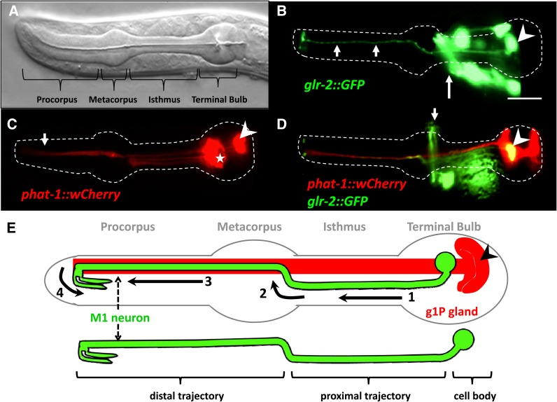
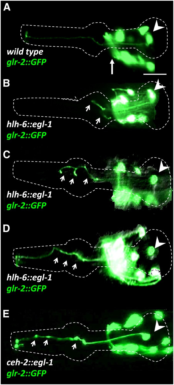
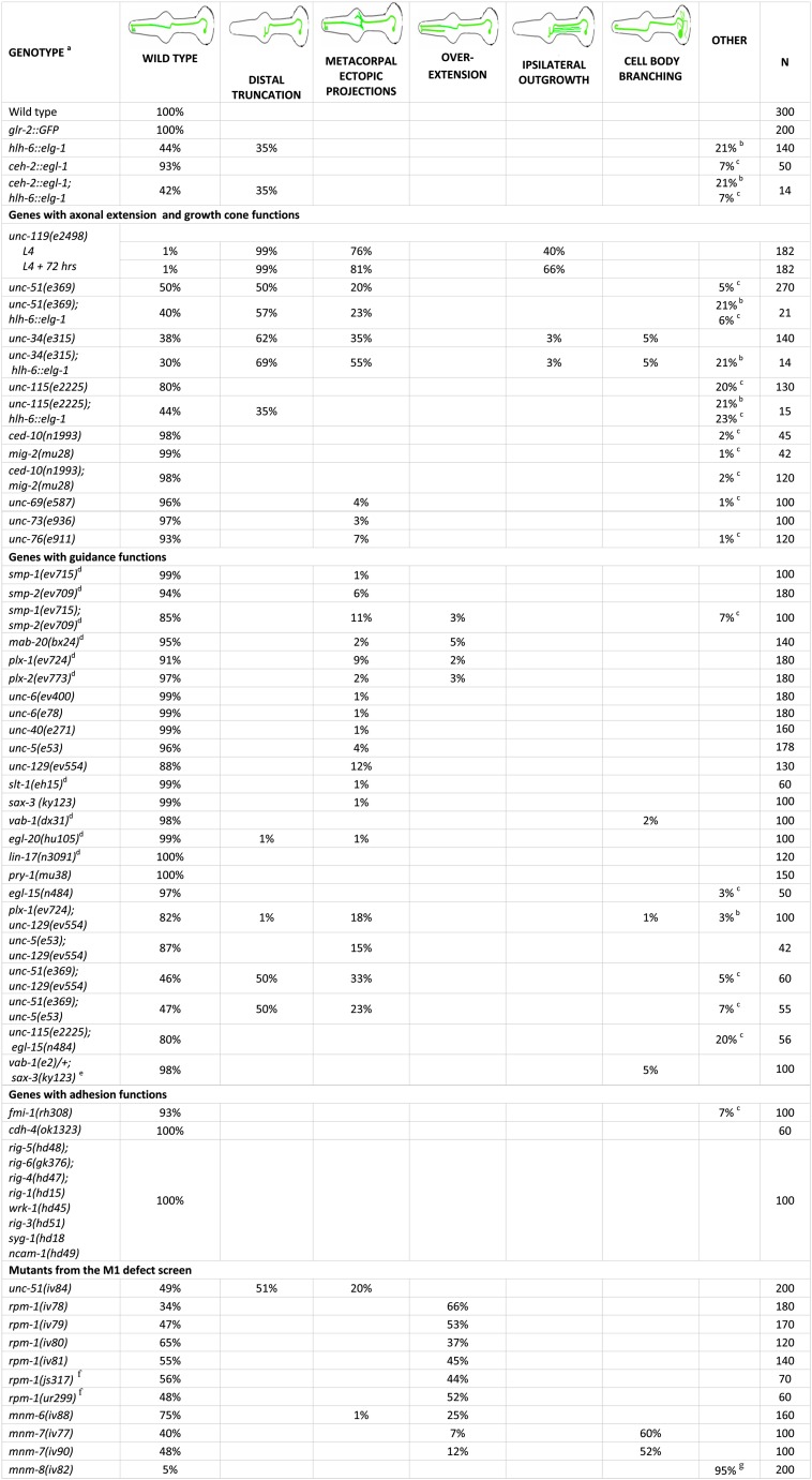
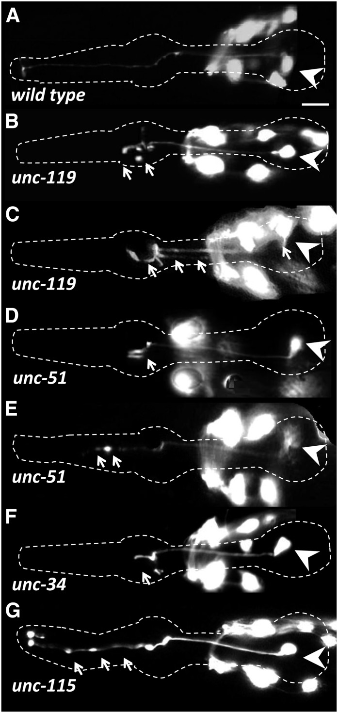
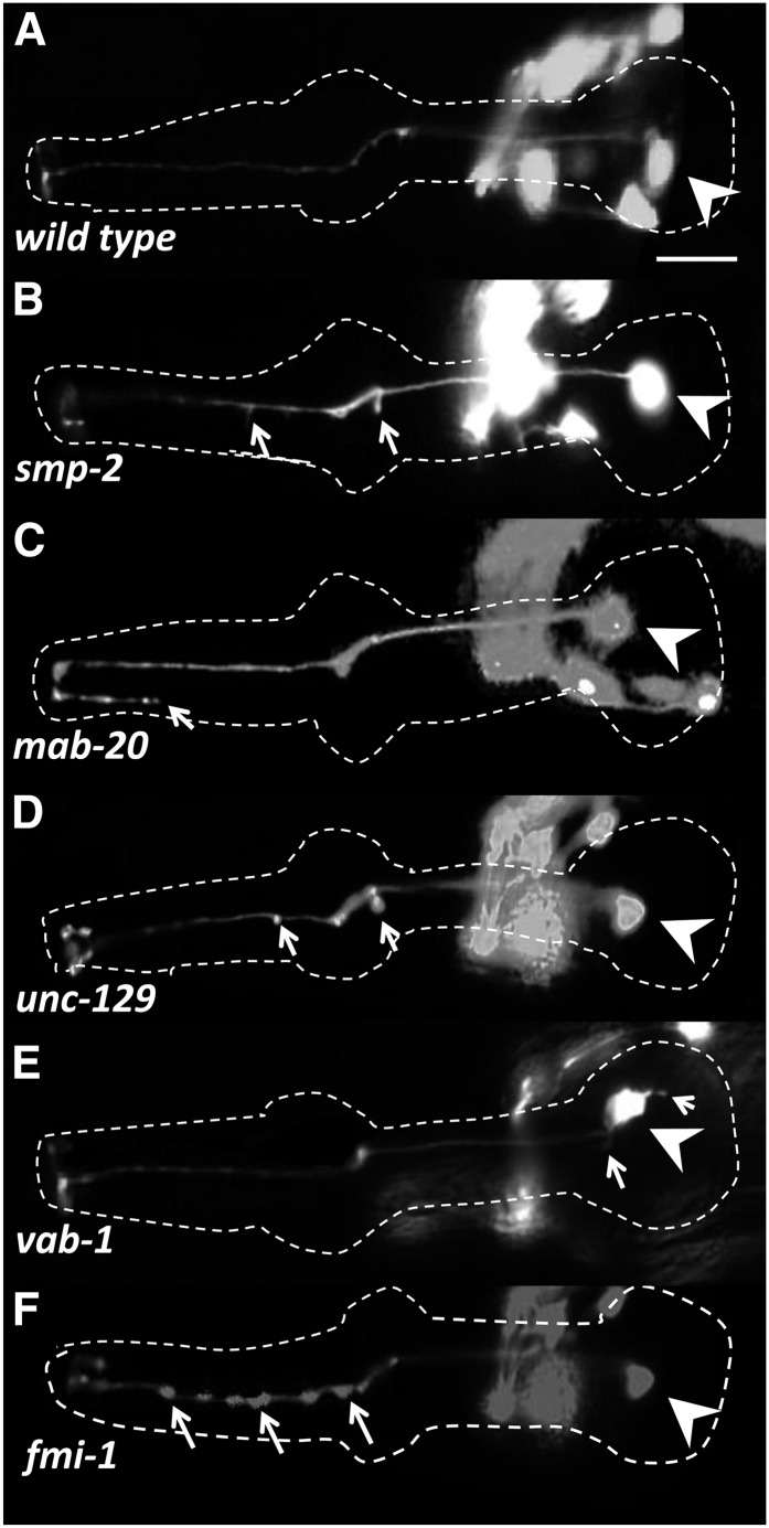
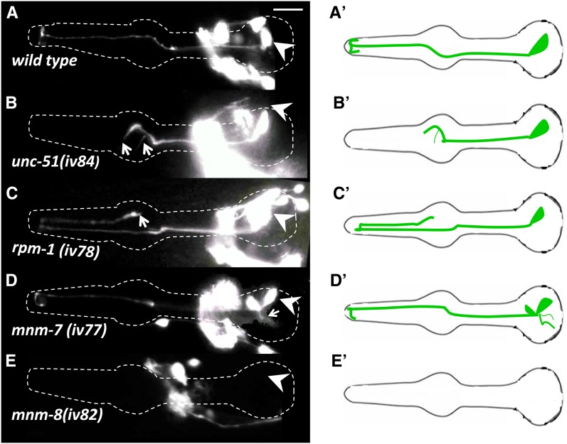
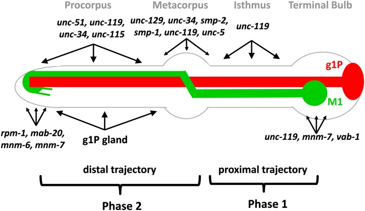
The guidance of axons to their correct targets is a critical step in development. The C. elegans pharynx presents an attractive system to study neuronal pathfinding in the context of a developing organ. The worm pharynx contains relatively few cells and cell types, but each cell has a known lineage and stereotyped developmental patterns. We found that extension of the M1 pharyngeal axon, which spans the entire length of the pharynx, occurs in two distinct phases. The first proximal phase does not require genes that function in axon extension (unc-34, unc-51, unc-115, and unc-119), whereas the second distal phase does use these genes and is guided in part by the adjacent g1P gland cell projection. unc-34, unc-51, and unc-115 had incompletely penetrant defects and appeared to act in conjunction with the g1P cell for distal outgrowth. Only unc-119 showed fully penetrant defects for the distal phase. Mutations affecting classical neuronal guidance cues (Netrin, Semaphorin, Slit/Robo, Ephrin) or adhesion molecules (cadherin, IgCAM) had, at best, weak effects on the M1 axon. None of the mutations we tested affected the proximal phase of M1 elongation. In a forward genetic screen, we isolated nine mutations in five genes, three of which are novel, showing defects in M1, including axon overextension, truncation, or ectopic branching. One of these mutations appeared to affect the generation or differentiation of the M1 neuron. We conclude that M1 axon extension is a robust process that is not completely dependent on any single guidance mechanism.

Keywords: Caenorhabditis elegans; axon; genetics; neuron; pharynx.


Figures

Figure 1
Figure 1
Anatomy of the pharynx showing the M1 neuron and the g1P gland in L4 or young adult. (A) Nomarski image of the C. elegans pharynx. (B) Fluorescence micrographs of M1 marked by the glr-2::gfp reporter (arrowhead). Other nonpharyngeal green cells are part of the nerve ring (long arrow) and can be distinguished from M1 by position. Note the M1 projection into the procorpus (short arrows). (C) Fluorescence micrographs of the pharyngeal gland cells marked by the phat-1::wCherry reporter. The g1P cell body (arrowhead) is located in the terminal bulb and extends a projection to the procorpus (arrow). Other gland cells are also marked with this reporter (★). (D) Double transgenic animals with a gland-expressed phat-1::wCherry and the glr-2::gfp. Arrowhead indicates the M1 cell body and the arrow denotes the nonpharyngeal cells in the nerve ring. (E) Diagram of the C. elegans pharynx (outlined in gray) with the M1 neuron (green) and g1P gland (red). The different portions of the M1 axon trajectory are numbered from 1 through 4 (see text). Modified from (Albertson and Thomson 1976). Scale bar in (B) = 10 µm.
Figure 2
Figure 2
M1 axon phenotypes (marked with glr-2::gfp) in wild-type (A) and gland-ablated hlh-6::egl-1 (B–D) or in I3 neuron-ablated ceh-2::egl-1 transgenics (E). Arrowhead denotes the M1 cell body and the long arrow in wild-type (A) indicates the nonpharyngeal cells in the nerve ring in these at L4 or young adults. Short arrows highlight M1 defects. (B–D) In 56% of gland-ablated transgenics, the M1 axon is either truncated (B) with abnormal branching in the metacorpus and procorpus (C) or follows an abnormal trajectory in the procorpus (D). (E) In 7% of I3 ablated animals, the M1 axon in the procorpus exhibits an abnormal shape with GFP swelling (arrows), although the axon always extended fully. Scale bar in (A) = 10 µm.
Figure 3
Figure 3
Quantification of M1 axon phenotypes are shown for mutants or after cell ablation. All values are in percents, and for each category a cartoon showing representative examples of the axonal phenotypes is shown. Footnotes on the figure are as follows. (A) All strains included glr-2::gfp, except as noted. Some neurons had multiple abnormalities and therefore some genotypes sum to >100%. (B) M1 showed abnormal trajectories in the procorpus. (C) Axons showed swelling or small ectopic projections in the procorpus. (D) Independent glr-2::gfp transgenes were made for each of these strains. (E) This was the maternal genotype as vab-1(e2); sax-3(ky123) homozygous segregants arrest before the stage when M1 can be scored. Note that two-thirds of scored progeny will be vab-1/+; sax-3. (F) These rpm-1 alleles were identified independently of this screen. (G) M1 neuron was absent as scored by the absence of glr-2::gfp in the pharynx.
Figure 4
Figure 4
Time course measuring the efficiency and timing of the gland killing in hlh-6::egl-1 transgenics. Examples of 1.5-fold (first row), two-fold (second row), and three-fold (third row) embryos are shown. First column (A, D, G) shows Nomarski images, second column (B, E, H) shows wild-type glands visualized using the hlh-6::yfp reporter, and the third column (C, F, I) represents hlh-6::egl-1 transgenics at similar developmental stages. Note the absence of the hlh-6::yfp reporter in the glandless embryos (C, F, I). Arrow indicates gland cell bodies and arrowheads indicate the g1P projection. Intestine expressing elt-2::tdCherry transgene marker (*). Scale bar in (G) = 5 µm.
Figure 5
Figure 5
M1 axon abnormalities in growth cone defective mutants in L4 or adult stages. The M1 neurons express the glr-2::gfp reporter. The pharynx is outlined in each panel, arrowheads indicate the M1 cell body, and arrows highlight M1 defects. Other neurons visible in the figure are nonpharyngeal. Note that with the exception of unc-119, all mutations show incomplete penetrance (Figure 3). (A) Wild-type M1 neuron. (B) The unc-119 young adult exhibits axon truncation in metacorpus with abnormal branching. (C) Older unc-119 animals (L4 + 72 hr) exhibit axon truncations, bilateral branches in the isthmus, and abnormal branches from the cell body. The unc-51 mutants exhibit axon truncations in metacorpus (D) or procorpus (E). (F) The unc-34 mutants with a truncation of the M1 axon at the metacorpus accompanied with short branches. (G) The unc-115 animals exhibit swelling of M1 axon throughout the procorpus and metacorpus. Scale bar in (A) = 10 µm.
Figure 6
Figure 6
M1 axon phenotypes in L4 or young adult guidance and adhesion mutants. The M1 neuron expresses the glr-2::gfp reporter. The pharynx is outlined in each panel; arrowheads indicate the M1 cell body and arrows highlight M1 defects. Other neurons visible in the figure are nonpharyngeal cells that are part of the nerve ring. Note that penetrance is low in all cases (Figure 3). (A) Wild-type (this is the same image as shown in Figure 4A). (B) The smp-2 exhibits small projections at the procorpus and metacorpus. (C) The mab-20 shows overextensions of M1 terminal branches beyond their normal targets. (D) The unc-129 has short projections and swelling of the M1 axon in the metacorpus. (E) The vab-1 mutant has an apparently normal M1 axon, but one that exhibits abnormal minor branches from the cell body. (F) The fmi-1 mutant shows abnormal swelling of the M1 axon in the procorpus. Scale bar in (A) = 10 µm.
Figure 7
Figure 7
Phenotypes of mutants isolated from the forward genetic screen visualized in L4 or young adults. M1 is marked with glr-2::gfp. Arrowheads indicate cell bodies and arrows denote defects, which are incompletely penetrant in all cases (Figure 3). Other neurons visible in the figure are part of the nonpharyngeal cells in the nerve ring. (A′–E′) Cartoons for each phenotypic class. (A) Wild-type. (B) The unc-51(iv84) shows axon truncation and an ectopic branch. (C) The rpm-1(iv78) results in overextension of M1 terminal branches beyond their normal targets. (D) The mnm-7(iv77) shows ectopic branching from the cell body. (E) The mnm-8(iv82) shows a 95% penetrant M1 missing phenotype, whereas the other neurons that express the glr-2::gfp reporter are still present. Scale bar in (A) = 10 µm.
Figure 8
Figure 8
Model for M1 axon guidance. M1 is shown in green and g1P is shown in red. M1 development is divided into two steps. During phase 1, M1 builds its proximal trajectory independent of genes affecting growth cones. In phase 2, M1 builds its distal trajectory, which is affected in growth cone–defective mutants and loss of the g1P cell. The regions where M1 axon phenotypes occur are shown for each gene. These genes are categorized in groups, which act in metacorpus, procorpus, and or at the anterior pharyngeal tip, where the axon terminates. The unc-119 (which also acts for distal extension), mnm-7, and vab-1 are necessary for preventing ectopic branching in the isthmus or terminal bulb, rather than axon extension in those regions.

References

    1. Albertson D. G., Thomson J. N., 1976. The pharynx of Caenorhabditis elegans. Philos. Trans. R. Soc. Lond. B Biol. Sci. 275: 299–325 - PubMed
    1. Altun-Gultekin Z., Andachi Y., Tsalik E. L., Pilgrim D., Kohara Y., et al. , 2001. A regulatory cascade of three homeobox genes, ceh-10, ttx-3 and ceh-23, controls cell fate specification of a defined interneuron class in C. elegans. Development 128: 1951–1969 - PubMed
    1. Araujo S. J., Tear G., 2003. Axon guidance mechanisms and molecules: lessons from invertebrates. Nat. Rev. Neurosci. 4: 910–922 - PubMed
    1. Aronoff R., Mellem J. E., Maricq A. V., Sprengel R., Seeburg P. H., 2004. Neuronal toxicity in Caenorhabditis elegans from an editing site mutant in glutamate receptor channels. J. Neurosci. 24: 8135–8140 - PMC - PubMed
    1. Arvanitis D., Davy A., 2008. Eph/ephrin signaling: networks. Genes Dev. 22: 416–429 - PMC - PubMed

Publication types

Substances

LinkOut - more resources