

The human cerebral cortex flattens during adolescence
- PMID: 24048830
- PMCID: PMC6618418
- DOI: 10.1523/JNEUROSCI.1459-13.2013
The human cerebral cortex flattens during adolescence
Abstract
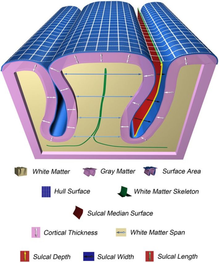
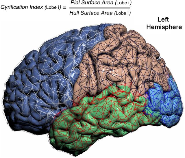
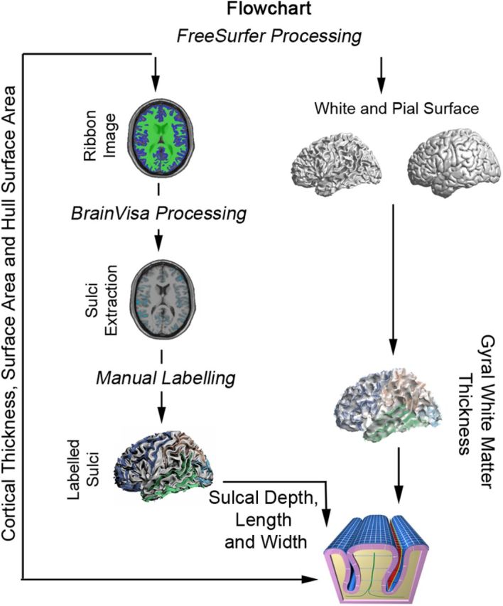
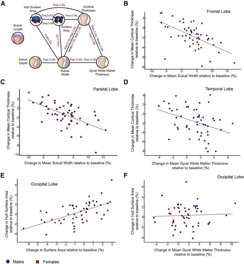
The human cerebral cortex appears to shrink during adolescence. To delineate the dynamic morphological changes involved in this process, 52 healthy male and female adolescents (11-17 years old) were neuroimaged twice using magnetic resonance imaging, approximately 2 years apart. Using a novel morphometric analysis procedure combining the FreeSurfer and BrainVisa image software suites, we quantified global and lobar change in cortical thickness, outer surface area, the gyrification index, the average Euclidean distance between opposing sides of the white matter surface (gyral white matter thickness), the convex ("exposed") part of the outer cortical surface (hull surface area), sulcal length, depth, and width. We found that the cortical surface flattens during adolescence. Flattening was strongest in the frontal and occipital cortices, in which significant sulcal widening and decreased sulcal depth co-occurred. Globally, sulcal widening was associated with cortical thinning and, for the frontal cortex, with loss of surface area. For the other cortical lobes, thinning was related to gyral white matter expansion. The overall flattening of the macrostructural three-dimensional architecture of the human cortex during adolescence thus involves changes in gray matter and effects of the maturation of white matter.
Figures
References
-
- American Psychiatric Association. Diagnostic and statistical manual of mental disorders. Washington, DC: American Psychiatric Association; 1994.
-
- Benjamini Y, Hochberg Y. Controlling the false discovery rate: A practical and powerful approach to multiple testing. J R Stat Soc B. 1995;57:289–300.
-
- Brouwer RM, Mandl RC, Schnack HG, van Soelen IL, van Baal GC, Peper JS, Kahn RS, Boomsma DI, Hulshoff Pol HE. White matter development in early puberty: a longitudinal volumetric and diffusion tensor imaging twin study. PLoS One. 2012;7:e32316. doi: 10.1371/journal.pone.0032316. - DOI - PMC - PubMed
Publication types
MeSH terms
LinkOut - more resources
Full Text Sources
Other Literature Sources