. 2013 Sep 18;33(38):15248-58.
doi: 10.1523/JNEUROSCI.6361-11.2013.

Cyclooxygenase-1-dependent prostaglandins mediate susceptibility to systemic inflammation-induced acute cognitive dysfunction

Affiliations

Cyclooxygenase-1-dependent prostaglandins mediate susceptibility to systemic inflammation-induced acute cognitive dysfunction

Éadaoin W Griffin et al. J Neurosci. .

Abstract

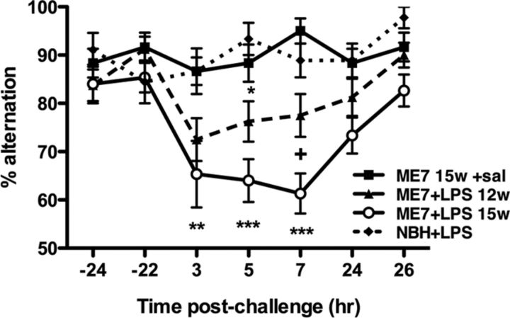
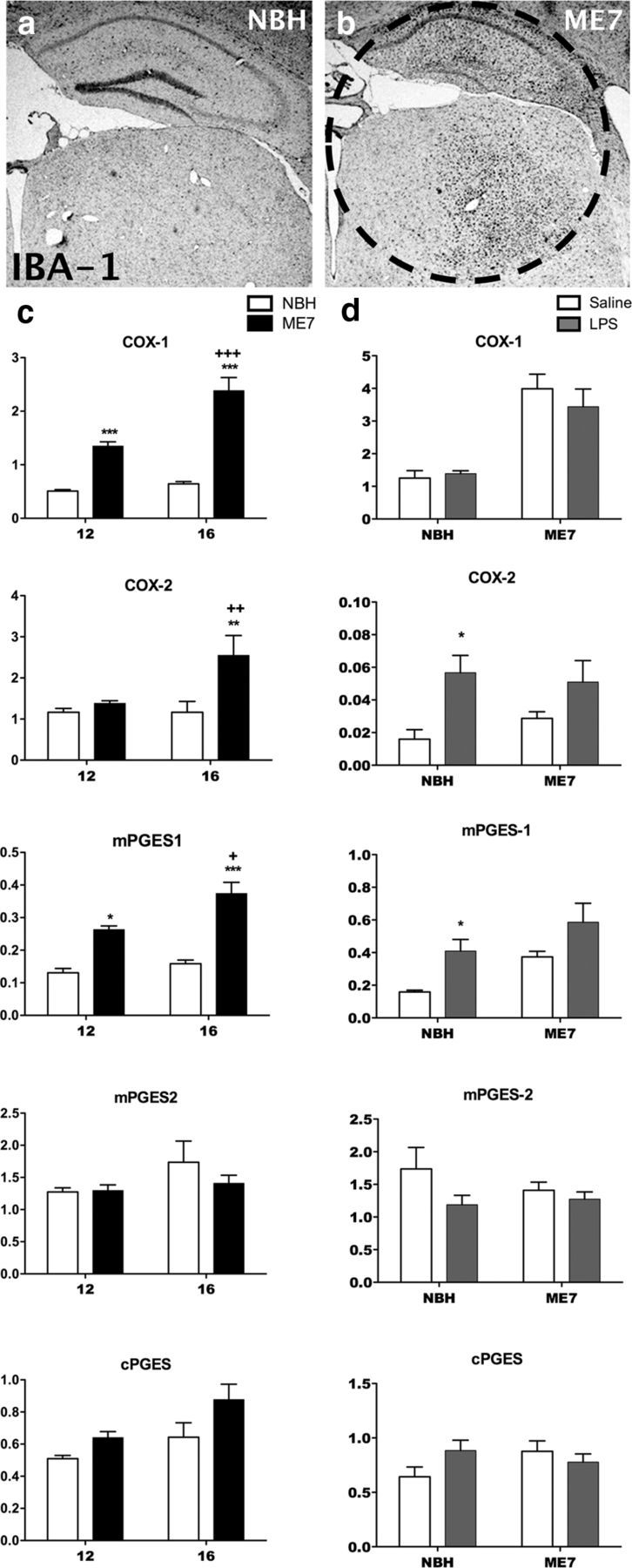
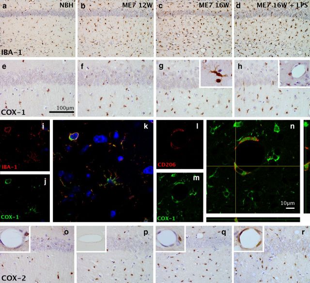
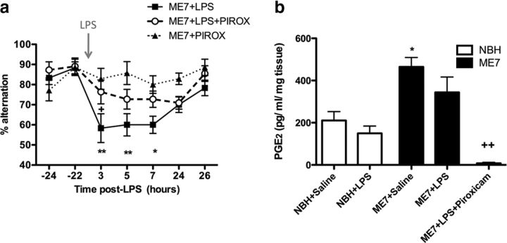
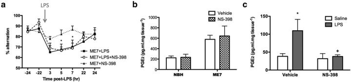
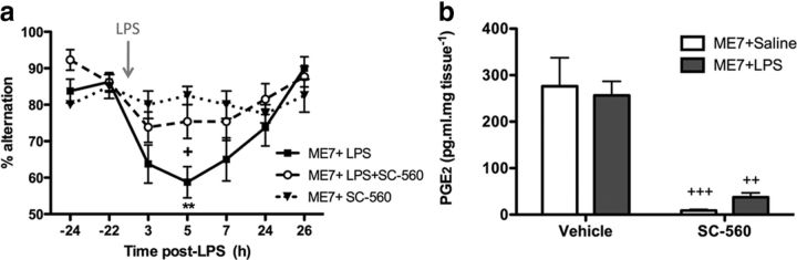
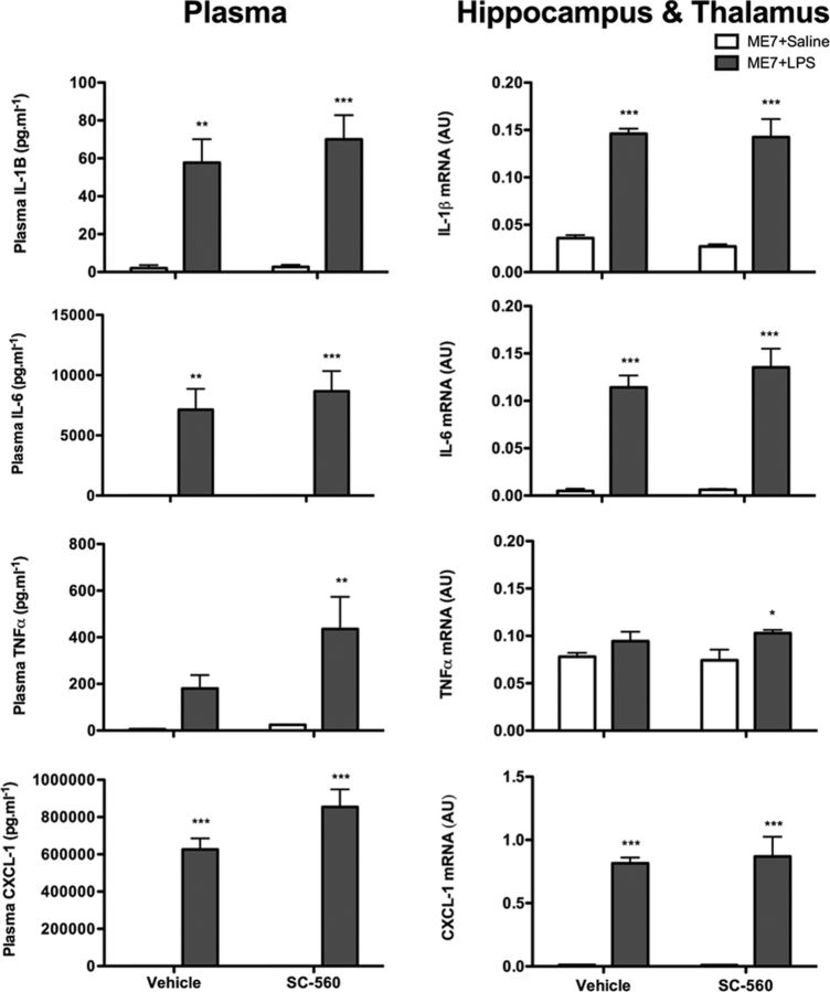
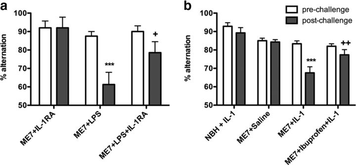
Systemic inflammatory events often precipitate acute cognitive dysfunction in elderly and demented populations. Delirium is a highly prevalent neuropsychiatric syndrome that is characterized by acute inattention and cognitive dysfunction, for which prior dementia is the major predisposing factor and systemic inflammation is a frequent trigger. Inflammatory mechanisms of delirium remain unclear. We have modeled aspects of delirium during dementia by exploiting progressive neurodegeneration in the ME7 mouse model of prion disease and by superimposing systemic inflammation induced by the bacterial endotoxin lipopolysaccharide (LPS). Here, we have used this model to demonstrate that the progression of underlying disease increases the incidence, severity, and duration of acute cognitive dysfunction. This increasing susceptibility is associated with increased CNS expression of cyclooxygenase (COX)-1 in microglia and perivascular macrophages. The COX-1-specific inhibitor SC-560 provided significant protection against LPS-induced cognitive deficits, and attenuated the disease-induced increase in hippocampal and thalamic prostaglandin E2, while the COX-2-specific inhibitor NS-398 was ineffective. SC-560 treatment did not alter levels of the proinflammatory cytokines interleukin (IL)-1β, tumor necrosis factor-α, IL-6, or C-X-C chemokine ligand 1 in blood or brain, but systemic IL-1RA blocked LPS-induced cognitive deficits, and systemic IL-1β was sufficient to induce similar deficits in the absence of LPS. Furthermore, the well tolerated COX inhibitor ibuprofen was protective against IL-1β-induced deficits. These data demonstrate that progressive microglial COX-1 expression and prostaglandin synthesis can underpin susceptibility to cognitive deficits, which can be triggered by systemic LPS-induced IL-1β. These data contribute to our understanding of how systemic inflammation and ongoing neurodegeneration interact to induce cognitive dysfunction and episodes of delirium.


Figures

Figure 1.
Figure 1.
Disease progression increases the incidence, severity, and duration of acute exacerbations of cognitive dysfunction. Working memory was assessed by alternation in the paddling T-maze. All animals were assessed 24 h before acute challenge, tested for 5–6 h postchallenge, and reassessed 24 h after challenge. T-maze alternation of NBH animals (n = 9) versus ME7 animals at 12 weeks (n = 16) or 15–16 weeks (n = 15) postinoculation challenged with LPS (100 μg/kg) and ME7 animals at 15–16 weeks challenged with sterile saline (n = 12) were compared by two-way ANOVA. Significant post hoc differences between ME7+LPS (12 weeks and 15–16 weeks) and NBH+LPS are denoted as follows: *p < 0.05; **p < 0.01; and ***p < 0.001. Significant post hoc differences between ME7+LPS (15–16 weeks) and ME7+LPS (12 weeks) animals are denoted as follows: +p < 0.05. All data have been presented as the mean ± SEM. sal, Saline; w, week; hr, hour.
Figure 2.
Figure 2.
Expression of prostaglandin biosynthetic enzymes during disease progression and post-LPS challenge. a, b, Low-magnification (2.5×) images of IBA-1 immunohistochemistry showing the pattern of microgliosis in the hippocampus and thalamus of NBH (a) and ME7 (16 week; b) animals. The circled region is the hippocampal and thalamic “punch” dissected from 2 mm coronal sections and used for PCR analysis. The transcription of mRNA species for the biosynthetic enzymes COX-1, COX-2, mPGES-1, mPGES-2, and cPGES were assessed in the hippocampal and thalamic region of the brain using quantitative PCR. c, NBH at 12 and 16 weeks postinoculation is shown by white bars; ME7 at 12 and 16 weeks is shown by black bars. Expression was assessed by two-way ANOVA with significant post hoc Bonferroni analyses denoted by **p < 0.01, ***p < 0.001 with respect to NBH controls and ++p < 0.01, +++ p < 0.001 with respect to ME7 animals at 12 weeks; n = 4 for all groups except for ME7 at 12 weeks (n = 8) for COX-1 and COX-2; and ME7 at 16 weeks (n = 7) for the PGE synthases. d, The effect of saline or LPS injection on NBH and ME7 at 16 weeks is shown by white and gray bars, respectively. Expression was assessed by two-way ANOVA with post hoc Bonferroni analyses: *p < 0.05 with respect to saline-treated control. n = 7 for all groups except NBH+saline (n = 6) for COX-1 and COX-2; NBH+saline (n = 4), NBH+LPS (n = 5), ME7+saline (n = 7), and ME7+LPS (n = 4) for the PGE synthases. All data have been presented as the mean ± SEM.
Figure 3.
Figure 3.
Immunohistochemical detection of microglia and cyclooxygenases. a–d, The distribution of microglia in the CA1 hippocampal region of normal and diseased brains was examined using the microglial marker IBA-1 (20×). e–h, Expression of COX-1 in the CA1 hippocampal region of normal and diseased brains shows increased density of positive cells at 16 weeks > 12 weeks > NBH (40×; scale bar, 100 μm), but no further increase post-LPS. g, Inset, The COX-1-positive cells show clear microglial morphology. h, Inset, Hippocampal blood vessels show a population of COX-1-positive cells that are clearly distinguishable from the endothelial layer. i–k, Double-immunofluoresence labeling for IBA-1 (i, red) and COX-1 (j, green) shows double-labeled microglia (k). l–n, Double-immunofluoresence labeling for CD206 (l, red) and COX-1 (m, green) shows double-labeled perivascular macrophages (n). Scale bar, 10 μm. o–r, Expression of COX-2 in the CA1 hippocampal region with inset magnifications of the endothelium. COX-2 increases at the endothelium following LPS (r, inset). COX-2 antibody preabsorbed with COX-2 peptide blocks LPS-induced endothelial labeling (p, inset).
Figure 4.
Figure 4.
Nonspecific COX inhibition protects against LPS-induced working memory deficits and attenuates disease-associated increases in hippocampal PGE2. a, Working memory was assessed by alternation in the paddling T-maze. ME7 animals were challenged with LPS (100 μg/kg, n = 11) or saline (n = 7) in the presence of the nonspecific COX inhibitor piroxicam administered at 10 mg/kg, i.p., 60 min before LPS injection. Control ME7 animals (ME7+LPS, n = 12) were challenged with LPS 60 min post-vehicle injection (0.2 m Tris-HCl). Data were analyzed by two-way ANOVA, and Bonferroni post hoc analyses followed significant main effects: **p < 0.01, *p < 0.05 with respect to ME7+piroxicam (PIROX); +p < 0.05 with respect to ME7+LPS+PIROX. b, Hippocampal and thalamic PGE2 was measured in NBH and ME7 animals 2 h postchallenge with LPS or saline, in the presence or absence of piroxicam. After a significant main effect by one-way ANOVA, Bonferroni post hoc tests revealed significant differences as denoted by *p < 0.05 with respect to NBH+saline, and +++p < 0.001 with respect to ME7+saline. NBH+saline, n = 7; NBH+LPS, n = 5; ME7+saline, n = 9; ME7+LPS, n = 9; ME7+LPS+piroxicam, n = 4. All data have been presented as the mean ± SEM.
Figure 5.
Figure 5.
COX-2-specific inhibition fails to protect against LPS-induced working memory deficits, and does not affect disease-associated elevated hippocampal and thalamic PGE2, but blocks LPS-induced increases in hypothalamic PGE2. a, Working memory was assessed by alternation in the paddling T-maze. ME7 animals were challenged with LPS (100 μg/kg, i.p.; n = 9) or saline (n = 9) in the presence of the COX-2-specific inhibitor NS-398 administered at 8 mg/kg, i.p., 60 min before LPS injection. Control ME7 animals (ME7+LPS, n = 8) were challenged with LPS 60 min post-vehicle injection (33% DMSO). Data were analyzed by two-way ANOVA, and Bonferroni post hoc analyses followed significant main effects: *p < 0.05 with respect to saline challenged animals (ME7+NS-398). b, Hippocampal and thalamic PGE2 levels were measured in NBH and ME7 animals in the presence or absence of NS-398 (8 mg/kg, i.p.). There was a significant main effect of disease by two-way ANOVA but no effect of COX-2 inhibition on PGE2 concentrations (NBH+vehicle, n = 3; NBH+NS-398, n = 4; ME7+vehicle, n = 4; ME7+NS-398, n = 5). c, Hypothalamic PGE2 was measured 2 h postchallenge with LPS or saline, in the presence (n = 5 per group) or absence (n = 4 per group) of NS-398 (4 mg/kg, i.p.). After a significant main effect by two-way ANOVA, Bonferroni post hoc tests revealed significant differences, as denoted by *p < 0.05 with respect to vehicle+saline, and +p < 0.05 with respect to vehicle+LPS. All data have been presented as the mean ± SEM.
Figure 6.
Figure 6.
COX-1-specific inhibition provides clear protection against LPS-induced working memory deficits, and decreases hippocampal and thalamic prostaglandin E2 concentrations. a, Working memory was assessed by alternation in the paddling T-maze. ME7 animals were challenged with LPS (100 μg/kg, i.p.; n = 13) or saline (n = 8) in the presence of the COX-1-specific inhibitor SC-560 administered at a dose of 30 mg/kg, i.p., 60 min before LPS injection. Control ME7 animals (ME7+LPS, n = 16) were challenged with LPS 60 min post-vehicle injection (24% DMSO). Data were analyzed by two-way ANOVA, and Bonferroni post hoc analyses followed significant main effects: **p < 0.01 denotes a significant decrease in ME7+LPS alternation versus ME7+SC-560; and +p < 0.05 versus ME7+LPS+SC-560. b, Hippocampal and thalamic PGE2 levels were measured in ME7 animals 2 h postchallenge with LPS or saline, in the presence or absence of SC-560 (30 mg/kg, i.p.). After a significant main effect by two-way ANOVA, Bonferroni post hoc tests revealed significant differences, as denoted by ++p < 0.01 and +++p < 0.001 with respect to vehicle-treated controls; n = 5 per group with the exception of ME7+saline+SC-560 (n = 4). All data have been presented as the mean ± SEM.
Figure 7.
Figure 7.
COX-1-specific inhibition does not attenuate inflammatory responses to LPS challenge. Expression of IL-1β, TNF-α, IL-6, and CXCL-1 was measured in the plasma and the hippocampus and thalamus of ME7 animals 2 h after challenge with saline or LPS (100 μg/kg), in the presence or absence of SC-560 (30 mg/kg). Plasma levels of proinflammatory cytokines were measured using DuoSet ELISA kits and brain transcription of proinflammatory cytokine transcripts using quantitative PCR. There was a significant main effect of challenge by two-way ANOVA, with post hoc Bonferroni tests indicating a significant effect of LPS: *p < 0.05, **p < 0.01, ***p < 0.001 with respect to saline-challenged control, n = 5 per group with the exception of ME7+saline+SC-560 (n = 4). All data have been presented as the mean ± SEM.
Figure 8.
Figure 8.
Systemic IL-1RA blocks LPS-induced impairments in T-maze alternation, systemic IL-1β induces working memory deficits, and ibuprofen is protective. a, ME7 animals were challenged with LPS (100 μg/kg, n = 7) or saline (n = 5) in the presence of human recombinant IL-1RA at a dose of 10 mg/kg, i.p. Control ME7 animals (ME7+LPS, n = 8) were challenged with LPS and placebo. Data were analyzed by two-way ANOVA, and Bonferroni post hoc analyses followed significant main effects: ***p < 0.001 denotes a significant decrease in ME7+LPS with respect to ME7+IL-1RA; +p < 0.05 with respect to ME7+LPS postchallenge. b, Working memory was assessed by alternation in the paddling T-maze at the earlier time point of 1 h post-IL-1 challenge. ME7 animals were challenged with recombinant IL-1β (15 μg/kg, i.p.; n = 10) in the presence of ibuprofen administered at (30 mg/kg, i.p.), 60 min before IL-1β injection. Control NBH and ME7 animals (NBH+IL-1, n = 14; ME7+IL-1, n = 15) were challenged with IL-1β or saline (ME7+Saline, n = 14) 60 min after vehicle (Saline) injection. Data were analyzed by two-way ANOVA, and Bonferroni post hoc analyses followed significant main effects: ***p < 0.001 denotes a significant decrease in ME7+IL-1 with respect to ME7+saline; ++p < 0.01 denotes a significant protection with respect to ME7+IL-1 postchallenge. All data have been presented as the mean ± SEM.

References

    1. Ajmone-Cat MA, Mancini M, De Simone R, Cilli P, L., M Microglial polarization and plasticity: evidence from organotypic hippocampal slice cultures. Glia. 2013 doi: 10.1002/glia.22550. doi: 10.1002/glia.22550. Advance online publication. Retrieved August 20, 2013. - DOI - DOI - PubMed
    1. American Psychiatric Association. Diagnostic and statistical manual of mental disorders. Ed 4. Washington, DC: American Psychiatric Association; 1994.
    1. Barrientos RM, Higgins EA, Biedenkapp JC, Sprunger DB, Wright-Hardesty KJ, Watkins LR, Rudy JW, Maier SF. Peripheral infection and aging interact to impair hippocampal memory consolidation. Neurobiol Aging. 2006;27:723–732. doi: 10.1016/j.neurobiolaging.2005.03.010. - DOI - PubMed
    1. Beloosesky Y, Hendel D, Weiss A, Hershkovitz A, Grinblat J, Pirotsky A, Barak V. Cytokines and C-reactive protein production in hip-fracture-operated elderly patients. J Gerontol A Biol Sci Med Sci. 2007;62:420–426. doi: 10.1093/gerona/62.4.420. - DOI - PubMed
    1. Blanco FJ, Guitian R, Moreno J, de Toro FJ, Galdo F. Effect of antiinflammatory drugs on COX-1 and COX-2 activity in human articular chondrocytes. J Rheumatol. 1999;26:1366–1373. - PubMed

Publication types

MeSH terms

LinkOut - more resources