. 2013 Oct 15;191(8):4358-66.
doi: 10.4049/jimmunol.1301170. Epub 2013 Sep 18.

The mitochondrial antiviral protein MAVS associates with NLRP3 and regulates its inflammasome activity

Affiliations

The mitochondrial antiviral protein MAVS associates with NLRP3 and regulates its inflammasome activity

Sangjun Park et al. J Immunol. .

Abstract

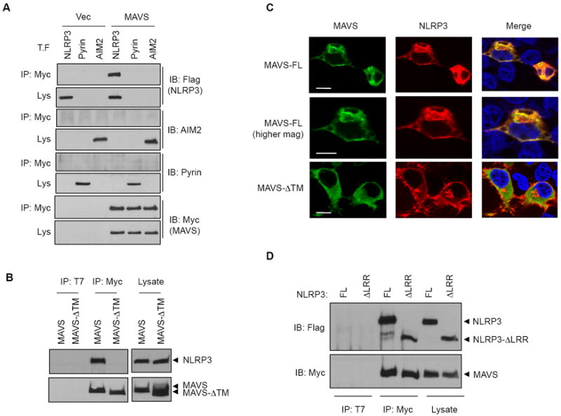
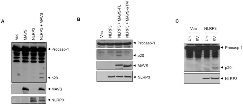
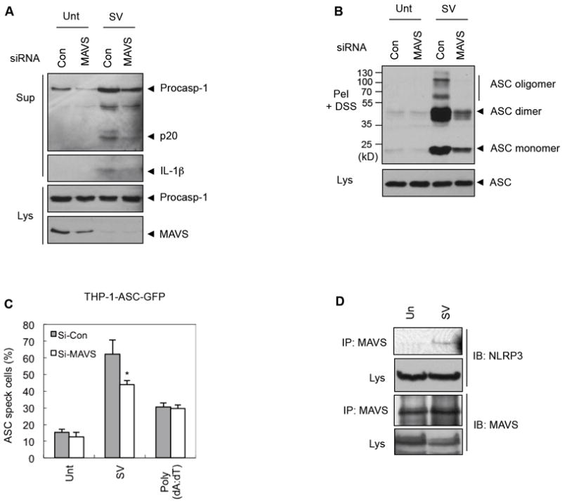
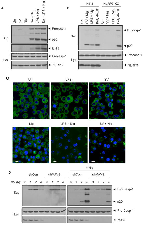
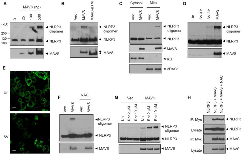
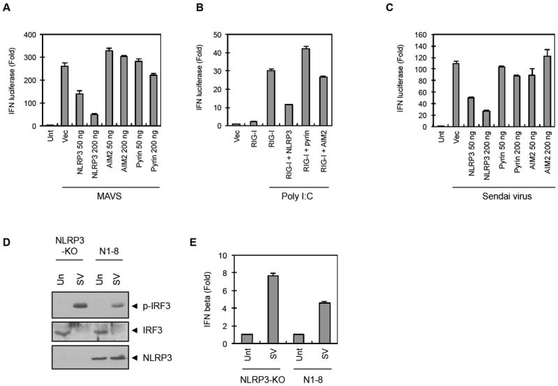
NLRP3 assembles an inflammasome complex that activates caspase-1 upon sensing various danger signals derived from pathogenic infection, tissue damage, and environmental toxins. How NLRP3 senses these various stimuli is still poorly understood, but mitochondria and mitochondrial reactive oxygen species have been proposed to play a critical role in NLRP3 activation. In this article, we provide evidence that the mitochondrial antiviral signaling protein MAVS associates with NLRP3 and facilitates its oligomerization leading to caspase-1 activation. In reconstituted 293T cells, full-length MAVS promoted NLRP3-dependent caspase-1 activation, whereas a C-terminal transmembrane domain-truncated mutant of MAVS (MAVS-ΔTM) did not. MAVS, but not MAVS-ΔTM, interacted with NLRP3 and triggered the oligomerization of NLRP3, suggesting that mitochondrial localization of MAVS and intact MAVS signaling are essential for activating the NLRP3 inflammasome. Supporting this, activation of MAVS signaling by Sendai virus infection promoted NLRP3-dependent caspase-1 activation, whereas knocking down MAVS expression clearly attenuated the activation of NLRP3 inflammasome by Sendai virus in THP-1 and mouse macrophages. Taken together, our results suggest that MAVS facilitates the recruitment of NLRP3 to the mitochondria and may enhance its oligomerization and activation by bringing it in close proximity to mitochondrial reactive oxygen species.


Figures

FIGURE 1
FIGURE 1
Association of MAVS with NLRP3. (A) 293T cells were transfected with empty vector (Vec) or Myc-MAVS expression plasmid together with Flag-NLRP3, T7-pyrin or T7-AIM2 (0.3 μg) expression plasmid as indicated. Cell lysates were immunoprecipitated using anti-Myc antibody, and the immunoprecipitates were then immunoblotted with anti-Flag (NLRP3), anti-AIM2, anti-pyrin or anti-Myc (MAVS) antibody, respectively. (B) 293T cells were transfected with Flag-NLRP3 and Myc-MAVS or Myc-MAVS-ΔTM plasmids (0.3 μg) as indicated. Cell lysates were immunoprecipitated with anti-T7 (negative control) or anti-Myc antibody. The immunoprecipitates were immunoblotted with anti-Flag (upper) or anti-Myc (lower) antibody. (C) 293T cells were transfected with NLRP3- and MAVS full length (FL) or ΔTM-expressing constructs (0.1 μg). MAVS or NLRP3 was visualized by immunofluorescence confocal microscopy as described in “Materials and Methods”. Bar: 10 μm. (D) 293T cells were transfected with Myc-MAVS and Flag-NLRP3 full-length or Flag-NLRP3-ΔLRR constructs (0.3 μg). Coimmunoprecipitation was performed as described in (B).
FIGURE 2
FIGURE 2
Activation of NLRP3 inflammasome by MAVS. (A, B) Immunoblots of caspase-1 in cell lysates of 293T-C1A cells transfected with empty vector (Vec) MAVS, NLRP3, NLRP3 plus MAVS full length, or NLRP3 plus MAVS-ΔTM constructs as indicated. (C) Immunoblots of caspase-1 in cell lysates of 293T-C1A cells transfected with empty vector (Vec) or NLRP3 followed by no treatment (Un) or infection with Sendai virus (SV, 9 HA/ml) for 8 h as indicated. Cell lysates were also immunoblotted with anti-Myc (MAVS) or anti-Flag (NLRP3) antibody (Lower panels, A and B) or anti-Flag (NLRP3) antibody (lower panel, C) as indicated.
FIGURE 3
FIGURE 3
Sendai virus-mediated activation of NLRP3 inflammasome in THP-1 cells. (A) Immunoblots of caspase-1 and IL-1β in culture supernatants (Sup) of PMA-differentiated THP-1 cells transfected with scrambled siRNA (Con) or human MAVS-specific (MAVS) siRNA oligonucleotides (50 nM) for 48 h, followed by infection with Sendai virus (10 HA/ml, 6 h). Cell lysates (Lys) were immunoblotted with anti-caspase-1 or anti-MAVS antibody as indicated. (B) Immunoblot of ASC in DSS-crosslinked NP-40 insoluble pellets of PMA-differentiated THP-1 cells transfected with scrambled siRNA (Con) or human MAVS-specific (MAVS) siRNA oligonucleotides followed by infection with Sendai virus (10 HA/ml, 6 h) as indicated. The lower panel shows an immunoblot of ASC in the soluble lysates from the same cells. (C) PMA-differentiated THP-1-ASC-GFP cells were transfected with siRNA as in (A), followed by treatment with Sendai virus (10 HA/ml, 6 h) or transfection with poly dA:dT (2 μg, 6 h). ASC pyroptosome-containing cells (ASC specks) were counted using fluorescence microscopy and represented as percentages of total cells counted. Asterisk indicates significant difference compared to Si-Con cells (*, p<0.05, n = 5). (D) PMA-differentiated THP-1 cells were left untreated or infected with Sendai virus (10 HA/ml) for 4 h. Cell lysates were fractionated and the mitochondria-enriched fractions were immunoprecipitated with anti-MAVS antibody. Immunoprecipitates and cytosolic lysates were immunoblotted with anti-NLRP3 or anti-MAVS antibody as indicated.
FIGURE 4
FIGURE 4
Sendai virus-mediated activation of NLRP3 inflammasome in mouse macrophages. (A) Immunoblots of caspase-1 and IL-1β in culture supernatants (Sup) of wild type mouse BMDMs treated with Sendai virus (SV, 10 HA/ml, 6 h), nigericin (Nig, 5 μM, 45 min), Sendai virus (6 h) followed with nigericin (45 min) (SV+Nig), LPS (0.25 μg/ml, 4 h) followed with nigericin (LPS+Nig), or Sendai virus (6 h) followed with LPS (4 h) and then nigericin (45 min) (LPS+SV+Nig) as indicated. Cell lysates (Lys) were immunoblotted with anti-caspase-1 or anti-NLRP3 antibody as indicated. (B) Immunoblot of caspase-1 in culture supernatants (Sup) and cell lysates (Lys) of NLRP3-deficient (NLRP3-KO) or NLRP3-restored (N1-8) BMDMs treated with Sendai virus (SV, 10 HA/ml, 6 h) plus nigericin (Nig, 5 μM, 45 min), LPS (0.25 μg/ml, 4 h) plus nigericin (LPS+Nig) or transfected with poly dA:dT (2 μg, 6 h) as indicated. (C) Confocal images of stable NLRP3-GFP-expressing BMDMs left untreated (Un) or treated with LPS, Sendai virus (SV), nigericin (Nig), LPS plus nigericin (LPS+Nig), or Sendai virus plus nigericin (SV+Nig) as described in A and B above. The green and blue signals represent NLRP3 and nuclear fluorescence, respectively. Bar: 10 μm. (D) Immunoblot of caspase-1 in culture supernatants (Sup) and cell lysates (Lys) of stable mouse BMDMs cell lines expressing Scrambled shRNA- (shCon) or mouse MAVS-specific shRNA (shMAVS) after treatment with Sendai virus alone (10 HA/ml, 0-4 h, left panels) or Sendai virus (10 HA/ml, 0-4 h) followed by nigericin (45 min, right panels). Cell lysates were immunoblotted as indicated.
FIGURE 5
FIGURE 5
NLRP3 oligomerization by MAVS. (A, B) 293T cells were transfected with a construct for NLRP3 (0.3 μg) and increasing amount of MAVS construct (A), or NLRP3 and MAVS or MAVS-ΔTM constructs (0.3 μg, B) as indicated. Cell lysates were immunoblotted with anti-Flag (NLRP3) or anti-Myc (MAVS) antibodies. (C) 293T cells were transfected with a NLRP3 construct together with an empty vector (Vec) or MAVS construct. Cell lysates were fractionated into cytosolic or mitochondrial fraction, and then immunoblotted with anti-Flag (NLRP3), anti-Myc (MAVS), anti-IκB (cytosol) or anti-VDAC1 (mitochondria) antibodies. (D) 293T cells were transfected with NLRP3 (0.3 μg) followed by infection with Sendai virus (SV, 10 HA/ml) for the indicated times, or transfected with NLRP3 together with MAVS (0.3 μg) as indicated. Cell lysates were immunoblotted for NLRP3 and MAVS. (E) Confocal images of NLRP-GFP-expressing BMDMs left untreated (Un) or infected with Sendai virus (SV, 10 HA/ml, 8 h) as indicated. Bar: 10 μm. (F-G) 293T cells were transfected with a NLRP3 construct (0.3 μg) together with an empty vector or MAVS construct (0.3 μg), in the presence or absence of NAC (15 mM, 18 h) (F) or followed by treatment with rotenone for 6 h as indicated. Cell lysates were immunoblotted for NLRP3 or MAVS as described above. (H) 293T cells were transfected with a Flag-NLRP3 (0.3 μg) plasmid alone, or together with a Myc-MAVS (0.3 μg) plasmid in the presence or absence of NAC (15 mM, 18 h) as indicated. Cell lysates were immunoprecipitated using anti-Myc antibody, and the immunoprecipitates were then immunoblotted with anti-Flag (NLRP3) or anti-Myc (MAVS) antibody, respectively.
FIGURE 6
FIGURE 6
Inhibition of MAVS-induced Type I IFN signaling by NLRP3. (A) 293T cells were transfected with IFN-β promoter-luciferase reporter plasmid (100 ng), pβ-gal (100 ng) and the indicated expression constructs for MAVS (200 ng) together with empty vector (Vec) NLRP3, AIM2 or pyrin. Reporter luciferase activity was then determined as in the “Materials and Methods”. (B) 293T cells were transfected as in A together with an expression construct for RIG-I with or without NLRP3, pyrin or AIM2 constructs (B) or with constructs for NLRP3, pyrin or AIM2 (C) as indicated. 24 h after transfection cells were transfected with poly I:C (10 μg, 20 h, B) or infected with Sendai virus (SV, 10 HA, 8 h, C) as indicated. Reporter luciferase activity was then determined as in A. (D, E) NLRP3-deficient BMDMs or NLRP3-restored (N1-8) BMDMs were left untreated or infected with Sendai virus (SV, 5 HA/ml) for 8 h. Cell lysates were immunoblotted for the indicated proteins (D) and mRNA levels of IFN-β were quantitated by real-time PCR as described in “Materials and Methods”. p-IRF3, phospho-IRF3.

Similar articles

Cited by

References

    1. Tschopp J, Schroder K. NLRP3 inflammasome activation: The convergence of multiple signalling pathways on ROS production? Nat Rev Immunol. 2010;10:210–215. - PubMed
    1. Franchi L, Munoz-Planillo R, Nunez G. Sensing and reacting to microbes through the inflammasomes. Nat Immunol. 2012;13:325–332. - PMC - PubMed
    1. Wen H, Ting JP, O’Neill LA. A role for the NLRP3 inflammasome in metabolic diseases--did Warburg miss inflammation? Nat Immunol. 2012;13:352–357. - PMC - PubMed
    1. Strowig T, Henao-Mejia J, Elinav E, Flavell R. Inflammasomes in health and disease. Nature. 2012;481:278–286. - PubMed
    1. Aksentijevich I, DP C, Remmers EF, Mueller JL, Le J, Kolodner RD, Moak Z, Chuang M, Austin F, Goldbach-Mansky R, Hoffman HM, Kastner DL. The clinical continuum of cryopyrinopathies: novel CIAS1 mutations in North American patients and a new cryopyrin model. Arthritis Rheum. 2007;56:1273–1285. - PMC - PubMed

Publication types

MeSH terms