. 2013 Dec;41(22):10345-57.
doi: 10.1093/nar/gkt804. Epub 2013 Sep 17.

DHX9 helicase is involved in preventing genomic instability induced by alternatively structured DNA in human cells

Affiliations

DHX9 helicase is involved in preventing genomic instability induced by alternatively structured DNA in human cells

Aklank Jain et al. Nucleic Acids Res. 2013 Dec.

Abstract

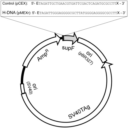
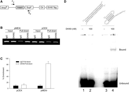
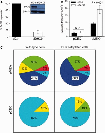
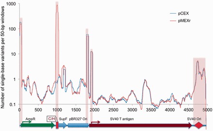
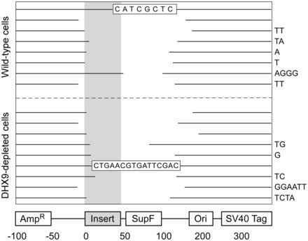
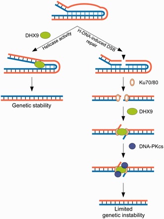
Sequences that have the capacity to adopt alternative (i.e. non-B) DNA structures in the human genome have been implicated in stimulating genomic instability. Previously, we found that a naturally occurring intra-molecular triplex (H-DNA) caused genetic instability in mammals largely in the form of DNA double-strand breaks. Thus, it is of interest to determine the mechanism(s) involved in processing H-DNA. Recently, we demonstrated that human DHX9 helicase preferentially unwinds inter-molecular triplex DNA in vitro. Herein, we used a mutation-reporter system containing H-DNA to examine the relevance of DHX9 activity on naturally occurring H-DNA structures in human cells. We found that H-DNA significantly increased mutagenesis in small-interfering siRNA-treated, DHX9-depleted cells, affecting mostly deletions. Moreover, DHX9 associated with H-DNA in the context of supercoiled plasmids. To further investigate the role of DHX9 in the recognition/processing of H-DNA, we performed binding assays in vitro and chromatin immunoprecipitation assays in U2OS cells. DHX9 recognized H-DNA, as evidenced by its binding to the H-DNA structure and enrichment at the H-DNA region compared with a control region in human cells. These composite data implicate DHX9 in processing H-DNA structures in vivo and support its role in the overall maintenance of genomic stability at sites of alternatively structured DNA.


Figures

Figure 1.
Figure 1.
Plasmid features. Schematic of the pSP189 shuttle vector map showing the location of the control (pCEX) and H-DNA-forming (pMEXr) inserts cloned at the EcoRI (E) and XhoI (X) sites 4 bp upstream of the supF gene.
Figure 2.
Figure 2.
Human DHX9 recognizes DNA secondary structures in plasmids. (A), representative agarose gel of CC pMEXr (lanes 1–5) and pCEX (lanes 6–10) incubated with 30 nM purified recombinant human DHX9 protein (lanes 3–5 and 8–10) in the presence of 5 mM ATP (lanes 3 and 4, and 8 and 9) or 5 mM AMP-PNP (lanes 5 and 10) followed by MBN cleavage. Lane M, 1 kb DNA marker. (B), plot of net percentage of OC and linear (L) DNA released from total DNA (OC + L + CC) (CC = closed circular DNA) at the end of the reaction from two experiments, as described in Panel (A). The net percentage was defined as F = fs – fc, were fs was the %[(OC + L)/(OC + L + CC)] for each sample, and fc was the average %[(OC + L)/(OC + L + CC)] for the two samples without MBN (i.e. lanes 1, 3 and 6, 8, respectively). (C), schematic of the diagnostic restriction sites (AhdI and EcoO109I) used to map MBN-specific cleavage in pMEXr. Grey arrows, lengths of restriction fragments released from pCEX and pMEXr when cleaved by AhdI, EcoO109I and EcoRI, which are located several bp from the cloned inserts; C/H, position of the control (C) and H-DNA-forming (H) inserts in pCEX and pMEXr, respectively. (D), MBN cleavage mapping. PhosphorImager scan of an agarose gel after electrophoresis of pCEX (lanes 1–4) and pMEXr (lanes 5–8) pre-incubated with 30 nM DHX9 (lanes 2 and 3, and 6 and 7) in the presence of 5 mM ATP, treated with 40 units MBN (lanes 3 and 4, and 7 and 8), end-labeled with T4 DNA polymerase and cleaved with Eco0109I and AhdI. Lane E, control lane containing pMEXr cleaved with EcoRI, labeled with T4 DNA polymerase, and then cleaved with AhdI and EcoO109I; E*, ethidium bromide staining of lane E.
Figure 3.
Figure 3.
Human DHX9 recognizes H-DNA-forming sequences in U2OS cells and in vitro. (A), schematic of the plasmids and the primer pairs used for PCR amplification in ChIP assays. Fp and Rp, forward and reverse primers, respectively. (B), representative agarose gel image of PCR products from the ChIP assays. (C), plot of the percentage enrichment relative to input DNA from three independent ChIP assays, as shown in (B). (D), top, cartoon of predicted duplex (left) and intra-molecular triplex (right) DNA structures formed by the oligonucleotides. We note that because the oligonucleotide used for the duplex DNA substrate is self-complementary, both intra-molecular hairpin structures and inter-molecular duplex DNA may be formed by the oligonucleotide; bottom, EMSA of DHX9 binding to the c-MYC sequence-related triplex structure (100 nM) or duplex DNA (100 nM) in the presence of 5 mM AMP-PNP. Lane 1, duplex DNA only; lane 2, duplex DNA and 100 nM DHX9; lane 3, triplex DNA only; lane 4, triplex DNA and 100 nM DHX9.
Figure 4.
Figure 4.
Mutation frequencies and spectra affecting supF gene function in wild-type and DHX9-depleted human U2OS cells. (A), representative western blot of DHX9 in U2OS cells treated with siRNA and plot of percentage DHX9 expression (average of three experiments). (B), frequencies of supF gene mutations for pCEX (control) and pMEXr (H-DNA) transfected into human U2OS cells treated with siRNA. N.S., not significant. (C), Pie charts displaying the mutation spectra of pCEX and pMEXr transfected into wild-type and DHX9-depleted human U2OS cells; 15 random mutant colonies per group were analyzed; percentages were rounded. Dark blue, large (>100 bp) deletions; green, medium size (50–100 bp) deletions; yellow, small (<50 bp) deletions; red, insertions; light blue, point mutations.
Figure 5.
Figure 5.
Spontaneous mutations in plasmid DNA. Landscape of single base substitutions present in the pCEX and pMEXr population following replication in human U2OS cells. Plasmid DNA was recovered from U2OS cells and single base mutants along the entire plasmid were detected by Illumina next-generation sequencing (see ‘Materials and Methods’ section). y-axis, total number of base variants in sliding 50-bp windows; x-axis, position along the plasmid map superimposed to plasmid genetic features. Pink highlight, selected regions with high numbers of variants.
Figure 6.
Figure 6.
Representation of deletion mutants. Gaps represent the deleted sequences aligned to the plasmid map (bottom) for 17 of the deletion mutants shown in Figure 3. Right-hand side nucleotides, micro-homologies at the junctions; shaded bar, triplex-forming region in pMEXr; boxed sequences, insertions.
Figure 7.
Figure 7.
Model depicting the involvement of DHX9 in the processing of H-DNA (intra-molecular triplexes) structures. The left panel represents the first pathway in which the helicase activity of DHX9 resolves the mutagenic H-DNA structure, thus preserving genetic stability. The right panel represents the second pathway in which DHX9 participates in micro-homology mediated end-joining of H-DNA-induced DSBs perhaps by protecting the free ends, and therefore limiting genomic instability. These two models are not mutually exclusive.

References

    1. Nambiar M, Raghavan SC. How does DNA break during chromosomal translocations? Nucleic Acids Res. 2011;39:5813–5825. - PMC - PubMed
    1. Wang G, Vasquez KM. Naturally occurring H-DNA-forming sequences are mutagenic in mammalian cells. Proc. Natl Acad. Sci. USA. 2004;101:13448–13453. - PMC - PubMed
    1. Wang G, Christensen LA, Vasquez KM. Z-DNA-forming sequences generate large-scale deletions in mammalian cells. Proc. Natl Acad. Sci. USA. 2006;103:2677–2682. - PMC - PubMed
    1. Hubert L, Jr, Lin Y, Dion V, Wilson JH. Topoisomerase 1 and single-strand break repair modulate transcription-induced CAG repeat contraction in human cells. Mol. Cell. Biol. 2011;31:3105–3112. - PMC - PubMed
    1. Belotserkovskii BP, De Silva E, Tornaletti S, Wang G, Vasquez KM, Hanawalt PC. A triplex-forming sequence from the human c-MYC promoter interferes with DNA transcription. J. Biol. Chem. 2007;282:32433–32441. - PubMed

Publication types