. 2013 Dec;87(23):12721-36.
doi: 10.1128/JVI.01942-13. Epub 2013 Sep 18.

Bromo- and extraterminal domain chromatin regulators serve as cofactors for murine leukemia virus integration

Affiliations

Bromo- and extraterminal domain chromatin regulators serve as cofactors for murine leukemia virus integration

Saumya Shree Gupta et al. J Virol. 2013 Dec.

Erratum in

Abstract

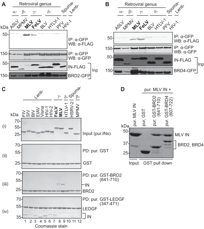
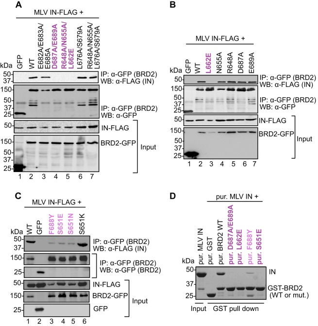
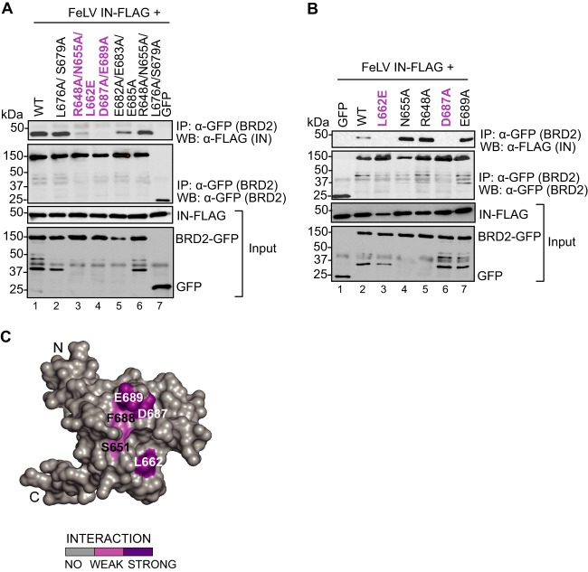
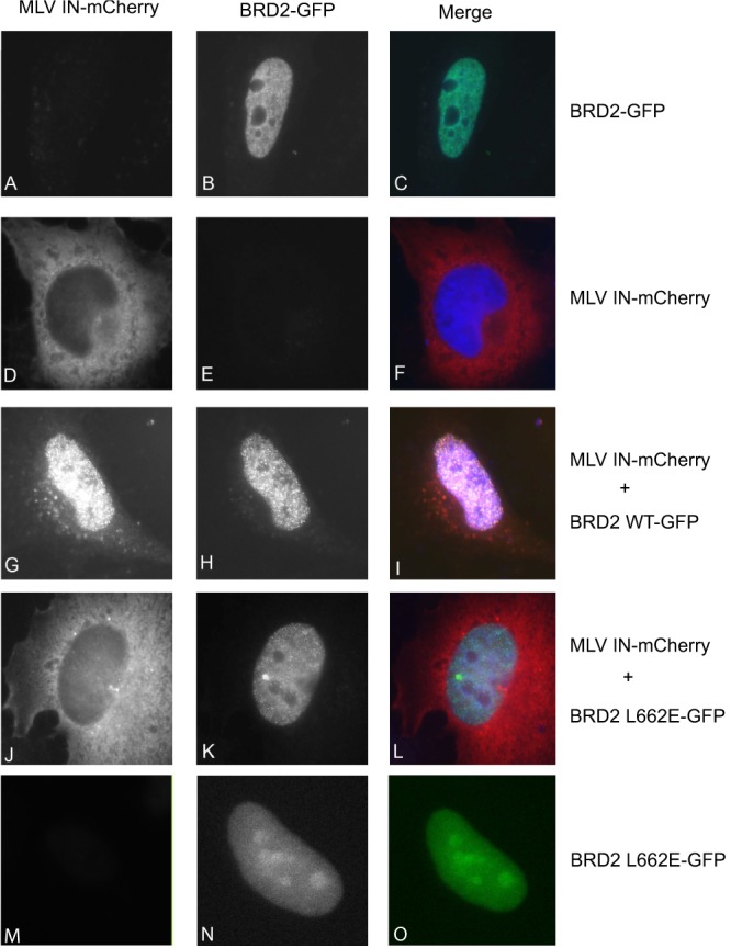
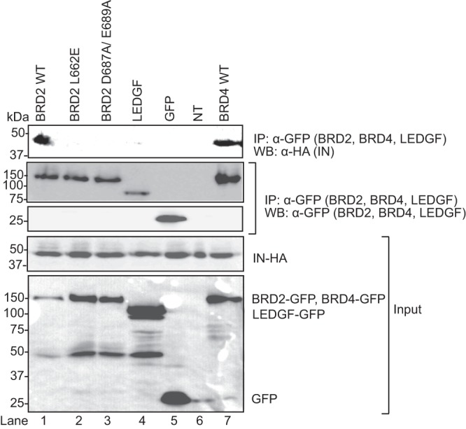
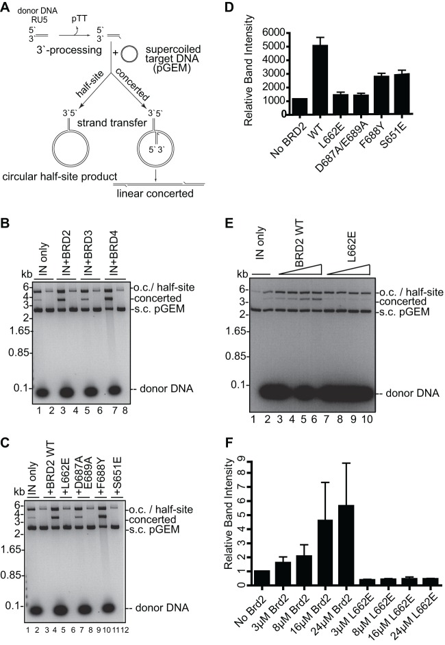
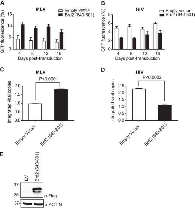
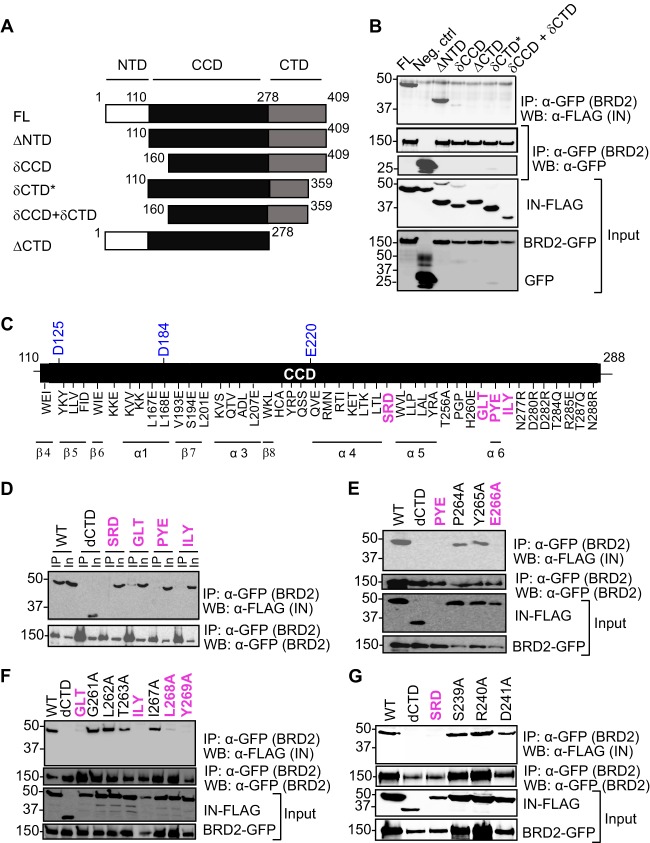
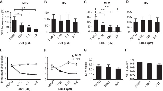
Retroviral integrase (IN) proteins catalyze the permanent integration of proviral genomes into host DNA with the help of cellular cofactors. Lens epithelium-derived growth factor (LEDGF) is a cofactor for lentiviruses, including human immunodeficiency virus type 1 (HIV-1), and targets lentiviral integration toward active transcription units in the host genome. In contrast to lentiviruses, murine leukemia virus (MLV), a gammaretrovirus, tends to integrate near transcription start sites. Here, we show that the bromodomain and extraterminal domain (BET) proteins BRD2, BRD3, and BRD4 interact with gammaretroviral INs and stimulate the catalytic activity of MLV IN in vitro. We mapped the interaction site to a characteristic structural feature within the BET protein extraterminal (ET) domain and to three amino acids in MLV IN. The ET domains of different BET proteins stimulate MLV integration in vitro and, in the case of BRD2, also in vivo. Furthermore, two small-molecule BET inhibitors, JQ1 and I-BET, decrease MLV integration and shift it away from transcription start sites. Our data suggest that BET proteins might act as chromatin-bound acceptors for the MLV preintegration complex. These results could pave a way to redirecting MLV DNA integration as a basis for creating safer retroviral vectors.


Figures

Fig 1
Fig 1
Interaction with BET proteins, BRD2 and BRD4, is limited to and conserved among gammaretroviral INs. (A and B) Extracts of HEK293T cells expressing IN-FLAG along with GFP-tagged BRD2 (A) or BRD4 (B) were immunoprecipitated (IP) with a GFP affinity matrix, and immunoprecipitates were analyzed by Western blotting (WB) using mouse anti-FLAG or mouse anti-GFP antibody as indicated. Migration positions of molecular mass standards (kDa) are shown alongside the gel. (C) GST pulldown experiments using GST alone (ii), GST-tagged BRD2(641–710) (iii), or LEDGF(347–471) (control) (iv). Proteins recovered on glutathione Sepharose beads were separated by SDS-PAGE and detected with Coomassie R-250. (i) Input samples of retroviral IN proteins; lanes 1 to 7, FIV, SIV, BIV, EIAV, visna virus, HIV-1, and HIV-2 (lentiviral IN proteins), respectively; lanes 8 and 9, FeLV and MLV (gammaretroviral IN), respectively; lane 10, HTLV-1 IN (δ-retroviral IN); lane 11, HSRV-2 (spumaretroviral IN); lane 12, MPMV (β-retroviral IN). (ii) GST pulldown using GST as negative control. (iii) GST pulldown with GST-BRD2(641–710); only IN proteins belonging to the gammaretroviral genus are specifically coprecipitated with the GST-tagged BRD2 fragment. (iv) GST pulldown using the C-terminal domain of LEDGF as bait; lentiviral IN specifically binds to LEDGF. Positions of the migration of GST-tagged proteins and the molecular mass markers are indicated on the right or left side of the gel. (D) GST pulldown assay. Purified GST-tagged BRD2(641–710) and BRD4(607–722) were incubated with purified MLV IN and glutathione Sepharose beads. Proteins bound to beads were separated by 12% denaturing PAGE and detected by staining with Coomassie blue. Inp, input; PD, pulldown; pur., purified.
Fig 2
Fig 2
Mapping the IN binding interface in the BRD2 ET domain. (A to C) Coimmunoprecipitation of FLAG-tagged MLV IN with GFP-tagged WT BRD2 or its point mutants. Proteins recovered after coimmunoprecipitation with a GFP affinity matrix were analyzed by Western blotting using mouse anti-GFP or anti-FLAG antibodies as indicated. (D) GST pulldown assay. GST-tagged BRD2(641–710) or its point mutants were incubated with MLV IN and glutathione Sepharose beads. Proteins bound to beads were separated by 12% denaturing PAGE and detected by staining with Coomassie blue. Residues whose mutations abrogate binding are colored dark purple; residues whose mutations displayed weaker binding to MLV IN are shown in light purple.
Fig 3
Fig 3
Mapping the FeLV IN binding interface on BRD2. (A and B) Coimmunoprecipitation of FLAG-tagged FeLV IN with GFP-tagged BRD2 wild type (WT) or GFP-tagged BRD2 point mutants. Proteins recovered after coimmunoprecipitation with a GFP affinity matrix were analyzed by Western blotting using mouse anti-GFP or anti-FLAG antibodies as indicated. (C) Homology model of BRD2 ET domain based on Protein Data Bank accession no. 2JNS. Homology model of the BRD2 ET domain based on Protein Data Bank accession no. 2JNS (76). The coimmunoprecipitation results are mapped on the surface of the BRD2 ET domain. Residues whose mutations abrogate binding are colored dark purple; residues whose mutations displayed weaker binding to MLV IN are shown in light purple.
Fig 4
Fig 4
Colocalization of MLV IN with BRD2. HeLa cells were transfected with EGFP-BRD2, EGFP-BRD2 L662E, or MLV IN-mCherry or the indicated combinations. At 48 h posttransfection, cells were fixed and the localization of the fluorescent fusion proteins was documented. The mCherry and EGFP signals and their overlays are shown in the left, middle, and right images of each panel, respectively. DNA was stained with TO-PRO-3. The merged images contain signals of IN-mCherry (red), GFP-BRD2 (green), and DNA (blue). Images were taken at ×63 magnification.
Fig 5
Fig 5
BRD2 mutants, L662E and D687A/E689A, fail to interact with MLV IN in vivo. HEK293T cells were transfected with the plasmids encoding the N-terminally GFP-tagged BRD2 WT (wild type) or BRD2 mutant L662E, D687A/E689A, or LEDGF or BRD4 and infected (MOI of 1) 24 h posttransfection with an MLV containing an HA-tagged IN. Extracts from the infected cells were immunoprecipitated with anti-GFP antibody coupled to protein A Sepharose. GFP-BRD2 WT, mutants, LEDGF, and BRD4 were detected by Western blotting using mouse monoclonal anti-GFP antibody, whereas MLV IN was detected with a rat anti-HA antibody.
Fig 6
Fig 6
BET proteins stimulate strand transfer activity of recombinant MLV IN in vitro. (A) Schematic of strand transfer reactions using a circular DNA target. Concerted integration results in a gapped linear product whereas the half-site integration yields tailed relaxed circles. (B) Concerted integration activity of MLV IN in the presence of BRD2, BRD3, and BRD4. The enzyme (8 μM) was incubated with supercoiled pGEM target DNA in the absence (lanes 2, 4, 6, and 8) or presence (lanes 1, 3, 5, and 7) of 3 μM preprocessed donor DNA and in the absence (lane 1) or presence of 24 μM BET proteins BRD2 ET (lane 3), BRD3 ET (lane 5), and BRD4 ET (lane 7). Donor DNA (32 bp) mimics the U5 MLV cDNA terminus. Deproteinized reaction products were separated in 1.5% agarose gels and detected by staining with ethidium bromide. Migration positions of concerted and half-site reaction products, supercoiled (s.c.) pGEM target DNA forms, donor DNA, and a DNA size ladder are indicated. (C) Comparison of strand transfer activities of MLV IN in the presence of WT and mutant BRD2 ET domain. The donor DNA was omitted in lanes 2, 4, 6, 8, 10, and 12. (D) Quantification using ImageJ software of the band intensities of the concerted integration products in the presence of WT or mutant BRD2 ET domain as shown in panel C. (E) Comparison of strand transfer activities of MLV IN in the presence of 3 to 24 μM WT or mutant BRD2 ET domain. The donor DNA was omitted in lane 1. (F) Quantification of relative band intensities corresponding to the concerted integration products as shown in panel E. Error bars represent standard deviations from at least two independent experiments. o.c., open circular (nicked) DNA always present in DNA preparations.
Fig 7
Fig 7
Overexpression of IN binding domain of BRD2 (residues 640 to 801) increases MLV integration. (A and B) HEK293T-based cell lines stably expressing BRD2(640–801) were challenged with MLV-based (A) and HIV-1-based (B) retroviral vectors expressing EGFP (MOI, 1). Cells were harvested at indicated time points for flow cytometry analysis. Overall GFP fluorescence over time is calculated as mean fluorescence intensity × % gated cells. Error bars reflect duplicate measurements in each experiment. (C and D) Integrated proviral vector copies were quantified by qPCR at 16 days postinfection in cells transduced with MLV-based (C) and HIV-based (D) vectors. Values represent the means ± standard errors of the means; n is >3 throughout. Results were analyzed by unpaired t tests. (E) Expression of the BRD2(640–801) fragment and control cell lines used in this experiment was analyzed by Western blotting using anti-FLAG antibody.
Fig 8
Fig 8
Mapping the BRD2 binding site on MLV IN. (A) Domains of MLV IN and its deletion mutants. The N-terminal HHCC zinc binding domain, the catalytic core containing the DDE motif, and the positively charged C-terminal domain are indicated. (B) BRD2 interacts with the catalytic core and the C-terminal domain of MLV IN. HEK293T cells transfected with FLAG-tagged IN constructs and GFP-tagged BRD2 were lysed. BRD2 precipitated with anti-GFP affinity beads and detected with mouse anti-GFP antibody after extensive washing with lysis buffer and MLV IN WT or deletion mutants were detected with mouse anti-FLAG antibody. Two percent of each sample was used to confirm the expression of GFP-BRD2 or IN-FLAG (input, lower panels). A result representative of several experiments is presented. (C) A schematic of the catalytic core domain showing the positions of IN mutants constructed for this study. IN mutants that are defective for binding to BRD2 are highlighted in pink. The three catalytically important residues, D125, D184, and E220, are highlighted in blue. All the residues are mutated to alanine, except where indicated otherwise. (D to G) Results of coimmunoprecipitation of FLAG-tagged MLV IN WT and its point mutants with GFP-tagged BRD2.
Fig 9
Fig 9
BET bromodomain inhibitors, JQ1 and I-BET, reduce integration of MLV, but not of HIV-1, in HEK293T cells. (A to D) HEK293T cells were transduced with MLV-based (A and C) and HIV-1-based (B and D) vectors carrying EGFP as a reporter gene at an MOI of 0.1 in the presence of bromodomain inhibitors, I-BET and JQ1, at the concentrations indicated. Cells were treated with inhibitor or DMSO (control) for 36 h. Reporter gene activity was determined by FACS at 3 days following vector transduction. Overall GFP fluorescence is plotted. (E and F) qPCR analysis for integrated viral copies at 20 days postinfection in inhibitor- or DMSO-treated cells transduced with MLV- and HIV-1-based vectors. The graph indicates the integrated vector relative to DMSO sample. (G and H) qPCR analysis of JQ1- and I-BET-treated or DMSO-treated HEK293T cells transduced with MLV vector. Graphs indicate the amounts of PCR products relative to DMSO-treated sample (control) at 3 h postinfection for MSSEs (G) and 8 h postinfection for late RT (H) products. All bars represent the means ± standard errors of the means; n = 3; *, P < 0.05; **, P < 0.01. Results were analyzed by unpaired t tests and are representative of at least three independent experiments.

References

    1. Engelman A, Mizuuchi K, Craigie R. 1991. HIV-1 DNA integration: mechanism of viral DNA cleavage and DNA strand transfer. Cell 67:1211–1221 - PubMed
    1. Hare S, Maertens GN, Cherepanov P. 2012. 3′-processing and strand transfer catalysed by retroviral integrase in crystallo. EMBO J. 31:3020–3028 - PMC - PubMed
    1. Fujiwara T, Craigie R. 1989. Integration of mini-retroviral DNA: a cell-free reaction for biochemical analysis of retroviral integration. Proc. Natl. Acad. Sci. U. S. A. 86:3065–3069 - PMC - PubMed
    1. Holman AG, Coffin JM. 2005. Symmetrical base preferences surrounding HIV-1, avian sarcoma/leukosis virus, and murine leukemia virus integration sites. Proc. Natl. Acad. Sci. U. S. A. 102:6103–6107 - PMC - PubMed
    1. Goodarzi G, Im GJ, Brackmann K, Grandgenett D. 1995. Concerted integration of retrovirus-like DNA by human immunodeficiency virus type 1 integrase. J. Virol. 69:6090–6097 - PMC - PubMed

Publication types

MeSH terms