. 2013 Sep 13:19:1925-36.
eCollection 2013.

Acquisition of RGC phenotype in human Müller glia with stem cell characteristics is accompanied by upregulation of functional nicotinic acetylcholine receptors

Affiliations

Acquisition of RGC phenotype in human Müller glia with stem cell characteristics is accompanied by upregulation of functional nicotinic acetylcholine receptors

Silke Becker et al. Mol Vis. .

Abstract

Purpose: Human Müller glia with stem cell characteristics (hMGSCs) can be induced to express genes and proteins of retinal ganglion cells (RGCs) upon in vitro inhibition of Notch-1 activity. However, it is not known whether expression of these markers is accompanied by acquisition of RGC function. This study investigated whether hMGSCs that express RGC markers also display neural functionality, as measured by their intracellular calcium concentration ([Ca(2+)]i) responsiveness following neurotransmitter stimulation in vitro.

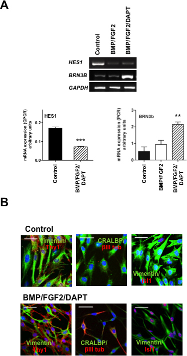
Methods: Changes in mRNA expression of RGC markers and neurotransmitter receptors were assessed either by conventional or quantitative reverse transcription PCR (RT-PCR), while changes in protein levels were confirmed by immunocytochemistry. The [Ca(2+)]i levels were estimated by fluorescence microscopy.

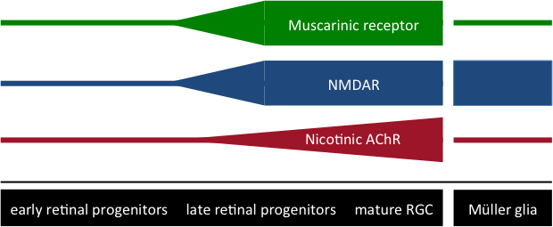
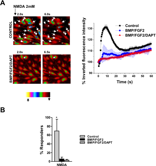
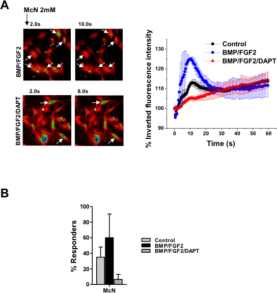
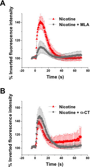
Results: We showed that while undifferentiated hMGSCs displayed a profound elevation of [Ca(2+)]i after stimulation with N-methyl-D-aspartate (NMDA), this was lost following Notch-1 inhibition. Conversely, untreated hMGSCs did not respond to muscarinic receptor stimulation, whereas [Ca(2+)]i was increased in differentiated hMGSCs that expressed RGC precursor markers. Differentiated hMGSC-derived RGCs, but not undifferentiated hMGSCs, responded to stimulation by nicotine with a substantial rise in [Ca(2+)]i, which was inhibited by the α4β2 and α6β2 nicotinic receptor antagonist methyllycaconitine. Notch-1 attenuation not only caused a decrease in the gene expression of the Notch effector HES1 and increased expression of RGC markers, but also an increase in the gene and protein expression of α4 and α6 nicotinic receptor subunits.

Conclusions: These observations suggest that in response to Notch-1 inhibition, hMGSCs differentiate into a population of RGCs that exhibit some of the functionality observed in differentiated RGCs.


Figures

Figure 1
Figure 1
Expression levels of neurotransmitter receptors differ in early and late retinal progenitors, as well as in Müller glia. Varying expression levels of N-methyl-D-aspartate (NMDA) receptors, muscarinic receptors and nicotinic acetylcholine receptors (AChR) are depicted throughout development in early and late retinal progenitors and mature retinal ganglion cells (RGCs), as well as in Müller glia.
Figure 2
Figure 2
Expression of retinal ganglion cell (RGC) markers following Notch-1 inhibition in human Müller glia with stem cell characteristics (hMGSCs) is consistent with the acquisition of a neural phenotype. A: Inhibition of Notch-1 by treatment with basement membrane protein (BMP), basic fibroblast growth factor-2 (FGF2) and N-[N-(3,5-Difluorophenacetyl)-L-alanyl]-S-phenylglycin t-butyl ester (DAPT) attenuated mRNA expression of HES1 (***p<0.001, n=4), while significantly upregulating BRN3b mRNA, a marker of RGC precursors (**p<0.01, n=4), as shown by quantitative and conventional reverse transcription PCR (RT-PCR), respectively. B: In untreated hMGSCs, the Müller cell markers vimentin and CRALBP (green) were highly expressed, while THY1, βIII-tubulin, and ISL-1 (red), which are characteristic of RGCs, were undetectable or found at low levels (upper panel). After differentiation of hMGSCs by Notch-1 inhibition, the expression of vimentin and CRALBP (green) was attenuated, while that of THY1, βIII-tubulin, and ISL-1 was augmented (red, lower panel).
Figure 3
Figure 3
Cytosolic calcium ([Ca2+]i ) response to N-methyl-D-aspartate (NMDA) receptor activation is consistent with the development of retinal ganglion cell (RGC) phenotype by Notch-1 inhibition in human Müller glia with stem cell characteristics (hMGSCs). A: Examplary heat map images are shown with Fura Red-loaded hMGSCs cultured under control conditions and after treatment with basement membrane protein (BMP), basic fibroblast growth factor-2 (FGF2) and N-[N-(3,5-Difluorophenacetyl)-L-alanyl]-S-phenylglycin t-butyl ester (DAPT). The images are recorded at 40× magnification and are representative of fluorescence intensity before (2 s) and at the maximum effect of receptor activation (6 s and 6.5 s, respectively). The color bar describes the intensity–color relationship, with yellow being the brightest and black being the dimmest. Control cells showing a rapid decrease in fluorescence intensity in response to NMDA exposure, which signifies an increase in [Ca2+]i, are marked with white arrows. Cells cultured with BMP, FGF2, and DAPT did not respond to NMDA with a reduction in fluorescence intensity (left panel). In response to NMDA (2 mM), untreated hMGSCs (control, n=41 cells from two experiments) showed a strong reduction in the inverted fluorescence intensity, displayed as a percentage of the value at 0 s, corresponding to an increase in [Ca2+]i,. This was absent in hMGSCs treated with either BMP or FGF2 alone (n=32 cells from two experiments) or a combination of BMP, FGF2, and DAPT (n=50 cells from 3 experiments), *** p<0.001 control versus BMP/FGF2 or BMP/FGF2/DAPT (right panel). B: The fraction of cells responding to NMDA with a rise in [Ca2+]i was greatly diminished after Notch-1 inhibition following differentiation without or with DAPT, in comparison to control cells (n=50 from four experiments, respectively, *p<0.05).
Figure 4
Figure 4
Cytosolic calcium ([Ca2+]i ) response to muscarinic receptor activation is consistent with the development of an retinal ganglion cell (RGC) phenotype by Notch-1 inhibition in human Müller glia with stem cell characteristics (hMGSCs). A: Examplary heat map images are shown with Fura Red-loaded hMSCs cultured under control conditions and after treatment with basement membrane protein (BMP), basic fibroblast growth factor-2 (FGF2) and N-[N-(3,5-Difluorophenacetyl)-L-alanyl]-S-phenylglycin t-butyl ester (DAPT). The images are recorded at 40× magnification and are representative of fluorescence intensity before (2 s) and at the maximum effect of receptor activation (10 s and 8 s, respectively). Control cells showing a small, slow decrease in fluorescence intensity in response to McN-A343 exposure, which signifies an increase in [Ca2+]i, are marked with white arrows. Cells cultured with BMP, FGF2 and DAPT showed a similar response to McN-A343 (left panel). Untreated hMGSCs (control, n=39 cells from three experiments) and hMGSCs treated with BMP, FGF2, and DAPT (n=32 cells from two experiments) responded to the muscarinic receptor agonist McN-A343 (2 mM) with a small reduction in the inverted fluorescence intensity, displayed as a percentage of the value at 0 s, corresponding to a minor rise in [Ca2+]i, which was greatly augmented after treatment with BMP and FGF2 alone (n=42 cells from two experiments, *p<0.05 and ***p<0.001, respectively). B: There was no significant alteration in the percentage of cells responding to the muscarinic receptor agonist McN-A343 with or without differentiation by Notch-1 inhibition (n=50 from four experiments).
Figure 5
Figure 5
Notch-1 inhibition leads to upregulation of nicotinic α4- and α6-receptor subunits in human Müller glia with stem cell characteristics (hMGSCs). A: Exemplary images show mRNA expression of nicotinic acetylcholine receptors (nAChR) α1–4, 6, and 7 in hMGSCs under control conditions, after differentiation with basement membrane protein (BMP) and basic fibroblast growth factor-2 (FGF2) alone or after addition of N-[N-(3,5-Difluorophenacetyl)-L-alanyl]-S-phenylglycin t-butyl ester (DAPT; left panel). Following differentiation of hMGSCs with the Notch-1 inhibitors BMP, FGF2, and DAPT (red bars), mRNA expression of the α4 and α6 nicotinic acetylcholine receptors (nAChR) subunits was significantly upregulated in comparison to untreated cells (control, black bars) or after treatment with BMP and FGF2 alone (blue bars), as shown by reverse transcription PCR (RT-PCR; *p<0.05, n=6, right panel). B: Immunohistochemical staining showed an increase in protein expression of the α4 and α6 nicotinic AChR subunits (green) with concomitant upregulation of THY1 by hMGSCs treated with BMP, FGF2, and DAPT, in comparison to control (40× magnification).
Figure 6
Figure 6
Increased cytosolic calcium ([Ca2+]i ) in response to nicotinic receptor activation is greatly attenuated by methyllycaconitine, but not α-conotoxin MII in retinal ganglion cell (RGC)-committed precursors. The effect of pharmacological inhibition of the α4β2 and α6β2 nicotinic acetylcholine receptors (AChRs) with methyllycaconitine (MLA, 100 nmoles/l) and of the α3β2 nicotinic AChR with α-conotoxin MII (α-CT, 100 nmoles/l) on the rise in [Ca2+]i triggered by nicotine (2 mM) was assessed. A: Methyllycaconitine caused a substantial reduction in the [Ca2+]i rise (n=99 cells from 3 experiments) in comparison to stimulation with nicotine alone (n=113 cells from 3 experiments , ***p<0.001). B: α-conotoxin MII did not significantly affect the rise in [Ca2+]i after stimulation with nicotine (n=254 cells from 4 experiments) in comparison to stimulation with nicotine alone (n=290 cells from 4 experiments).

References

    1. Coles BL, Angenieux B, Inoue T, Del Rio-Tsonis K, Spence JR, McInnes RR, Arsenijevic Y, van der Kooy D. Facile isolation and the characterization of human retinal stem cells. Proc Natl Acad Sci USA. 2004;101:15772–7. - PMC - PubMed
    1. Tropepe V, Coles BL, Chiasson BJ, Horsford DJ, Elia AJ, McInnes RR, van der Kooy D. Retinal stem cells in the adult mammalian eye. Science. 2000;287:2032–6. - PubMed
    1. Gu P, Harwood LJ, Zhang X, Wylie M, Curry WJ, Cogliati T. Isolation of retinal progenitor and stem cells from the porcine eye. Mol Vis. 2007;13:1045–57. - PMC - PubMed
    1. MacNeil A, Pearson RA, MacLaren RE, Smith AJ, Sowden JC, Ali RR. Comparative analysis of progenitor cells isolated from the iris, pars plana, and ciliary body of the adult porcine eye. Stem Cells. 2007;25:2430–8. - PubMed
    1. Lawrence JM, Singhal S, Bhatia B, Keegan DJ, Reh TA, Luthert PJ, Khaw PT, Limb GA. MIO-M1 cells and similar Müller glial cell lines derived from adult human retina exhibit neural stem cell characteristics. Stem Cells. 2007;25:2033–43. - PubMed

Publication types

MeSH terms