Case Reports
doi: 10.4047/jap.2013.5.3.359.
Epub 2013 Aug 31.
Minimally invasive treatment for esthetic enhancement of white spot lesion in adjacent tooth
Affiliations
- PMID: 24049579
- PMCID: PMC3774952
- DOI: 10.4047/jap.2013.5.3.359
Item in Clipboard
Case Reports
Minimally invasive treatment for esthetic enhancement of white spot lesion in adjacent tooth
J Adv Prosthodont.
2013 Aug.
Abstract
This article describes the treatment provided to a patient with the maxillary anterior teeth exhibiting severe secondary caries beneath the previous restoration and a white spot lesion on the adjacent incisor. Two implants were placed after extraction of hopeless teeth with the guided bone regeneration technique. A white spot lesion of the adjacent incisor was treated with minimally invasive treatment. This clinical report describes the multidisciplinary treatment for the white spot lesion and esthetic restoration of missing anterior teeth.
Keywords: Bleaching; Clinical report; Resin infiltration; White spot.
Figures
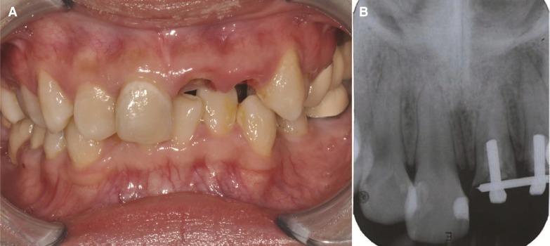
(A) Pre-treatment clinical view, (B) Pre-treatment radiograph. Note the cervical white spot lesion with poor oral hygiene.
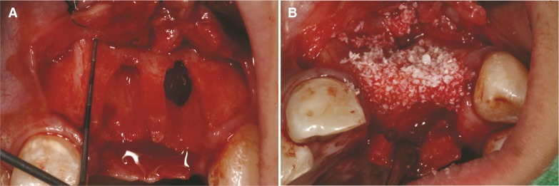
(A) Extraction socket after 6-week-healing, (B) Defects covered with resorbable bilayer collagen membrane.
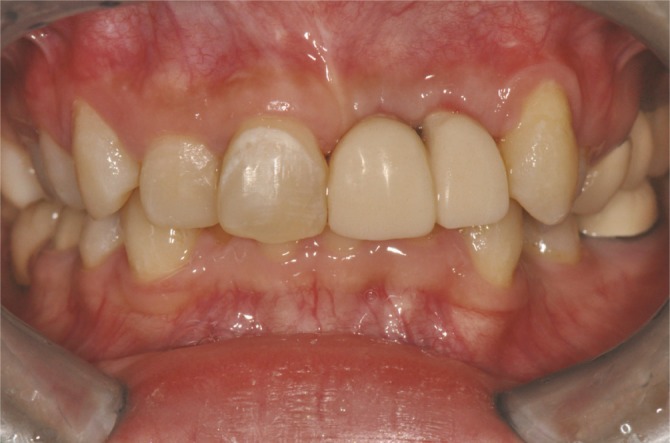
Placement of provisional restoration.
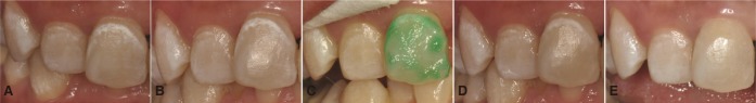
(A) Left central incisor after bleaching, (B) 2 weeks after bleaching, (C) Hydrochloric acid etching for resin infiltration technique, (D) After resin infiltration technique, (E) Composite resin restoration. Note the disappearance of general white bandlike area in the middle portion and distal part of white spot lesion after resin infiltration.
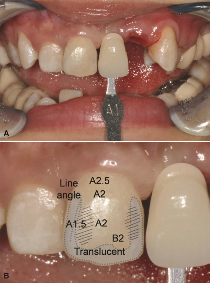
(A) Clinical photograph with shade guide, (B) Shade mapping on clinical photograph.
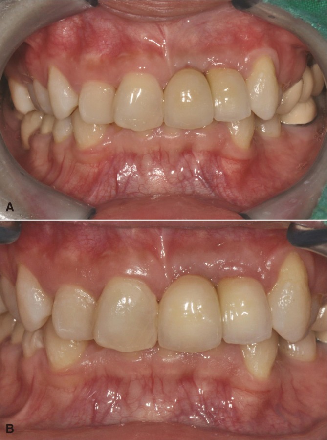
(A) Post-treatment clinical view after 1 month, (B) Post-treatment clinical view after 6 months.
References
-
- Paris S, Meyer-Lueckel H. Masking of labial enamel white spot lesions by resin infiltration-a clinical report. Quintessence Int. 2009;40:713–718. - PubMed
-
- Loesche WJ. Chemotherapy of dental plaque infections. Oral Sci Rev. 1976;9:65–107. - PubMed
-
- Kidd EA, Fejerskov O. What constitutes dental caries? Histopathology of carious enamel and dentin related to the action of cariogenic biofilms. J Dent Res. 2004;83:C35–C38. - PubMed
-
- Suzuki M, Jordan RE, Skinner DH, Boksman L. Clinical management of non-carious enamel defects. Int Dent J. 1982;32:148–158. - PubMed
-
- Ellwood RP, O'Mullane D. Enamel opacities and dental esthetics. J Public Health Dent. 1995;55:171–176. - PubMed
Publication types
LinkOut - more resources
Full Text Sources
Other Literature Sources