. 2013 Aug 8;4(9):1595-609.
doi: 10.1364/BOE.4.001595. eCollection 2013.

Static and dynamic crystalline lens accommodation evaluated using quantitative 3-D OCT

Affiliations

Static and dynamic crystalline lens accommodation evaluated using quantitative 3-D OCT

Enrique Gambra et al. Biomed Opt Express. .

Abstract

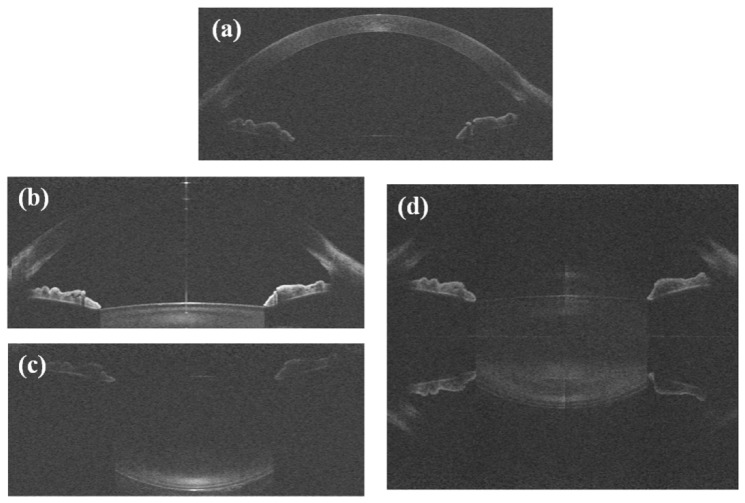
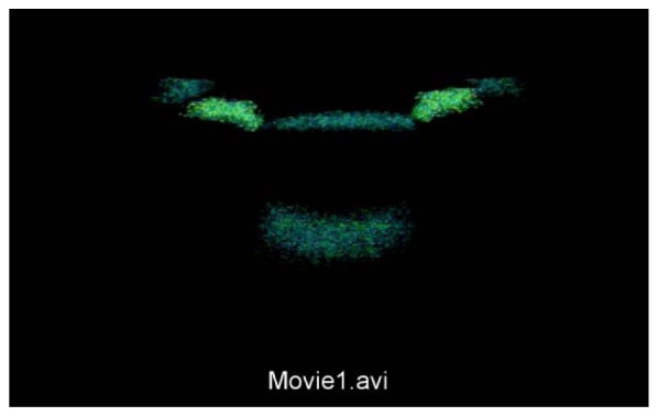
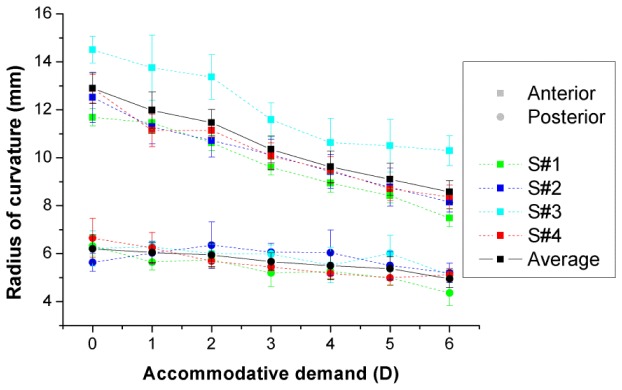
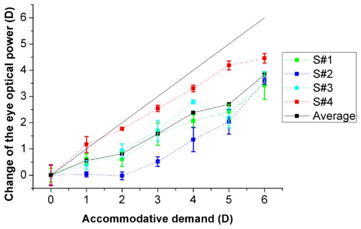
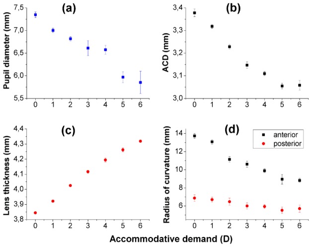
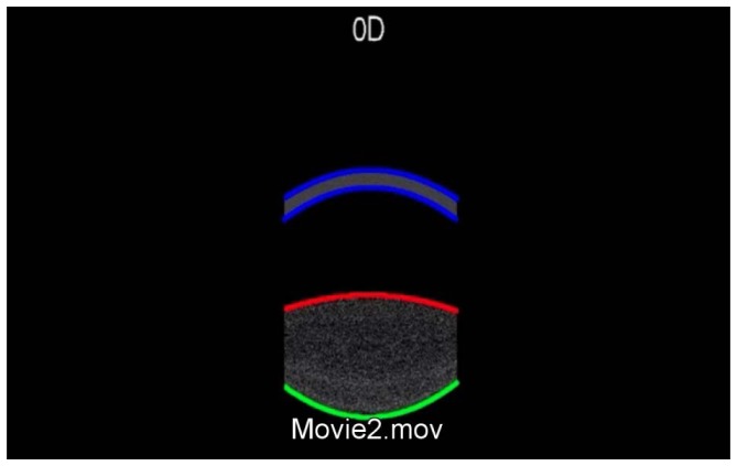
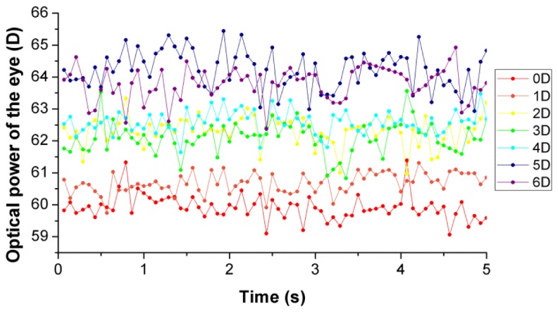
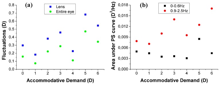
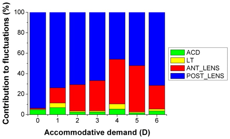
Custom high-resolution high-speed anterior segment spectral domain Optical Coherence Tomography (OCT) provided with automatic quantification and distortion correction algorithms was used to characterize three-dimensionally (3-D) the human crystalline lens in vivo in four subjects, for accommodative demands between 0 to 6 D in 1 D steps. Anterior and posterior lens radii of curvature decreased with accommodative demand at rates of 0.73 and 0.20 mm/D, resulting in an increase of the estimated optical power of the eye of 0.62 D per diopter of accommodative demand. Dynamic fluctuations in crystalline lens radii of curvature, anterior chamber depth and lens thickness were also estimated from dynamic 2-D OCT images (14 Hz), acquired during 5-s of steady fixation, for different accommodative demands. Estimates of the eye power from dynamical geometrical measurements revealed an increase of the fluctuations of the accommodative response from 0.07 D to 0.47 D between 0 and 6 D (0.044 D per D of accommodative demand). A sensitivity analysis showed that the fluctuations of accommodation were driven by dynamic changes in the lens surfaces, particularly in the posterior lens surface.

Keywords: (110.4500) Optical coherence tomography; (110.6880) Three-dimensional image acquisition; (120.4640) Optical instruments; (120.6650) Surface measurements, figure; (330.7322) Visual optics, accommodation; (330.7327) Visual optics, ophthalmic instrumentation.


Figures

Fig. 1
Fig. 1
Typical raw sOCT images: (a) cornea; (b) anterior crystalline lens surface; (c) posterior crystalline lens surface, imaged separately in the 3-D protocol; (d) entire crystalline lens, imaged simultaneously using the complex conjugate images removal technique, in the dynamic 2-D image acquisition. After the unfolding procedure, a mirror image of some elements of the eye can still be seen.
Fig. 2
Fig. 2
(Media 1): Distortion-corrected lateral view of the 3-D rendering of the crystalline lens, from data images acquired at accommodative demands ranging from 0 to 6 D in 1-D steps.
Fig. 3
Fig. 3
Radii of curvature of the anterior (squares) and posterior (circles) surfaces of the lens for the individual subjects (color symbols with dashed lines) and the average across subjects (black symbols with solid lines). Data for each subject are average of 5 repeated measurements. Error bars stand for standard deviation of repeated measurements. The radius of curvature of the posterior surface of the lens is negative, but has been depicted positive for illustration purposes.
Fig. 4
Fig. 4
Change in the optical power of the eye with accommodative demand, with respect to the value obtained for the unaccommodated condition (0D). Data are average of 5 repeated measurements for each subject. Solid line corresponds to the ideal response. Error bars stand for half of the standard deviation of repeated measurements (for clarity).
Fig. 5
Fig. 5
Anterior segment geometry as a function of accommodative demand: (a) Pupil diameter, (b) anterior chamber depth, (c) lens thickness, and (d) lens radii of curvature. Error bars stand for the standard deviation of data from 58 to 70 images acquired during 5 seconds of sustained accommodation (at each accommodative demand).
Fig. 6
Fig. 6
(Media 2): Dynamic fluctuations of the horizontal section of the anterior segment (5-second sequences for each accommodative stimulus, ranging from 0 to 6 D). Data are following image processing, including merging and distortion corrections. The superimposed lines are circumference section fittings to the anterior and posterior cornea and lens.
Fig. 7
Fig. 7
Optical power of the eye during 5 seconds of sustained accommodation, for different accommodative demands.
Fig. 8
Fig. 8
(a) Fluctuations of the optical power of the lens and of the entire eye calculated as the standard deviation of the power estimates during 5-seconds of sustained accommodation, as a function of the accommodative demand. (b) Area under the power spectrum density curve of the optical power of the eye in two different frequency bands (0-0.6 Hz and 0.9-2.5 Hz), as a function of accommodative demand.
Fig. 9
Fig. 9
Relative contribution of the anterior chamber depth (ACD), lens thickness (LT) and anterior and posterior lens radii to the fluctuations of the optical power of the eye.

References

    1. Locke L. C., Somers W., “A comparison study of dynamic retinoscopy techniques,” Optom. Vis. Sci. 66(8), 540–544 (1989).10.1097/00006324-198908000-00009 - DOI - PubMed
    1. Mallen E. A. H., Wolffsohn J. S., Gilmartin B., Tsujimura S., “Clinical evaluation of the Shin-Nippon SRW-5000 autorefractor in adults,” Ophthalmic Physiol. Opt. 21(2), 101–107 (2001).10.1016/S0275-5408(00)00049-1 - DOI - PubMed
    1. He J. C., Burns S. A., Marcos S., “Monochromatic aberrations in the accommodated human eye,” Vision Res. 40(1), 41–48 (2000).10.1016/S0042-6989(99)00156-X - DOI - PubMed
    1. Hofer H., Artal P., Singer B., Aragón J. L., Williams D. R., “Dynamics of the eye’s wave aberration,” J. Opt. Soc. Am. A 18(3), 497–506 (2001).10.1364/JOSAA.18.000497 - DOI - PubMed
    1. Plainis S., Ginis H. S., Pallikaris A., “The effect of ocular aberrations on steady-state errors of accommodative response,” J. Vis. 5(5), 466–477 (2005), http://www.journalofvision.org/5/5/710.1167/5.5.7 - DOI - PubMed