

Efficacy and Complications of Intravitreal Rituximab Injection for Treating Primary Vitreoretinal Lymphoma
- PMID: 24049708
- PMCID: PMC3763885
- DOI: 10.1167/tvst.1.3.1
Efficacy and Complications of Intravitreal Rituximab Injection for Treating Primary Vitreoretinal Lymphoma
Abstract
Purpose: To assess the long-term clinical outcomes of intravitreal injections of rituximab (IVR), an anti-CD20 monoclonal antibody, to treat CD20-positive primary vitreoretinal lymphoma (PVRL).
Methods: Twenty eyes of 13 women (mean age, 66.2 ± 9.9 years) with CD20-positive PVRL were included in this prospective, interventional case series. All patients had discontinued previous intravitreal methotrexate (IVM) treatment because of severe corneal epitheliopathy. Weekly IVR injections (1 mg/0.1 ml) for 4 weeks were administered as a one-course protocol. Additional injections were administered when the PVRL recurred. The effects and the adverse events associated with IVR injections were evaluated.
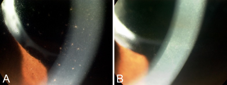
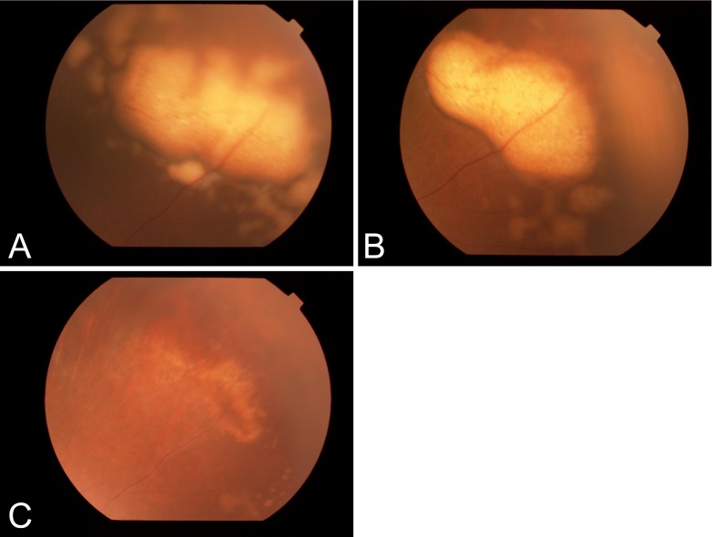
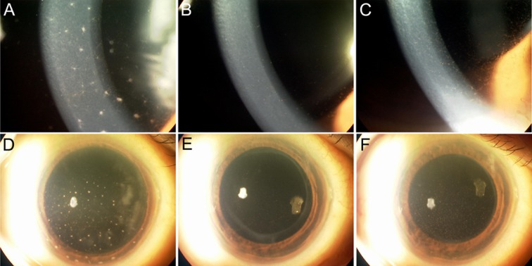
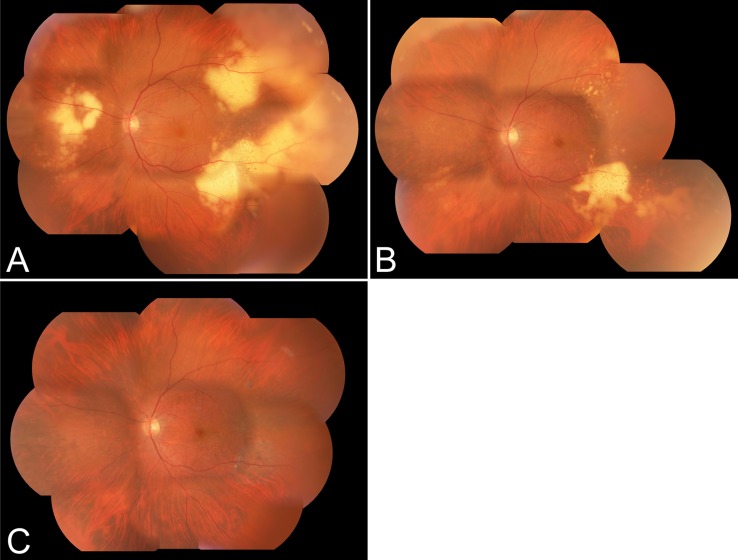
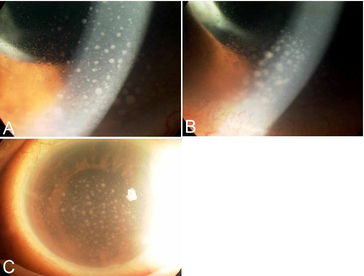
Results: All patients completed a 1-year follow-up (mean observation after IVR injections, 24.7 ± 6.3 months). Before treatment, diffuse keratic precipitates (KPs), anterior vitreous cells, or both were observed in 18 (90%) eyes of 11 patients, and typical subretinal infiltrates were seen in eight (40%) eyes of six patients; all improved with one treatment course. The anterior segment lesions recurred in 11 (55%) eyes of nine patients and resolved with another course of injections. Transient IOP elevations occurred in 12 (60%) eyes of 10 patients within 3.8 ± 1.9 weeks after the first treatment course; iridocyclitis with mutton-fat KPs developed in seven (35%) eyes of six patients with elevated IOP and resolved with topical treatment. No other significant ocular complications or systemic side effects developed.
Conclusions: Injections of IVR were shown to be an efficacious alternative treatment for PVRL, although the disease recurred in approximately half of the eyes. Complications included transient IOP elevations and iridocyclitis with mutton-fat KPs that were managed topically.
Translational relevance: The results of this trial support IVR as one element of combined modality therapy for treating PVRL patients without CNS involvement, particularly for those who respond poorly and have side effects with IVM. (http://www.umin.ac.jp/ctr/ number, UMIN000005604).
Keywords: CD20; complication; intravitreal injection; primary vitreoretinal lymphoma; rituximab.
Figures
References
-
- Commins DL. Pathology of primary central nervous system lymphoma. Neurosurg Focus. 2006;21:E2. - PubMed
-
- Schabet M. Epidemiology of primary CNS lymphoma. J Neurooncol. 1999;43:199–201. - PubMed
-
- Hochberg FH, Baehring JM, Hochberg EP. Primary CNS lymphoma. Nat Clin Pract Neurol. 2007;3:24–35. - PubMed
-
- Bataille B, Delwail V, Menet E, et al. Primary intracerebral malignant lymphoma: report of 248 cases. J Neurosurg. 2000;92:261–266. - PubMed
-
- Kadoch C, Treseler P, Rubenstein JL. Molecular pathogenesis of primary central nervous system lymphoma. Neurosurg Focus. 2006;21:E1. - PubMed
LinkOut - more resources
Full Text Sources
