. 2013 May;2(4):2.
doi: 10.1167/tvst.2.4.2. Epub 2013 May 29.

Considering Apical Scotomas, Confusion, and Diplopia When Prescribing Prisms for Homonymous Hemianopia

Affiliations

Considering Apical Scotomas, Confusion, and Diplopia When Prescribing Prisms for Homonymous Hemianopia

Henry L Apfelbaum et al. Transl Vis Sci Technol. 2013 May.

Abstract

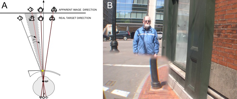
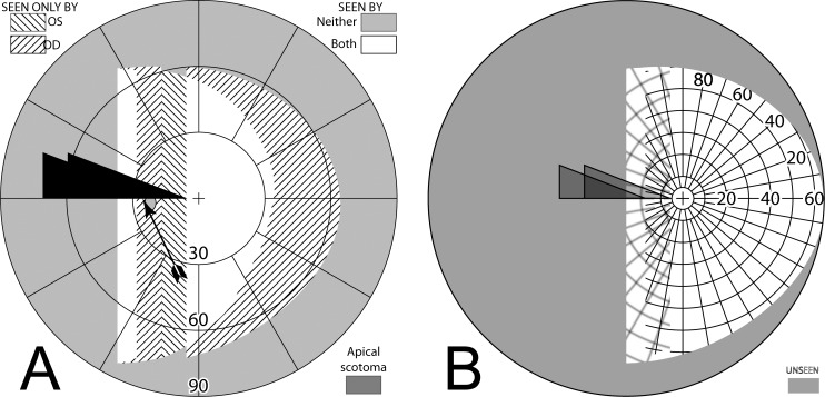
Purpose: While prisms are commonly prescribed for homonymous hemianopia to extend or expand the visual field, they cause potentially troubling visual side effects, including nonveridical location of perceived images, diplopia, and visual confusion. In addition, the field behind a prism at its apex is lost to an apical scotoma equal in magnitude to the amount of prism shift. The perceptual consequences of apical scotomas and the other effects of various designs were examined to consider parameters and designs that can mitigate the impact of these effects.

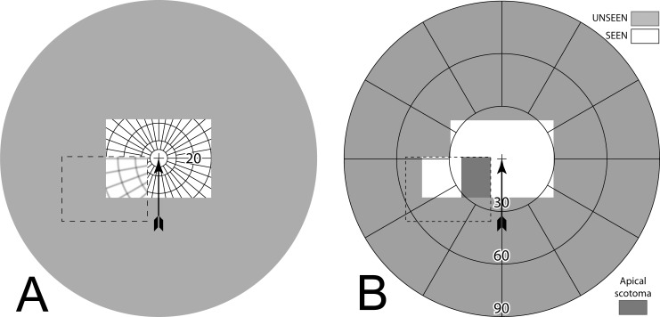
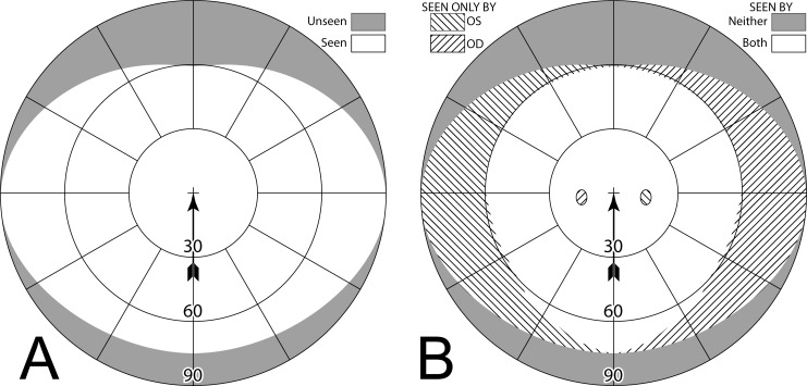
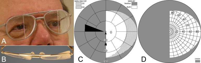
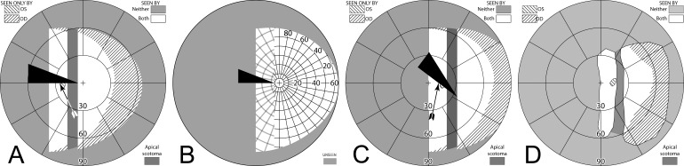
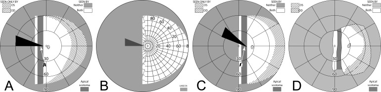
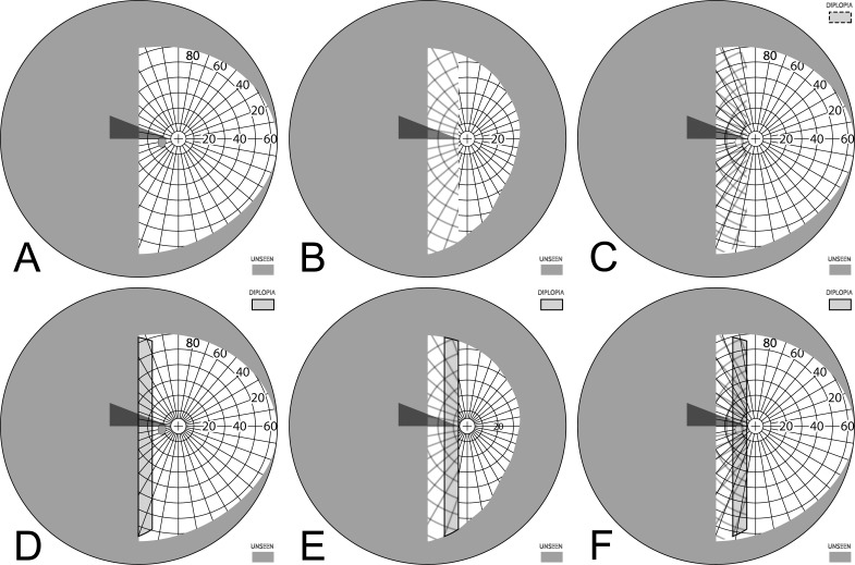
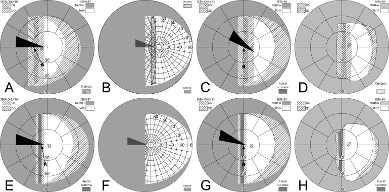
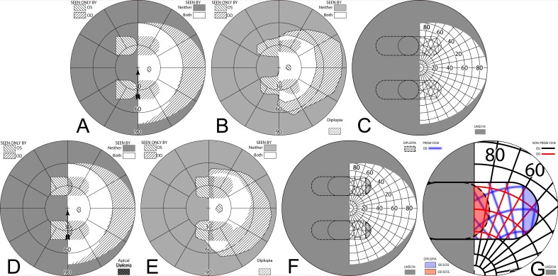
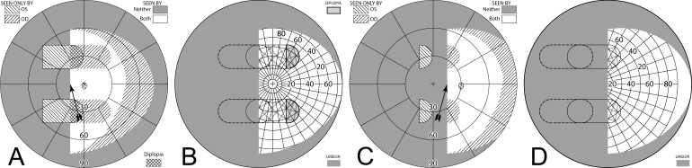
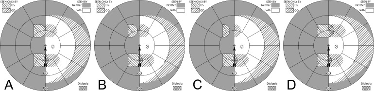
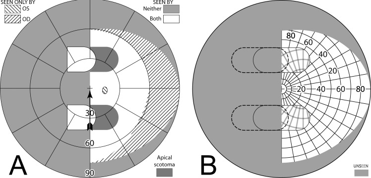
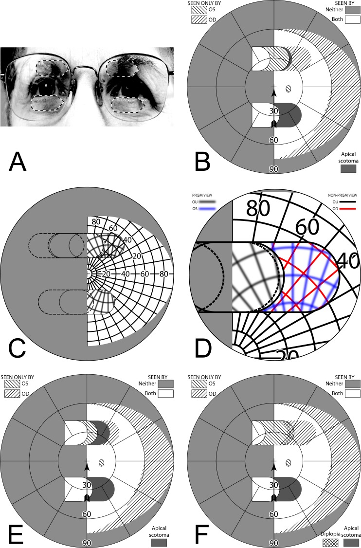
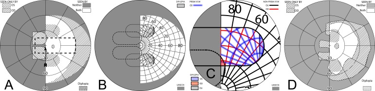
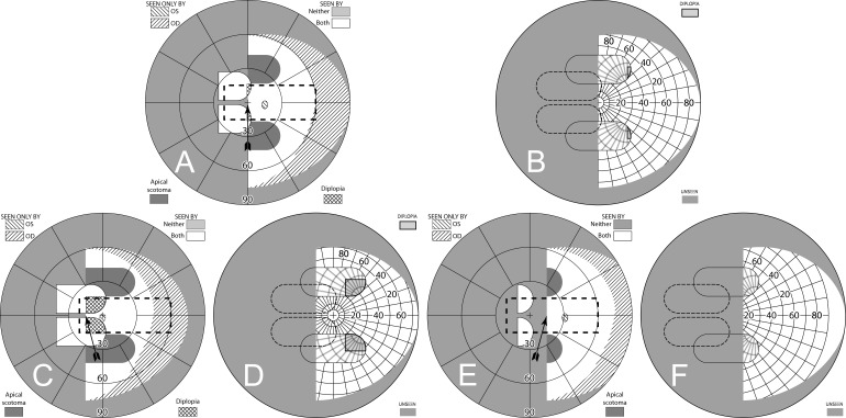
Methods: Various configurations of sector and peripheral prisms were analyzed, in various directions of gaze, and their visual effects were illustrated using simulated perimetry. A novel "percept" diagram was developed that yielded insights into the patient's view through the prisms. The predictions were verified perimetrically with patients.

Results: The diagrams distinguish between potentially beneficial field expansion via visual confusion and the pericentrally disturbing and useless effect of diplopia, and their relationship to prism power and gaze direction. They also illustrate the nonexpanding substitution of field segments of some popular prism designs.

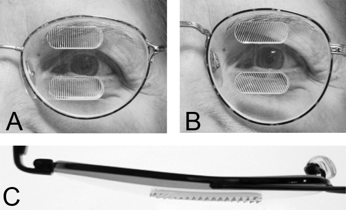
Conclusions: Yoked sector prisms have no effect at primary gaze or when gaze is directed toward the seeing hemifield, and they introduce pericentral field loss when gaze is shifted into them. When fitted unilaterally, sector prisms also have an effect only when the gaze is directed into the prism and may cause a pericentral scotoma and/or central diplopia. Peripheral prisms are effective at essentially all gaze angles. Since gaze is not directed into them, they avoid problematic pericentral effects. We derive useful recommendations for prism power and position parameters, including novel ways of fitting prisms asymmetrically.

Translational relevance: Clinicians will find these novel diagrams, diagramming techniques, and analyses valuable when prescribing prismatic aids for hemianopia and when designing new prism devices for patients with various types of field loss.

Keywords: TBI; hemianopia; low vision; perimetry; prism treatment; rehabilitation; stroke; traumatic brain injury; visual field loss.


Figures

Figure 1.
Figure 1.
Apical prism scotoma. (A) Schematic ray diagram of the apical scotoma of a sector prism. The apical scotoma occurs between the last undeviated ray and the first light ray, which is deviated by the prism (prism apex highlighted by yellow star). The angle between these two light rays is equivalent to the prism power in degrees. In this example, the cat is not visible through the prism, since it is located within the prism's apical scotoma, indicated by the gray shading. (B) Photograph taken with a 30Δ prism with base to the left covering most of the lower left quadrant of the frame. The prism extends the camera's field of view to the left, revealing crosswalk left of the lamppost, but loses field at its apex, as the lower half of the man is now missing. Had the prism extended the full height of the image, the man would be lost completely to the apical scotoma. The aperture of the camera lens creates vignetting at the prism edges.
Figure 2.
Figure 2.
(A) This percept diagram represents the way a Goldmann grid would be captured by a camera with a 30Δ prism interposed as in Figure 1B. The area outside the camera's field of view is shaded. The arrow points to the center of the field of view (the fixation or “gaze point”). The arrow is not included in subsequent percept diagrams, as fixation is always in the center, although the grid cross will appear off center when diagramming averted gaze views. A dashed line (not part of the percept) outlines the prism location. Its left and lower edges are outside of the camera's field. The base-left prism has allowed a portion of the grid otherwise outside the camera's field on the left to be visible to the camera. The lines in the prism view have been blurred slightly, not just to represent the loss of quality that occurs through prisms, but also as a diagramming aid to differentiate the shifted and unshifted areas. The wider spacing in that area is due to its view of more peripheral grid, not magnification. A 16.7° wide rectangular patch of grid is missing at the prism apex. That is the apical scotoma. (B) The Goldmann diagram that would result if the camera could respond to Goldmann stimuli with a prism similarly interposed. The simulated Goldmann diagram clearly shows the effect of the apical scotoma, shaded in darker gray for emphasis. The prism has substituted the field of view to the left, with a resulting loss of pericentral view.
Figure 3.
Figure 3.
Simulated normal Goldmann fields. (A) Binocular (B) Dichoptic (or superimposed separate OD and OS).
Figure 4.
Figure 4.
Bilateral sector prisms. (A) and (B) The prism bases are placed toward the blind side, shown here with base left. The prism apices are placed left of primary gaze by 2.1 mm. (C) Dichoptic simulated Goldmann diagram at primary gaze with these bilateral 20Δ sector prisms. The prisms have no effect, as they are entirely in the blind hemifield, so this is simply the same as a diagram of a left hemianope without prisms. The black triangle is included to indicate the prism orientation and apex location, but not its size. The arrowhead indicates the gaze point. (D) Corresponding percept diagram. It, too, is exactly the same as a diagram of a left hemianope without prisms.
Figure 5.
Figure 5.
Gazing 20° into bilateral 20Δ sector prisms. (A) Simulated Goldmann diagram with the prism configuration of Figure 4 and the patient facing the fixation target, but gazing left into the prisms by 20°, for a total gaze shift of 26°. The arrow is rotated counterclockwise at the gaze point into the prism to indicate the 26° angle of incidence (from perpendicular) of the gaze. A 20°-wide region of field from 17° into the blind hemifield is visible (6° to the prism apex plus 11° of prism shift), but the 11° just left of the prism apex is missing due to the apical scotomas. Since the gaze is shifted left, some of the right far periphery has been lost from view. (B) The corresponding percept diagram shows that the patient sees 20° of field shifted from 11° farther left, but there is a discontinuity where 11° of pericentral view has been lost to the apical scotomas. The triangle is also shown here to indicate the location of the prism apex. Fixation is at the lower intersection of the triangle and the hemifield margin. (C) In order to provide a fixation reference when gazing into the prism, it is necessary to perform Goldmann perimetry with the patient's head turned right so that the gaze falls on the shifted view of the perimeter's fixation target. A simulation of a field diagram taken that way is shown in (C). We have rotated the prism symbol clockwise about the gaze point to indicate the magnitude of head rotation to the right needed to see the fixation target with a 20° gaze into the prism (37°; 6° to the prism, plus 20° into the prism, plus 11° of prism shift). The arrow rotation indicates the gaze incidence at the prism. (D) An actual Goldmann visual field diagram taken this way. This patient has overall restricted peripheral fields on the seeing side in addition to the hemianopia. The measured apical scotoma is highlighted in dark gray.
Figure 6.
Figure 6.
Diagrams corresponding to those in Figure 5, but with a gaze of only 5° into the prisms. (A) Simulated Goldmann with head straight. (B) Percept diagram. (C) Simulated Goldmann with head turned 22° (6° to the prism, plus 5° into the prism, plus 11° of prism shift), so that a gaze shift of 11° (6° to the prism and 5° into the prism) enables the Goldmann fixation target foveation through the prism. (D) Actual Goldmann visual field measured that way. Although the gaze had to be averted 11° to achieve a 5° sliver of field substitution, 11° are still missed pericentrally (the apical scotoma).
Figure 7.
Figure 7.
Building a percept diagram with confusion and diplopia. (A) The OD view, when the patient is gazing 20° into a 20Δ OS-only sector prism, is unaffected by the prism. (B) OS view. The view shifted through the prism brings 11° of the grid from the blind hemifield into view, while losing view of an 11° section at the apical scotoma. (C) Combined percept. There is visual confusion in the 20° section where OS sees through the prism and OD does not. Although OS views 20° of field through the prism, only the leftmost 11° provide expansion. The remaining 9° seen by OS at the prism apex side are visible to OD, but are seen in a different apparent direction, and thus diplopic. At the prism apex OD sees the 11° of field lost to OS by the apical scotoma. (The OD optic nerve head is outlined for clarity, but not likely perceived.) The OD-only view in (D) shades the portion of the OD field behind the OS prism that is seen diplopically, and the OS-only view in (E) shades the corresponding portion of the OS field. The field of view covered by the outlined areas in (D) and (E) is identical, but separated in the patient's view by the 11° prism power. In the final diagram (F), the outline for the diplopic region of only the prism view is shown, as that is the area in which diplopia is present without adding any expansion benefit, while the remaining areas of visual confusion are a necessary consequence of true field expansion. Central diplopia and confusion are annoying and confusing and, thus, poorly tolerated.
Figure 8.
Figure 8.
Unilateral sector prism. With a sector prism (of the type in Fig. 5) fitted on just one eye, the fellow eye sees some of or the entire region lost to the apical scotoma, albeit with visual confusion. (A–D) The calculated head straight Goldmann diagram, percept (from Fig. 7), and calculated head turned and actual Goldmann diagrams, respectively, when gazing 20° into the prism. 20° of field is shifted into view by the prism. The 11° prism scotoma region is visible to the fellow eye, but with 20° of visual confusion and 9° (20°–11°) of diplopia. In the Goldmann diagrams the diplopic area is crosshatched. (E–H) Corresponding diagrams when gazing just 5° into the prism (yet 11° away from primary gaze, so 11° of field at the right periphery is lost); 5° of shifted field are viewed through the prism, and there is 5° of visual confusion, but no diplopia and only 5° of the apical scotoma has become visible to the nonprism eye.
Figure 9.
Figure 9.
Offset placement of bilateral sector prisms can avoid diplopia. This is illustrated for a gaze 20° into 20Δ sector prisms. The OD prism has the conventional 6° offset from primary gaze, while the OS prism is offset the additional 11° of the prism power. Although field extent is identical, the pericentral scotoma of the conventional bilateral fitting (Fig. 5) is gone, as is the diplopic area of unilateral fitting (Fig. 8), though central confusion remains. (A) Simulated Goldmann (with head straight). (B) The percept diagram shows that a region of visual confusion exists where OD is viewing through a prism and OS is not. The apex of the leftmost triangle in each diagram identifies the location of the OS prism apex.
Figure 10.
Figure 10.
57Δ horizontal EP Prism glasses (A) have a horizontal apex-base axis, while 57Δ oblique EP Prism glasses (B) tilt the apex-base axis approximately 25°. (C) A bird's-eye view of the horizontal EP.
Figure 11.
Figure 11.
(A–C) Forward gaze with unilateral 57Δ EP Prism glasses. (D–F) The corresponding diagrams with 40Δ prisms. (A) Simulated Goldmann visual field. The left eye sees regions shifted from 30° left, while the right eye maintains a view of the regions blocked by the left eye's prisms' apical scotomas. Since the monocularly-viewed regions do not overlap, there is confusion but no diplopia, and there is a small gap between the expansion area and the normal seeing hemifield. (B) Actual composite field diagram from a patient. Imprecision in measurements or different fitting parameters result in a small region of apparent diplopia. (C) The corresponding percept diagram shows that there is visual confusion in the peripheral prism regions (where it is reasonably tolerated). The patient does not intentionally gaze into the prisms, but activity or objects seen there can alert the user and cause a gaze shift into the blind hemifield, possibly adjusting head direction slightly to view the area of interest centrally and not through the prisms. Dashed outlines indicate the source of the prism views. For simplicity and clarity, we plot rounded rectangles as the projected shape of the prisms. In actuality, they would appear slightly trapezoidal, with slightly curved horizontal edges. (D–F) With the lower-power prisms, the diplopic area, where the prism view includes portions of the normal seeing hemifield, is larger, since the prism power is less than the angular size of the visible portion of the prisms. Diplopia crosshatching in the clinical perimetry is based on the overlap found in monocular OS and OD diagrams, but was not actually reported by the subjects, which we take as another indication of its inconsequence peripherally. Diplopia in the percept diagram is outlined and lightly shaded in prism view locations where it offers no expansion benefit. (G) Annotated detail of the upper prism area of (F), with color coding to identify the contribution of each eye to the visual confusion and diplopia. The area behind the prism shaded in red (and its red grid lines) is seen directly by OD, while the same area (blue shading and blurred blue grid lines) is seen by OS shifted to a different location by the prism (diplopia).
Figure 12.
Figure 12.
Unilateral horizontal 57Δ peripheral prisms, with gaze shifted. (A) With gaze shifted left 15°, more of the blind hemifield is visible in the prisms. Since the prism shift is less than the visible width of the prisms, there is diplopia in the overlap regions (crosshatched). (B) The corresponding percept diagram shows the visual confusion in the prism regions, combined with diplopia (outlined and lightly shaded in the prism view location). (C–D) Unlike sector prisms, when gaze is shifted 15° away from the blind side, the prisms still provide field expansion, providing some continued access to activity in the important region ahead of the wearer. Diagrams for 40Δ prisms are included in Supplementary Figure S1. They show more diplopia during gaze to the left and a smaller gap with gaze to the right.
Figure 13.
Figure 13.
Examples of altering the upper 40Δ prism position of Figure 11D. (A–C) Temporal shifts of 5°, 10°, and 15°, respectively, open an increasing gap between the expansion area and the midline, decreasing, not increasing, the expansion. The Table of Contents icon animates this effect. (D) A 5° nasal shift only increases the amount of peripheral diplopia.
Figure 14.
Figure 14.
Bilateral horizontal 40Δ peripheral prisms. Since both eyes have the same prism views, the prism apical scotomas remain in the binocular view. (A) Simulated Goldmann diagram shows field substitution, not expansion. (B) The corresponding percept diagram shows no visual confusion, as the view from each eye is the same. Images from the blind side are merely shifted to displace areas of the seeing hemifield.
Figure 15.
Figure 15.
Offset bilateral horizontal 40Δ peripheral prisms. (A) Spectacles with the upper prisms offset nasally (and outlined for clarity), shown for left hemianopia, though only the direction of the base (illustrated with the sharp points) will be different for right hemianopia. While the lower prisms could also be shifted, many spectacle frames do not provide much room for that, as the lens shape accommodates the flaring of the nose at the bottom. (B) Moving each of the upper prisms 10° (3.5 mm) nasally on its carrier lens (for a difference between them of 20°) essentially eliminates the 22° scotomas in the binocular field. (C) The percept diagram shows that even though the upper prisms are placed differently for each eye, they provide the same view where they overlap, since they have the same power. The view lost to the upper OD apical scotoma is seen in the OS prism, while the area of the OS apical scotoma is seen in the OD nonprism view. (D) Magnified detail of the upper prism area has the monocular views coded in color to more clearly identify the area of visual confusion possibly subject to suppression or rivalry. (E) Shifting each prism only 5° nasally (1.8 mm) still leaves significant scotomas. (F) Shifting each prism 15° (5.4 mm) introduces diplopia and needlessly extends the region of visual confusion. A total relative shift equal to the prism power is optimal.
Figure 16.
Figure 16.
Unilateral oblique 57Δ peripheral prisms, ±30° apex-base angle at 12 mm interprism separation, provide access to pericentral regions. (A) Simulated Goldmann visual field diagram shows true expansion with no remaining apical scotomas. The dashed rectangle outlines the typical view area through a car's windshield. When driving, the prisms' expansion regions fall outside the direct road view, lessening any effect of visual confusion against the bland interior background. (B) The percept diagram shows the areas of visual confusion associated with unilateral fitting. (C) The magnified view of the upper prism area of (B) color-codes the monocular portions and shades the diplopic correspondence as in Figure 11. A portion of the small region of interocular diplopia is also seen as monocular diplopia by OS; inconsequential, but an interesting wrinkle. (Monocular diplopia occurs when the visual angle of the effective prism base-apex distance is less than the prism power.) When gaze is directed to the left (Supplementary Figs. S3A, S3B) the diplopic region increases. Although partially seen foveally, patients have not reported noticing it. (D) A measured patient visual field diagram.
Figure 17.
Figure 17.
Bilateral oblique 57Δ peripheral prisms, ±30° apex-base angle. With gaze forward (A, B) there is a bit of diplopia and significant peripheral apical binocular scotomas. With gaze shifted left (C, D), since the prism views include larger portions of the seeing hemifield, there is a larger area of pericentral diplopia, while areas behind the prisms are still lost to the apical scotomas. With gaze shifted right (E, F) there is some field substitution (but not expansion) without confusion or diplopia. The dashed rectangles in (A), (C), and (E) outline the typical view area through a car's windshield. When driving, most of the peripheral scotoma is inside the car, not detracting from the view of the road.

References

    1. Zhang X, Kedar S, Lynn MJ, Newman NJ, Biousse V. Natural history of homonymous hemianopia. Neurology. 2006;66:901–905. - PubMed
    1. Gall C, Franke GH, Sabel BA. Vision-related quality of life in first stroke patients with homonymous visual field defects. Health Qual Life Outcomes. 2010;8:1–28. - PMC - PubMed
    1. Bowers AR, Mandel AJ, Goldstein RB, Peli E. Driving with hemianopia: 1. detection performance in a driving simulator. Invest Ophthalmol Vis Sci. 2009;50:5137–5147. - PMC - PubMed
    1. Papageorgiou E, Hardiess G, Schaeffel F, et al. Assessment of vision-related quality of life in patients with homonymous visual field defects. Graefe's Arch Clin Exp Ophthalmol. 2007;245:1749–1758. - PubMed
    1. Dickinson C. Low Vision: Principles and Practice. Oxford, England: Butterworth;; 1998. pp. 189–190.