. 2014 Jan;62(1):70-84.
doi: 10.1369/0022155413507453. Epub 2013 Sep 18.

Distribution of MT1 melatonin receptor promoter-driven RFP expression in the brains of BAC C3H/HeN transgenic mice

Affiliations

Distribution of MT1 melatonin receptor promoter-driven RFP expression in the brains of BAC C3H/HeN transgenic mice

E B Adamah-Biassi et al. J Histochem Cytochem. 2014 Jan.

Abstract

The pineal hormone melatonin activates two G-protein coupled receptors (MT1 and MT2) to regulate in part biological functions. The MT1 and MT2 melatonin receptors are heterogeneously distributed in the mammalian brain including humans. In the mouse, only a few reports have assessed the expression of the MT1 melatonin receptor expression using 2-iodomelatonin binding, in situ hybridization and/or polymerase chain reaction (PCR). Here, we described a transgenic mouse in which red fluorescence protein (RFP) is expressed under the control of the endogenous MT1 promoter, by inserting RFP cDNA at the start codon of MTNR1a gene within a bacterial artificial chromosome (BAC) and expressing this construct as a transgene. The expression of RFP in the brain of this mouse was examined either directly under a fluorescent microscope or immunohistochemically using an antibody against RFP (RFP-MT1). RFP-MT1 expression was observed in many brain regions including the subcommissural organ, parts of the ependyma lining the lateral and third ventricles, the aqueduct, the hippocampus, the cerebellum, the pars tuberalis, the habenula and the habenula commissure. This RFP-MT1 transgenic model provides a unique tool for studying the distribution of the MT1 receptor in the brain of mice, its cell-specific expression and its function in vivo.

Keywords: C3H/HeN mice; MT1 melatonin receptors; RFP-MT1 promoter expression.


Conflict of interest statement

Declaration of Conflicting Interests: The author(s) declared no potential conflicts of interest with respect to the research, authorship, and/or publication of this article.

Figures

Figure 1.
Figure 1.
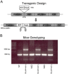
Transgenic design and mouse genotyping. (A) In the BAC clone (RP23-234B11) that contains the genomic locus of MT1 gene, the translational start codon was replaced with sequences encoding RFP and a poly A tail such that only RFP would be transcribed from the modified transgene. (1) and (2) indicate the positions of primers used for checking the RFP insert. (B) Using primers flanking the translational start codon, the PCR reaction detected a ~500-bp band in wild type (WT) and negative littermates (NEG). In the fA hemizygous transgenic mice, both a ~1500-bp band and a ~500-bp band were found, indicating the expression of RFP in these mice.
Figure 2.
Figure 2.
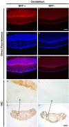
Direct fluorescence and direct immunohistochemistry in cerebellar sections of fA transgenic mice and their negative littermates. RFP expression was found in cerebellar sections of fA transgenic mice (A) but not in the cerebellum of their negative littermates (F) using direct fluorescence microscopy. DAPI nuclear staining of fA mice cerebellar sections (B) showed a merge pattern with RFP expression (C). DAPI nuclear staining for negative littermates (G) did not show any merge pattern (H). The presence of RFP-MT1 was confirmed by immunohistochemistry in the cerebellar sections of fA transgenic (E). A region of the cerebellum was taken at higher magnification (20×) (D). Negative littermate cerebellar tissues were used as a control for the RFP antibody and showed no staining in any region of the cerebellum (J and I). Scale bars are 100 µm (F) and 1 mm (J).
Figure 3.
Figure 3.
Expression of RFP-MT1 in the pars tuberalis and third ventricle. Direct fluorescence of RFP was detected in the median eminence of the pars tuberalis in fA mice (A) but not in negative littermates (G). DAPI nuclear staining in fA mice sections (B) merged with RFP expression (C), indicating the presence of RFP-MT1 in the median eminence of the pars tuberalis. DAPI muclear staining in negative littermate sections (H) did not show any pattern of merge expression with the red fluorescence image (I). The expression of RFP-MT1 was confirmed using direct immunohistochemistry in the median eminence of the pars tuberalis (E and J) and the ependyma lining the dorsal part of the third ventricle (F and K). Scale bars are 200 µm (G) and 100 µm (K).
Figure 4.
Figure 4.
Expression of RFP-MT1 in the aqueduct and the dorsomedial periaqueductal gray. Direct fluorescence microscopy revealed expression of RFP-MT1 in the aqueduct (A) and the dorsomedial periaqueductal gray in sections of fA mice (I) but not in negative littermate brain sections (E and M). DAPI nuclear staining in negative littermate sections (F and N) and fA mice sections (B and J) showed no merging (G and O) and merging (C and K), respectively, with RFP expression. The expression of RFP-MT1 was confirmed using direct immunohistochemistry in the aqueduct (D and H) and the dorsomedial periaqueductal gray (L and P). Scale bars are 100 µm (E and P) and 200 µm (M).
Figure 5.
Figure 5.
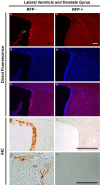
RFP-MT1 expression in the lateral ventricle and the hippocampus. RFP-MT1 was expressed in the ependymal lining the lateral ventricle. Direct fluorescence was observed in sections of fA mice (A) compared with negative littermates that did not show any RFP-MT1 expression (F); DAPI nuclear staining in fA mice (B) and wild type (G) sections, respectively, merged (C) and did not merge with RFP expression (H). The expression of RFP-MT1 in the lateral ventricle of fA mice was confirmed using immunohistochemistry (D and I). Isolated cells in the dentate gyrus of the hippocampus also expressed RFP-MT1 (E and J). Scale bars are 100 µm (F and J) and 200 µm (I).
Figure 6.
Figure 6.
Expression of RFP-MT1 in the subcommissural organ of the dorsal third ventricle. Direct fluorescence microscopy in negative littermate tissue (E) showed no RFP expression whereas fA mice tissues (A) expressed RFP in the subcommissural organ of the third ventricle. DAPI nuclear staining in fA mice (B) merged with RFP expression (C) but did not merge in negative littermate tissues (F and G). The expression of RFP-MT1 was confirmed using direct immunohistochemistry in the subcommissural organ of the third ventricle using antibody targeted against RFP (D and H). Scale bar is 100 µm (H).
Figure 7.
Figure 7.
Expression of RFP-MT1 in the habenula commissure and the habenula. Direct fluorescence of RFP was detected in the habenula commissure of fA mice sections (A) but not in negative littermates sections (E). DAPI nuclear staining in fA mice (B) and negative littermate (F) sections merged (C) or did not merge with RFP expression (G), respectively. The expression of RFP-MT1 was confirmed with immunohistochemistry in the habenula commissure using an antibody targeted against RFP (D and H). Direct fluorescence of RFP-MT1 was also expressed in the habenular tissues of fA mice (I) but not in negative littermate habenular tissues (M). The expression of RFP was restricted to the medial habenula (MHb). DAPI nuclear staining in habenular sections of negative (N) and fA (J) littermates did not merge (O) or merged with RFP expression (K), respectively. The expression of RFP-MT1 was confirmed using immunohistochemistry in the habenula (L and P). Scale bars are 100 µm (E and P) and 200 µm (M).
Figure 8.
Figure 8.
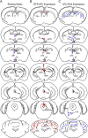
2-[125I]-Iodomelatonin binding, RFP-MT1 expression and MT1 RNA expression in different areas of the mouse brain. Lucida images of brain sections were drawn rostral to caudal showing areas where 2-[125I]-iodomelatonin binding (A), RFP-MT1 expression (B) and MT1 RNA expression (C) have been found according to data from our laboratory or in the literature. The purple, red and blue dots respectively indicate 2-iodomelatonin binding, MT1 expression using the fA transgenic mice and MT1 RNA expression using PCR or in situ hybridization. Acronyms used are: Cpu (Caudate Putamen or Striatum); Lv (Lateral ventricle); 3V (Third ventricle); PVN (Paraventricular Nucleus); SCN (Suprachiasmatic Nucleus); SPZ (Subparaventricular Zone); MHb (Medial Habenula); LHb (Lateral Habenula); PT (Pars Tuberalis); SCO (Subcommisural Organ); HbC (Habenula Commissure); D3V (Dorsal Third Ventricle); Aq (Aqueduct); DMPAG (Dorsomedial Periaqueductal Gray); SNC (Substantia Nigra); VTA (Ventral Tegmentum); GCL (Granular Cell Layer); MCL (Molecular Cell Layer); PCL (Purkinje Cell Layer).

References

    1. Aggelopoulos NC, Brown SJ, Demaine C, Harris NG, Jones HC. (1997). An abnormal distribution of melatonin receptors in the newborn rat with inherited prenatal hydrocephalus. Neurosci 80:669-673 - PubMed
    1. Al-Ghoul WM, Herman MD, Dubocovich ML. (1998). Melatonin receptor subtype expression in human cerebellum. Neuroreport 9:4063-4068 - PubMed
    1. Ambriz-Tututi M, Rocha-González HI, Cruz SL, Granados-Soto V. (2009). Melatonin: A hormone that modulates pain. Life Sci 84:489-498 - PubMed
    1. Andres KH, von During M, Veh RW. (1999). Subnuclear organization of the rat habenular complexes. J Comp Neurol 407:130-150 - PubMed
    1. Ayoub MA, Levoye A, Delagrange P, Jockers R. (2004). Preferential Formation of MT1/MT2 Melatonin Receptor Heterodimers with Distinct Ligand Interaction Properties Compared with MT2 Homodimers. Mol Pharmacol 66:312-321 - PubMed

Publication types

LinkOut - more resources