. 2013 Oct;123(10):4364-74.
doi: 10.1172/JCI70064. Epub 2013 Sep 24.

Dendritic epidermal T cells regulate skin antimicrobial barrier function

Dendritic epidermal T cells regulate skin antimicrobial barrier function

Amanda S MacLeod et al. J Clin Invest. 2013 Oct.

Abstract

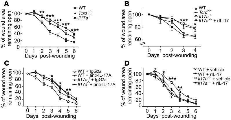
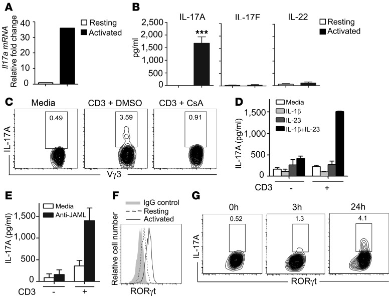
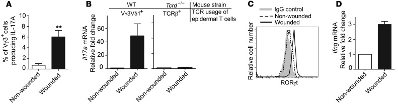
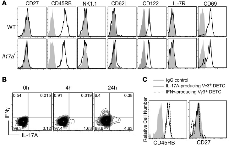
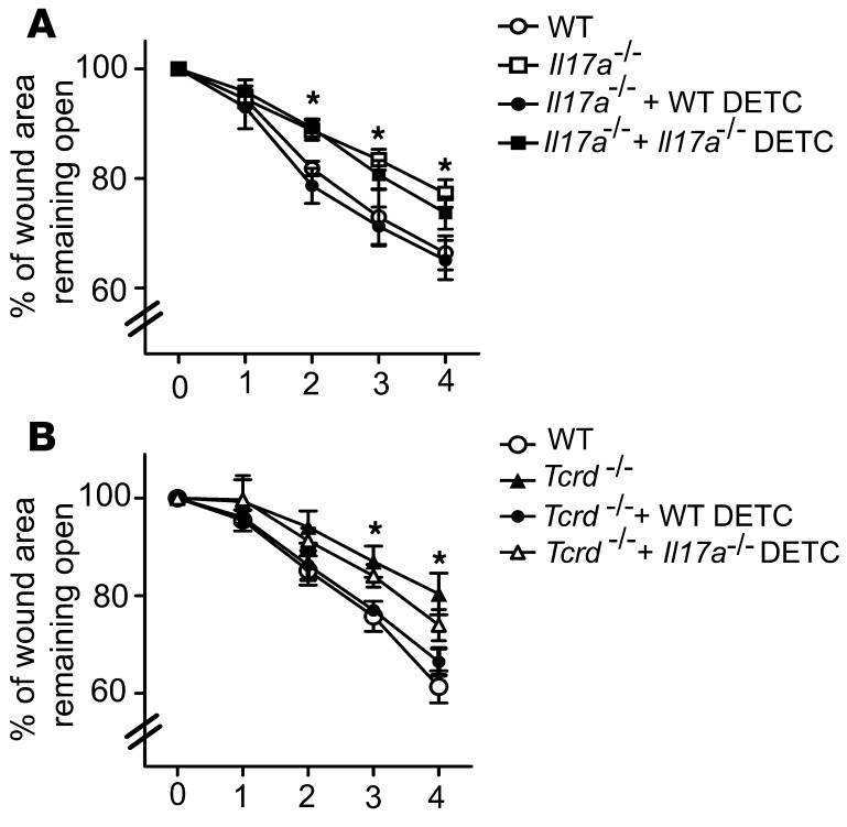
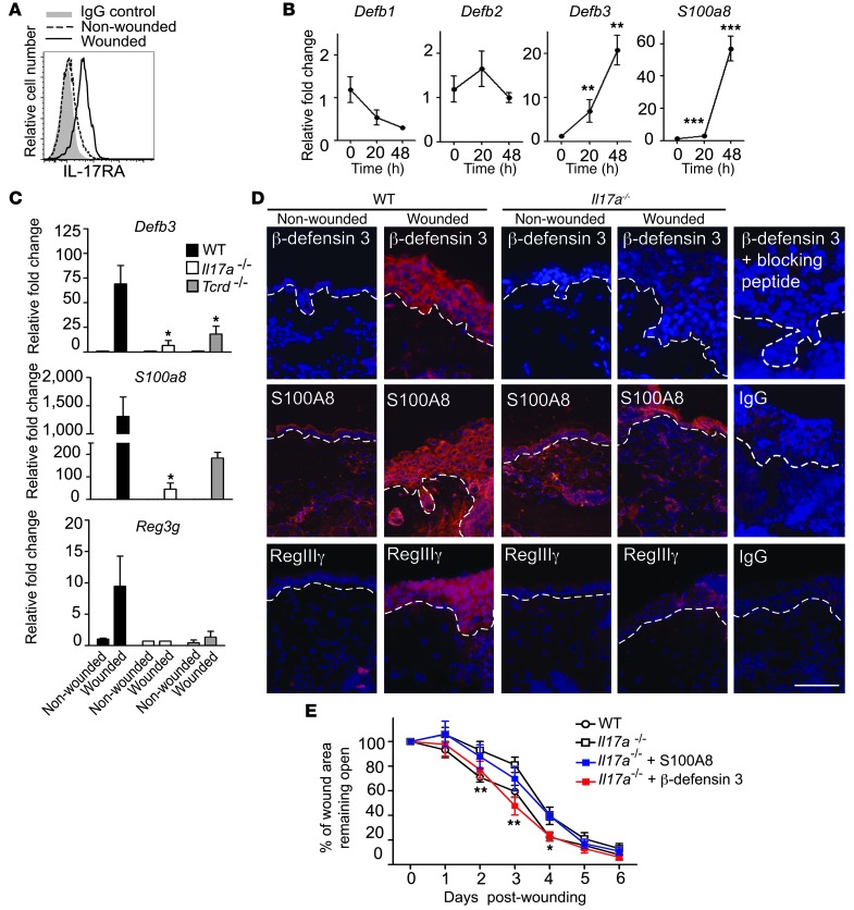
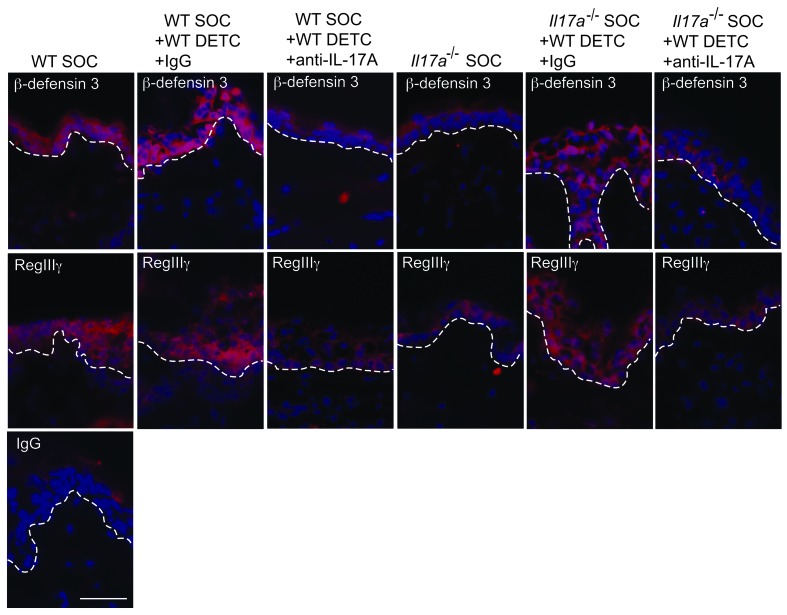
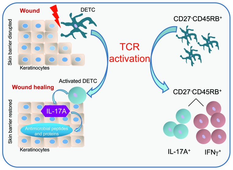
The epidermis, the outer layer of the skin, forms a physical and antimicrobial shield to protect the body from environmental threats. Skin injury severely compromises the epidermal barrier and requires immediate repair. Dendritic epidermal T cells (DETC) reside in the murine epidermis where they sense skin injury and serve as regulators and orchestrators of immune responses. Here, we determined that TCR stimulation and skin injury induces IL-17A production by a subset of DETC. This subset of IL-17A-producing DETC was distinct from IFN-γ producers, despite similar surface marker profiles. Functionally, blocking IL-17A or genetic deletion of IL-17A resulted in delayed wound closure in animals. Skin organ cultures from Tcrd-/-, which lack DETC, and Il17a-/- mice both exhibited wound-healing defects. Wound healing was fully restored by the addition of WT DETC, but only partially restored by IL-17A-deficient DETC, demonstrating the importance of IL-17A to wound healing. Following skin injury, DETC-derived IL-17A induced expression of multiple host-defense molecules in epidermal keratinocytes to promote healing. Together, these data provide a mechanistic link between IL-17A production by DETC, host-defense, and wound-healing responses in the skin. These findings establish a critical and unique role of IL-17A-producing DETC in epidermal barrier function and wound healing.


Figures

Figure 1
Figure 1. Local IL-17A production is critical for wound healing.
(A) Il17a–/– mice have delayed wound closure in vivo. Wound-closure kinetics in WT, Tcrd–/–, and Il17a–/– mice were measured over time. Data shown represent percentage of wound area remaining open ± SEM from 4 to 8 wounds. **P ≤ 0.01; ***P ≤ 0.001, Il17a–/– versus WT. (B) Wound-healing kinetics were measured in SOCs from WT, Tcrd–/–, and Il17a–/– mice. In some conditions, wounded skin was supplemented with rIL-17A or vehicle control. Data are presented as mean ± SEM from 3 wounds per condition. *P ≤ 0.05; ***P ≤ 0.001, WT versus Il17a–/–. (C) Blockade of IL-17A inhibits wound closure in vivo. Neutralizing IL-17 Ab or control Ab (IgG2a) was applied into the wound bed of WT and Il17a–/– mice. Data are presented as the mean percentage of the wound area remaining open ± SEM from 4 to 8 wounds. *P ≤ 0.05; **P ≤ 0.01. (D) Recombinant IL-17A restores defective wound healing in Il17a–/– skin in vivo. Wounded skin was supplemented with rIL-17A or vehicle control, and wound-healing kinetics were measured over time. Data shown represent mean ± SEM of 4 to 8 wounds per condition. *P ≤ 0.05;**P ≤ 0.01; ***P ≤ 0.001, Il17a–/– versus WT.
Figure 2
Figure 2. IL-17A production by a subset of DETC requires TCR stimulation.
(A) DETC upregulate Il17a upon TCR stimulation in vitro. Sorted Vγ3+ DETC express Il17a following activation by plate-bound anti-CD3ε (10 μg/ml). (B) Freshly isolated and sorted Vγ3+ DETC were stimulated with plate-bound anti-CD3ε (10 μg/ml), and IL-17A, IL-17F, and IL-22 protein was measured by ELISA. Data are shown as mean ± SEM from quadruplicate measurements and are representative of 3–4 independent experiments. ***P ≤ 0.001. (C) CsA inhibits anti-CD3ε–mediated IL-17A production in DETC. Cells are gated on Vγ3+Thy1.2+. (D) Stimulation with IL-1β and IL-23 alone does not induce IL-17A in DETC. Sorted Vγ3+ DETC were stimulated with IL-1β, IL-23, or the combination thereof in the presence or absence of suboptimal anti-CD3ε, and IL-17A secretion was assayed by ELISA. Data are shown as mean ± SEM from duplicates. (E) JAML costimulation increases IL-17A in DETC. ELISA analysis for IL-17A from culture supernatants of DETC stimulated with antibody against suboptimal doses of CD3 alone, CD3 and JAML, or JAML alone. Data are shown as mean ± SEM from duplicates. (F) DETC upregulate RORγt upon TCR stimulation. Freshly isolated DETC were stimulated with anti-CD3ε (10 μg/ml) for 3 hours, and RORγt levels were measured by flow cytometry. (G) A subset of RORγt+ DETC produces IL-17A. RORγt and IL-17A expression were assessed by flow cytometry in freshly isolated epidermal cell suspensions that were stimulated with anti-CD3ε (10 μg/ml). Cells are gated on Vγ3+Thy1.2+.
Figure 3
Figure 3. DETC produce IL-17A upon skin injury in vivo.
(A) Increased frequency of IL-17A–producing Vγ3+ DETC in wounded skin. Epidermal cells were isolated from skin areas directly surrounding wounds or from nonwounded skin, 18 hours after wounding. Intracellular IL-17A production was analyzed by flow cytometry. Data are pooled and shown as the mean ± SEM of Vγ3 cells producing IL-17A from independent analyses of 4 mice and are expressed as percentage of Vγ3+ cells producing IL-17A. **P < 0.01. (B) Signaling through the Vγ3Vδ1 TCR is required for DETC-mediated IL-17A expression in vivo. Vγ3+ DETC from WT and epidermal TCRβ+ cells from Tcrd–/– mice were isolated and sorted from the epidermis of wounded and nonwounded skin areas 18 hours following skin injury. IL-17A was measured by qPCR. Data are pooled and shown as mean ± SEM from 3 independent experiments with at least 4 mice per genotype per experiment. (C) RORγt increases in DETC upon skin injury. DETC were isolated 12 hours following skin injury from nonwounded and wounded skin areas and were stained for RORγt. Cells are gated on Vγ3+Thy1.2+. (D) Ifng is upregulated upon skin injury. Vγ3+ DETC were isolated and sorted from the epidermis of wounded and nonwounded skin areas 18 hours following skin injury. Ifng was measured by qPCR. Data are pooled and shown as mean ± SEM from 3 independent experiments with at least 4 mice per genotype per experiment.
Figure 4
Figure 4. IL-17A– and IFN-γ–producing DETC subsets maintain their CD2745RB+ phenotype.
(A) Phenotypic characterization of freshly isolated WT and Il17a–/– DETC. Cells are gated on Vγ3+Thy1.2+ and CD45RB, CD27, NK1.1, CD62L, CD122, IL-7R, and CD69 expression (solid lines), and their appropriate IgG controls (gray) were measured by flow cytometry. (B) Distinct DETC subsets produce IL-17A and IFN-γ. Intracellular staining on DETC short-term cell lines stimulated with anti-CD3ε (10 μg/ml) for IL-17A and IFN-γ production. Cells are gated on Vγ3+Thy1.2+. (C) IL-17A– and IFN-γ–producing DETC are CD27CD45RB+. Fresh epidermal cell suspensions were stimulated with anti-CD3ε (10 μg/ml), and expression of CD27, CD45RB, and intracellular IL-17A and IFN-γ were measured by flow cytometry. Cells are gated on IL-17A+Vγ3+Thy1.2+ and IFN-γ+Vγ3+Thy1.2+, respectively.
Figure 5
Figure 5. WT but not Il17a–/– DETC restore defective wound healing.
Preactivated WT or Il17a–/– DETC were added into the wound bed of wounded skin explants from Il17a–/– (A) or Tcrd–/– (B) mice, and wound-healing kinetics in SOC were measured over time. Data presented are mean ± SEM from 3 wounds per condition. *P < 0.05, Il17a–/–+WT DETC vs. Il17a–/– (A) and Tcrd–/– +WT DETC vs. Tcrd–/– (B).
Figure 6
Figure 6. IL-17A is critical for induction of epidermal host-defense molecules to mediate wound repair.
(A) IL-17RA expression increases upon wounding. Epidermal cells isolated from nonwounded and wounded sites were stained for IL-17RA 18 hours following wounding. Cells are gated on CD45. (B) IL-17A induces murine Defb3 and S100a8 in primary murine keratinocytes. **P < 0.01; ***P < 0.001. (C and D) Induction of AMP upon wounding is impaired in Il17a–/– mice. Levels of epidermal β-defensin 3, S100A8, and RegIIIγ were analyzed (C) by qPCR of epidermal sheets from nonwounded and wounded skin or (D) by immunofluorescence staining of wounded and nonwounded skin from WT and Il17a–/– animals 24 hours after wounding. (C) For qPCR, data are pooled from 4–6 wounds and expressed as mean ± SEM from 3 independent experiments. Data are expressed as relative fold change compared with nonwounded controls. *P ≤ 0.05. (D) Antibodies recognizing β-defensin 3, S100A8, or RegIIIγ were used (red staining). DAPI was used to visualize cell nuclei (blue staining). Dotted white lines represent the epidermal-dermal border. Scale bar: 50 μm. (E) β-Defensin 3 but not S100A8 ameliorates defective wound healing in Il17a–/– mice in vivo. Skin wounds were treated with recombinant β-defensin 3 or S100A8, and wound-healing kinetics were measured over time. Data shown represent mean ± SEM of 4 to 8 wounds per condition. *P ≤ 0.05; **P ≤ 0.01, Il17a–/– versus Il17a–/–+ β-defensin 3.
Figure 7
Figure 7. Robust restoration of antimicrobial peptides in Il17a–/– skin wounds by WT DETC via an IL-17A–dependent mechanism.
Wounded SOCs from WT or Il17a–/– mice were supplemented with preactivated WT DETC. Neutralizing anti–IL-17A antibody together with preactivated DETC was added into the wound bed of SOCs. Skin directly adjacent to the wound bed was analyzed for β-defensin 3 and RegIIIγ expression (red staining). DAPI was used to visualize cell nuclei (blue staining). Dotted white lines represent the epidermal-dermal border. Scale bar: 50 μm.
Figure 8
Figure 8. Schematic model for DETC-regulated immune functions during wound healing.
Upon skin injury, DETC become activated and a subset of DETC produces IL-17A. IL-17A is critical to inducing several AMP in keratinocytes, thus helping to reestablish the skin barrier during wound healing. DETC-mediated IL-17A production is highly dependent on TCR stimulation. DETC activation via TCR mediates production of IL-17A and IFN-γ in distinct subsets despite a homogenous CD27CD45RB+ phenotype. This is in contrast with previous observations in peripheral γδ T cells and thymic DETC precursors, suggesting that DETC cytokine commitment is uniquely regulated.

References

    1. Elias PM. Skin barrier function. Curr Allergy Asthma Rep. 2008;8(4):299–305. doi: 10.1007/s11882-008-0048-0. - DOI - PMC - PubMed
    1. Braff MH, Gallo RL. Antimicrobial peptides: an essential component of the skin defensive barrier. Curr Top Microbiol Immunol. 2006;306:91–110. - PubMed
    1. Buchau AS, Gallo RL. Innate immunity and antimicrobial defense systems in psoriasis. Clin Dermatol. 2007;25(6):616–624. doi: 10.1016/j.clindermatol.2007.08.016. - DOI - PMC - PubMed
    1. Buchau AS, et al. The host defense peptide cathelicidin is required for NK cell-mediated suppression of tumor growth. J Immunol. 2010;184(1):369–378. doi: 10.4049/jimmunol.0902110. - DOI - PMC - PubMed
    1. Baum CL, Arpey CJ. Normal cutaneous wound healing: clinical correlation with cellular and molecular events. Dermatol Surg. 2005;31(6):674–686. doi: 10.1111/j.1524-4725.2005.31612. - DOI - PubMed

Publication types

MeSH terms

Substances