. 2014 Apr;19(2):208-14.
doi: 10.1177/2211068213504296. Epub 2013 Sep 19.

Transfer, imaging, and analysis plate for facile handling of 384 hanging drop 3D tissue spheroids

Affiliations

Transfer, imaging, and analysis plate for facile handling of 384 hanging drop 3D tissue spheroids

Stephen P Cavnar et al. J Lab Autom. 2014 Apr.

Abstract

Three-dimensional culture systems bridge the experimental gap between in vivo and in vitro physiology. However, nonstandardized formation and limited downstream adaptability of 3D cultures have hindered mainstream adoption of these systems for biological applications, especially for low- and moderate-throughput assays commonly used in biomedical research. Here we build on our recent development of a 384-well hanging drop plate for spheroid culture to design a complementary spheroid transfer and imaging (TRIM) plate. The low-aspect ratio wells of the TRIM plate facilitated high-fidelity, user-independent, contact-based collection of hanging drop spheroids. Using the TRIM plate, we demonstrated several downstream analyses, including bulk tissue collection for flow cytometry, high-resolution low working-distance immersion imaging, and timely reagent delivery for enzymatic studies. Low working-distance multiphoton imaging revealed a cell type-dependent, macroscopic spheroid structure. Unlike ovarian cancer spheroids, which formed loose, disk-shaped spheroids, human mammary fibroblasts formed tight, spherical, and nutrient-limited spheroids. Beyond the applications we describe here, we expect the hanging drop spheroid plate and complementary TRIM plate to facilitate analyses of spheroids across the spectrum of throughput, particularly for bulk collection of spheroids and high-content imaging.

Keywords: 3D tissue spheroids; and hanging drop spheroids; high-throughput imaging.


Conflict of interest statement

Declaration of Conflicting Interests

The authors declared the following potential conflicts of interest with respect to the research, authorship, and/or publication of this article: S.T. has licensed the 384 hanging drop array technology to 3D Biomatrix and also has stock options.

Figures
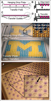
Figure 1
Figure 1
Transfer and imaging of 384 hanging drop spheroids. (A) Schematic representation of the hanging drop plate and complementary transfer and imaging (TRIM) plate. The circle in A defines the region detailed in B. (C) Schematic depiction of hanging drop plate transfer with user-independent alignment guides. The circle in C defines the region detailed in D, showing contact-dependent drop transfer. (E) Example images of the residual dye in the hanging drop plate (top) after transfer to the TRIM plate (bottom). The inset shows the bottom view of the hanging droplets prior to transfer. (F) Example image of 500-µm spheroids transferred from the hanging drop plate to the TRIM plate. Spheroids were stained with trypan blue for 4 h prior to transfer to improve image contrast.
Figure 2
Figure 2
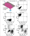
Spheroid collection for analysis by flow cytometry. (A) We co-seeded spheroids with MDA-MB-231 cells at ratios of 9:1 and 1:1 of cells expressing FP650 or green fluorescent protein (GFP), respectively. Schematic of spheroid collection by directing pipette flow into each well and collecting the overflow. After collection, centrifugation, and spheroid dissociation, we performed flow cytometry. FACS, fluorescence-activated cell sorting. (B–F) Flow cytometry scatter plots show MDA-MB-231 cells with no fluorescent protein (B), FP650 only (C), GFP only (D), and 9:1 (E) and 1:1 (F) ratios of FP650 and GFP cells, respectively. Note that we compare the top two quartiles with the bottom quartiles to estimate the relative seeding of FP650 and GFP cells to capture the heterogeneity of the FP650 cells.
Figure 3
Figure 3
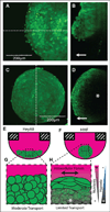
Three-dimensional imaging with the transfer and imaging (TRIM) plate. (A) Example two-photon z-stack image of a large spheroid of HeyA8 ovarian cancer cells expressing green fluorescent protein (GFP) and (B) the corresponding cross section with the location denoted by the dotted line in A. (C) Example two-photon z-stack image of a large spheroid of HMF-expressing GFP spheroid and (D) the corresponding cross section with the location denoted by the dotted line in A. Arrows (B, D) denote the upward direction toward the objective. Unlike the ovarian cancer spheroid, the HMF spheroid is dense and nutrient limited, resulting in lower internal fluorescence intensity, denoted by an asterisk (D, H). Schematics of the spheroid morphology show HeyA8 spheroids to curve with the droplet radius (E) and the HMF spheroid to be spherical (F). Loose interactions between HeyA8 cells are permissive to nutrient transport within the spheroid (G), but tight intracellular forces limit transport into the spheroid (H).

References

    1. Mehta G, Hsiao AY, Ingram M, et al. Opportunities and Challenges for Use of Tumor Spheroids as Models to Test Drug Delivery and Efficacy. J. Control. Release. 2012;164:192–204. - PMC - PubMed
    1. Zhou Y, Arai T, Horiguchi Y, et al. Multi-Parameter Analyses of Three-Dimensionally Cultured Tumor Spheroids Based on Respiratory Activity and Comprehensive Gene-Expression Profiles. Anal. Biochem. 2013;439:187–193. - PubMed
    1. Hsiao AY, Tung YC, Qu X, et al. 384 Hanging Drop Arrays Give Excellent Z-Factors and Allow Versatile Formation of Co-culture Spheroids. Biotechnol. Bioeng. 2012;109:1293–1304. - PMC - PubMed
    1. Tung YC, Hsiao AY, Allen SG, et al. High-Throughput 3D Spheroid Culture and Drug Testing Using a 384 Hanging Drop Array. Analyst. 2011;136:473–478. - PMC - PubMed
    1. Kelm JM, Fussenegger M. Microscale Tissue Engineering Using Gravity-Enforced Cell Assembly. Trends Biotechnol. 2004;22:195–202. - PubMed

Publication types

MeSH terms