. 2013 Oct;8(10):1950-60.
doi: 10.1038/nprot.2013.120. Epub 2013 Sep 19.

Large-scale identification of ubiquitination sites by mass spectrometry

Affiliations

Large-scale identification of ubiquitination sites by mass spectrometry

Namrata D Udeshi et al. Nat Protoc. 2013 Oct.

Abstract

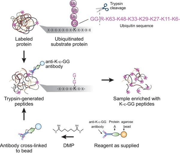
Ubiquitination is essential for the regulation of cellular protein homeostasis. It also has a central role in numerous signaling events. Recent advances in the production and availability of antibodies that recognize the Lys-ɛ-Gly-Gly (K-ɛ-GG) remnant produced by trypsin digestion of proteins having ubiquitinated lysine side chains have markedly improved the ability to enrich and detect endogenous ubiquitination sites by mass spectrometry (MS). The following protocol describes the steps required to complete a large-scale ubiquitin experiment for the detection of tens of thousands of distinct ubiquitination sites from cell lines or tissue samples. Specifically, we present detailed, step-by-step instructions for sample preparation, off-line fractionation by reversed-phase chromatography at pH 10, immobilization of an antibody specific to K-ɛ-GG to beads by chemical cross-linking, enrichment of ubiquitinated peptides using these antibodies and proteomic analysis of enriched samples by LC-tandem MS (MS/MS). Relative quantification can be achieved by performing stable isotope labeling by amino acids in cell culture (SILAC) labeling of cells. After cell or tissue samples have been prepared for lysis, the described protocol can be completed in ∼5 d.


Figures

Figure 1
Figure 1
Enrichment of K-ε-GG peptides using anti-K-ε-GG antibody. After the anti-K-ε-GG antibody has been chemically cross-linked to a Protein A beads using DMP, each of the eight bRP fractions are individually enriched for K-ε-GG peptides using the anti-K-ε-GG antibody which recognizes the di-glycyl remnant remaining on modified lysine residues following trypsin digestion.
Figure 2
Figure 2
Workflow for preparing samples for K-ε-GG enrichment. Samples are lysed in urea buffer, digested with LysC and Trypsin, and fractionated off-line using bRP chromatography. Fractions are pooled to 8 final fractions (see text for details) for enrichment of K-ε-GG peptides using anti-K-ε-GG antibody.
Figure 3
Figure 3
Example of an SDS-PAGE gel used to evaluate the efficiency of antibody cross-linking to Protein A beads. Note this figure has been previously published by Udeshi et al. in ref. 6. TFA eluates from ~30 ug of pre- and post-cross-linked anti-K-ε-GG antibody were analyzed by SDS-PAGE. More than a 10-fold decrease in staining density for heavy and light chains of the antibody is required.
Figure 4
Figure 4
Analysis of K-ε-GG peptide data. A) Bar plot showing the average number of distinct K-ε-GG and non-K-ε-GG peptides identified across 8 bRP fractions for three biological replicates. For this plot, proteins were derived from SILAC-labeled Jurkat cells that were treated with proteasome inhibitor MG-132 or deubiquitinase inhibitor PR-619, or were left untreated. Approximately 5 mg of protein was input into the K-ε-GG workflow per SILAC state. B) Pie chart showing the average percentage of K-ε-GG peptides identified in 1, 2, or >2 bRP fractions. Note that approximately 79% of K-ε-GG peptides are found in only 1 bRP fraction. C) Bar plot showing the average number of distinct K-ε-GG and non-K-ε-GG peptides found in total for the SILAC-based study.
Figure 5
Figure 5
MS/MS spectrum of a K-ε-GG peptide. An HCD MS/MS spectrum recorded on the [M+2H]+2 ion at m/z 586.81 of the modified Eukaryotic translation initiation factor 3 subunit C peptide LCKYIYAK harboring one K-ε-GG site (denoted by lower case k) and one carbamidomethylated cysteine residue (denoted by upper case C). Predicted b- and y-type ions are listed above and below the peptide sequence, respectively. Ions observed are labeled in the spectrum and indicate that K554 of the protein is modified with the di-glycine remnant.

References

    1. Ye Y, Rape M. Building ubiquitin chains: E2 enzymes at work. Nature reviews Molecular cell biology. 2009;10:755–764. - PMC - PubMed
    1. Dikic I, Wakatsuki S, Walters KJ. Ubiquitin-binding domains [mdash] from structures to functions. Nat Rev Mol Cell Biol. 2009;10:659–671. - PMC - PubMed
    1. Peng J, et al. A proteomics approach to understanding protein ubiquitination. Nat Biotechnol. 2003;21:921–926. - PubMed
    1. Danielsen JMR, et al. Mass Spectrometric Analysis of Lysine Ubiquitylation Reveals Promiscuity at Site Level. Mol. Cell. Proteomics. 2010;10 M110.003590. - PMC - PubMed
    1. Udeshi ND, et al. Methods for quantification of in vivo changes in protein ubiquitination following proteasome and deubiquitinase inhibition. Mol. Cell. Proteomics. 2012;11:148–159. - PMC - PubMed

Publication types