Neuronal STAT3 activation is essential for CNTF- and inflammatory stimulation-induced CNS axon regeneration
- PMID: 24052073
- PMCID: PMC3789169
- DOI: 10.1038/cddis.2013.310
Neuronal STAT3 activation is essential for CNTF- and inflammatory stimulation-induced CNS axon regeneration
Abstract
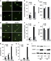
CNS neurons, such as retinal ganglion cells (RGCs), do not normally regenerate injured axons, but instead undergo apoptotic cell death. Regenerative failure is due to inhibitory factors in the myelin and forming glial scar as well as due to an insufficient intrinsic capability of mature neurons to regrow axons. Nevertheless, RGCs can be transformed into an active regenerative state upon inflammatory stimulation (IS) in the inner eye, for instance by lens injury, enabling these RGCs to survive axotomy and to regenerate axons into the lesioned optic nerve. The beneficial effects of IS are mediated by various factors, including CNTF, LIF and IL-6. Consistently, IS activates various signaling pathways, such as JAK/STAT3 and PI3K/AKT/mTOR, in several retinal cell types. Using a conditional knockdown approach to specifically delete STAT3 in adult RGCs, we investigated the role of STAT3 in IS-induced neuroprotection and axon regeneration. Conditional STAT3 knockdown in RGCs did not affect the survival of RGCs after optic nerve injury compared with controls, but significantly reduced the neuroprotective effects of IS. STAT3 depletion significantly compromised CNTF-stimulated neurite growth in culture and IS-induced transformation of RGCs into an active regenerative state in vivo. As a consequence, IS-mediated axonal regeneration into the injured optic nerve was almost completely abolished in mice with STAT3 depleted in RGCs. In conclusion, STAT3 activation in RGCs is involved in neuroprotection and is a necessary prerequisite for optic nerve regeneration upon IS.
Figures


Similar articles
-
Neuroprotective and axon growth-promoting effects following inflammatory stimulation on mature retinal ganglion cells in mice depend on ciliary neurotrophic factor and leukemia inhibitory factor.J Neurosci. 2009 Nov 11;29(45):14334-41. doi: 10.1523/JNEUROSCI.2770-09.2009. J Neurosci. 2009. PMID: 19906980 Free PMC article.
-
Interleukin-6 contributes to CNS axon regeneration upon inflammatory stimulation.Cell Death Dis. 2013 Apr 25;4(4):e609. doi: 10.1038/cddis.2013.126. Cell Death Dis. 2013. PMID: 23618907 Free PMC article.
-
CXCL12/SDF-1 facilitates optic nerve regeneration.Neurobiol Dis. 2013 Jul;55:76-86. doi: 10.1016/j.nbd.2013.04.001. Epub 2013 Apr 8. Neurobiol Dis. 2013. PMID: 23578489
-
Stimulating axonal regeneration of mature retinal ganglion cells and overcoming inhibitory signaling.Cell Tissue Res. 2012 Jul;349(1):79-85. doi: 10.1007/s00441-011-1302-7. Cell Tissue Res. 2012. PMID: 22293973 Review.
-
Promoting optic nerve regeneration.Prog Retin Eye Res. 2012 Nov;31(6):688-701. doi: 10.1016/j.preteyeres.2012.06.005. Epub 2012 Jul 7. Prog Retin Eye Res. 2012. PMID: 22781340 Review.
Cited by
-
Neuroinflammation, Microglia and Implications for Retinal Ganglion Cell Survival and Axon Regeneration in Traumatic Optic Neuropathy.Front Immunol. 2022 Mar 4;13:860070. doi: 10.3389/fimmu.2022.860070. eCollection 2022. Front Immunol. 2022. PMID: 35309305 Free PMC article. Review.
-
Central nervous system regeneration.Cell. 2022 Jan 6;185(1):77-94. doi: 10.1016/j.cell.2021.10.029. Cell. 2022. PMID: 34995518 Free PMC article. Review.
-
Thrombospondin-1 Mediates Axon Regeneration in Retinal Ganglion Cells.Neuron. 2019 Aug 21;103(4):642-657.e7. doi: 10.1016/j.neuron.2019.05.044. Epub 2019 Jun 26. Neuron. 2019. PMID: 31255486 Free PMC article.
-
The progress in optic nerve regeneration, where are we?Neural Regen Res. 2016 Jan;11(1):32-6. doi: 10.4103/1673-5374.175038. Neural Regen Res. 2016. PMID: 26981073 Free PMC article. Review.
-
Boosting CNS axon regeneration by harnessing antagonistic effects of GSK3 activity.Proc Natl Acad Sci U S A. 2017 Jul 3;114(27):E5454-E5463. doi: 10.1073/pnas.1621225114. Epub 2017 Jun 19. Proc Natl Acad Sci U S A. 2017. PMID: 28630333 Free PMC article.
References
-
- Diekmann H, Fischer D. Glaucoma and optic nerve repair. Cell Tissue Res. 2013;353:327–337. - PubMed
-
- Wohl SG, Schmeer CW, Witte OW, Isenmann S. Proliferative response of microglia and macrophages in the adult mouse eye after optic nerve lesion. Invest Ophthalmol Vis Sci. 2010;51:2686–2696. - PubMed
-
- Berry M, Ahmed Z, Lorber B, Douglas M, Logan A. Regeneration of axons in the visual system. Restor Neurol Neurosci. 2008;26:147–174. - PubMed
-
- Schwab ME, Kapfhammer JP, Bandtlow CE. Inhibitors of neurite growth. Ann Rev Neurosci. 1993;16:565–595. - PubMed
-
- Silver J, Miller JH. Regeneration beyond the glial scar. Nat Rev Neurosci. 2004;5:146–156. - PubMed
Publication types
MeSH terms
Substances
LinkOut - more resources
Full Text Sources
Other Literature Sources
Research Materials
Miscellaneous

