. 2013 Nov 1;288(44):31963-70.
doi: 10.1074/jbc.M113.509984. Epub 2013 Sep 19.

Crystal structures of β-1,4-galactosyltransferase 7 enzyme reveal conformational changes and substrate binding

Affiliations

Crystal structures of β-1,4-galactosyltransferase 7 enzyme reveal conformational changes and substrate binding

Yuko Tsutsui et al. J Biol Chem. .

Abstract

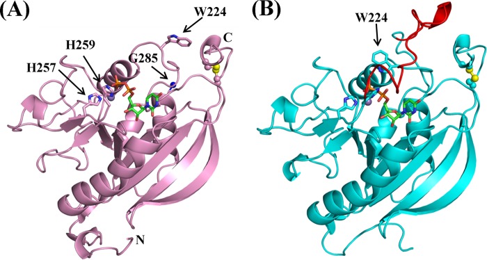
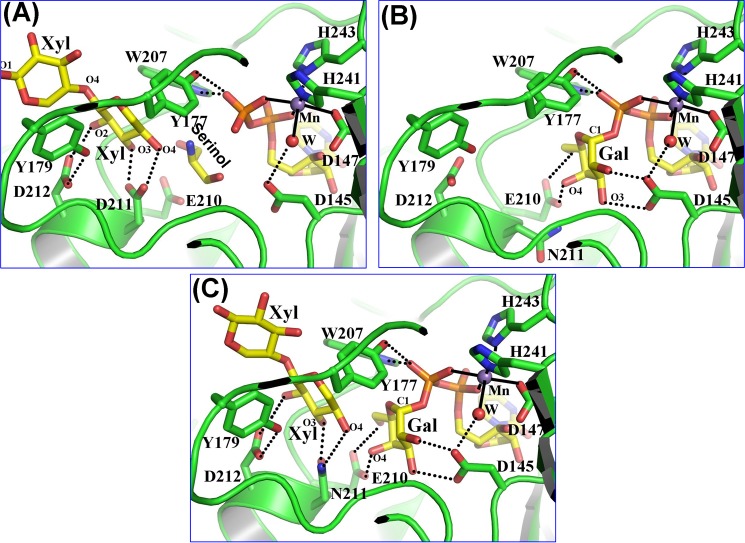
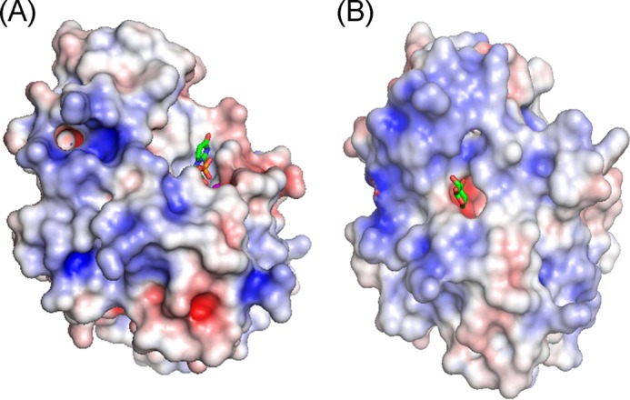
The β-1,4-galactosyltransferase 7 (β4GalT7) enzyme is involved in proteoglycan synthesis. In the presence of a manganese ion, it transfers galactose from UDP-galactose to xylose on a proteoglycan acceptor substrate. We present here the crystal structures of human β4GalT7 in open and closed conformations. A comparison of these crystal structures shows that, upon manganese and UDP or UDP-Gal binding, the enzyme undergoes conformational changes involving a small and a long loop. We also present the crystal structures of Drosophila wild-type β4GalT7 and D211N β4GalT7 mutant enzymes in the closed conformation in the presence of the acceptor substrate xylobiose and the donor substrate UDP-Gal, respectively. To understand the catalytic mechanism, we have crystallized the ternary complex of D211N β4GalT7 mutant enzyme in the presence of manganese with the donor and the acceptor substrates together in the same crystal structure. The galactose moiety of the bound UDP-Gal molecule forms seven hydrogen bonds with the protein molecule. The nonreducing end of the xylose moiety of xylobiose binds to the hydrophobic acceptor sugar binding pocket created by the conformational changes, whereas its extended xylose moiety forms hydrophobic interactions with a Tyr residue. In the ternary complex crystal structure, the nucleophile O4 oxygen atom of the xylose molecule is found in close proximity to the C1 and O5 atoms of the galactose moiety. This is the first time that a Michaelis complex of a glycosyltransferase has been described, and it clearly suggests an SN2 type catalytic mechanism for the β4GalT7 enzyme.

Keywords: Crystal Structure; Enzyme Mechanisms; Glycosaminoglycan; Glycosyltransferases; Protein Structure; Proteoglycan Synthesis.


Figures

FIGURE 1.
FIGURE 1.
The crystal structures of human β4GalT7 in the open (A) and the closed (B) conformation. A manganese ion and a UDP molecule are shown as a purple sphere and a green-and-orange stick figure, respectively. In the open conformation (A), the electron density was visible up to His259, and the clear electron density appeared again at Gly285 (blue sphere). The His257-X-His259 motif, Trp224, and the N and C termini are indicated. A disulfide bond is shown in a ball-and-stick format, with the yellow spheres indicating sulfur atoms. In B, the long loop region, residues 260–285, invisible in the open conformation, is highlighted in red.
FIGURE 2.
FIGURE 2.
Substrate binding to the Drosophila β4GalT7 molecule. The protein molecule is represented by the green graphic, with the interacting residues shown as colored sticks. The substrate molecules UDP-Gal and xylobiose are shown as yellow sticks. The manganese and water molecules are shown as magenta and red spheres. The hydrogen bonds are shown in black dotted lines, and the manganese coordination bond is shown in solid black lines. A, binding of a xylobiose molecule to the Drosophila β4GalT7 molecule in the closed conformation. The acceptor substrate forms four hydrogen bonds with the protein molecule. The C5 and O5 atoms of the acceptor xylose molecule form strong hydrophobic interactions with the aromatic side chain of the Tyr177 residue. Thus, the presence of this residue determines the enzyme acceptor specificity for xylose instead of glucose. The extended xylose moiety forms stacking interactions with the aromatic side chain of the Tyr179 residue. When the PG acceptor substrate binds to the enzyme, with its O-linked β-xylose moiety bound in the acceptor substrate binding site, this extended binding site is likely to facilitate the binding of the protein moiety of the PG acceptor molecule. B, UDP-gal binding to the D211N β4GalT7 mutant protein. The binding of the UDP moiety in the present crystal structure and in the human β4GalT7 closed conformation crystal structure (Fig. 1B) is quite similar (supplemental Fig. S3). The Gal moiety of the bound UDP-Gal forms seven hydrogen bonds with the protein molecule. These hydrogen-bonding interactions are similar to those that occur when UDP-Gal binds to the β4GalT1 molecule. C, the binding of the xylobiose together with UDP-Gal to Drosophila D211N β4GalT7 molecule. The molecular interactions of the substrates with the protein molecule are similar to their individual binding to the enzyme (panels A and B). In the present structure, the O4 oxygen atom of the xylobiose molecule forms a hydrogen bond with the Nϵ2 amino group of the Asn211 residue, which is in contrast to the panel A where the same O4 oxygen atom forms a hydrogen bond with the Oϵ2 atom of the Asp211 residue. The binding of the xylobiose does not seem to affect the binding of the UDP-Gal molecule or vice versa.
FIGURE 3.
FIGURE 3.
The electrostatic potentials of the open (A) and the closed (B) conformations of human β4GalT7 are shown, with the positively and negatively charged surfaces colored blue and red, respectively. The sticks represent UDP and a modeled xylose molecule in A and B, respectively.

References

    1. Varki A., Cummings R., Esko J., Freeze H., Hart G., Marth J. (1999) Essentials of Glycobiology, Cold Spring Harbor Laboratory Press, Cold Spring Harbor, NY - PubMed
    1. Ruoslahti E. (1989) Proteoglycans in cell regulation. J. Biol. Chem. 264, 13369–13372 - PubMed
    1. Chiodoni C., Colombo M. P., Sangaletti S. (2010) Matricellular proteins: from homeostasis to inflammation, cancer, and metastasis. Cancer Metastasis Rev. 29, 295–307 - PubMed
    1. Bishop J. R., Schuksz M., Esko J. D. (2007) Heparan sulphate proteoglycans fine-tune mammalian physiology. Nature 446, 1030–1037 - PubMed
    1. Afratis N., Gialeli C., Nikitovic D., Tsegenidis T., Karousou E., Theocharis A. D., Pavão M. S., Tzanakakis G. N., Karamanos N. K. (2012) Glycosaminoglycans: key players in cancer biology and treatment. FEBS J. 279, 1177–1197 - PubMed

Publication types

MeSH terms

Substances