. 2013 Oct 8;110(41):16544-9.
doi: 10.1073/pnas.1308331110. Epub 2013 Sep 19.

STING-IRF3 pathway links endoplasmic reticulum stress with hepatocyte apoptosis in early alcoholic liver disease

Affiliations

STING-IRF3 pathway links endoplasmic reticulum stress with hepatocyte apoptosis in early alcoholic liver disease

Jan Petrasek et al. Proc Natl Acad Sci U S A. .

Abstract

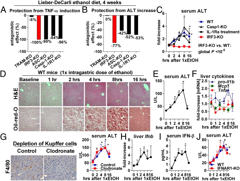
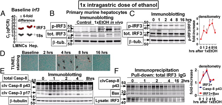
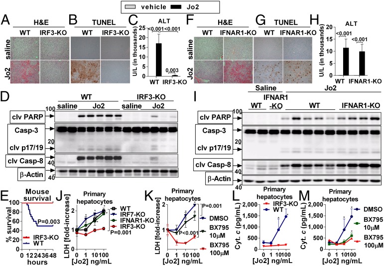
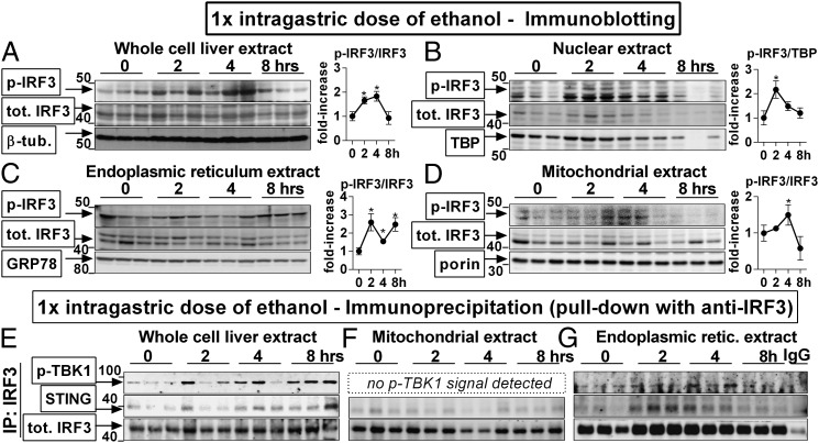
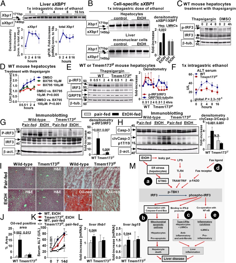
Emerging evidence suggests that innate immunity drives alcoholic liver disease (ALD) and that the interferon regulatory factor 3 (IRF3),a transcription factor regulating innate immune responses, is indispensable for the development of ALD. Here we report that IRF3 mediates ALD via linking endoplasmic reticulum (ER) stress with apoptotic signaling in hepatocytes. We found that ethanol induced ER stress and triggered the association of IRF3 with the ER adaptor, stimulator of interferon genes (STING), as well as subsequent phosphorylation of IRF3. Activated IRF3 associated with the proapoptotic molecule Bax [B-cell lymphoma 2 (Bcl2)-associated X protein] and contributed to hepatocyte apoptosis. Deficiency of STING prevented IRF3 phosphorylation by ethanol or ER stress, and absence of IRF3 prevented hepatocyte apoptosis. The pathogenic role of IRF3 in ALD was independent of inflammation or Type-I interferons. Thus, STING and IRF3 are key determinants of ALD, linking ER stress signaling with the mitochondrial pathway of hepatocyte apoptosis.

Keywords: Kupffer cells; interferon regulatory factor 3; steatohepatitis; stimulator of interferon genes.


Conflict of interest statement

The authors declare no conflict of interest.

Figures

Fig. 1.
Fig. 1.
The pathogenic role of IRF3 in ALD is independent of inflammation and Type-I IFN signaling. Mice of indicated genotypes were fed an alcohol diet and killed 4 wk later. Liver inflammation was assessed by TNF-α mRNA (A), and liver injury was evaluated by serum alanine aminotransferase (ALT) (B). The numbers indicate the extent by which deficiency of a specific gene protects from alcohol-induced increase in TNF-α or ALT; −100% indicates full protection. Source data are shown in SI Appendix, Figs. S1 A and C and S2 C and E, and ref. . (C) A single dose of ethanol (5 g/kg) was administered to WT, Casp1-KO, IRF3-KO, or IL-1R1-KO mice or to WT mice pretreated i.p. with IL-1 receptor antagonist (IL-1Ra), and serum ALT was measured. (DF) WT mice received a single dose of ethanol, and liver injury, steatosis, and inflammation were evaluated using histology (D), ALT (E), and expression of Tnfa, pro-Il-1b, and Mcp1 (F). WT mice received i.v. clodronate. Two days later, depletion of KC in the liver was verified by F4/80, ethanol was administered, and serum ALT was measured (G). WT mice received a single dose of ethanol, and liver Ifnb and serum IFN-β were evaluated (H and I). WT or IFNAR1-KO mice received a single dose of ethanol. Liver injury was evaluated by ALT (J). n = 5–8 mice per group (CJ). *P < 0.05 vs. baseline. (Magnification, 200×.)
Fig. 2.
Fig. 2.
IRF3 is activated in hepatocytes and associates with components of apoptotic signaling in ALD. (A) Mononuclear cells (LMNCs) or hepatocytes (Hep.) were isolated from the livers of WT mice, and expression of the Irf3 was evaluated. (B) WT mice were administered a single dose of ethanol. Two hours afterward, hepatocytes were isolated, and phospho-IRF3 was analyzed. (CF) WT mice were administered a single dose of ethanol and phosphorylation of IRF3 (C), apoptosis (D), and cleavage of Casp-8 and Casp-3 (E), and association of IRF3 with Casp-8 or Bax (F) was analyzed in the liver. n = 11 mice per cell type (A); n = 3 mice per treatment (B); and n = 6 mice per time point (CF). Arrows in D indicate TUNEL+ nuclei. *P < 0.05 vs. baseline. (Original magnification, 400×.)
Fig. 3.
Fig. 3.
IRF3 is required for apoptosis of hepatocytes. WT or IRF3-KO mice received a single i.p. dose of Jo2 (0.5 mg/kg) or saline and were killed after 8 h (AD) or were evaluated for survival (E). In another experiment, 0.5 mg/kg Jo2 or saline was administered to WT or IFNAR1-KO mice (FI). Liver injury was evaluated by H&E (A and F) and by serum ALT (C and H). Apoptosis was evaluated by TUNEL (B and G) and by cleavage of PARP, Casp-3, and Casp-8 in the liver (D and I). Primary hepatocytes were isolated from WT, IRF7-KO, IFNAR1-KO, or IRF3-KO mice and ex vivo treated with Jo2 for 8 h (JM). Some WT hepatocytes were pretreated for 30 min with BX795 (K and M). Hepatocyte death was evaluated by lactate dehydrogenase (LDH) (J and K) or cytochrome c in the supernatant (L and M). n = 7 (Jo2-treated, per genotype), and n = 3 (saline-treated, per genotype) in AD; n = 5–6 (Jo2-treated, per genotype), and n = 3 (saline treated, per genotype) in FI; and n = 24 (WT), and n = 13 (IRF3-KO) in E. Experiments in JM were performed in triplicate. Numbers in the graphs indicate P values. *P < 0.05 vs. baseline. Densitometric analysis for Fig. 3 D and I is shown in SI Appendix, Fig. S4 A and B. (Original magnification, 200×.)
Fig. 4.
Fig. 4.
IRF3 associates with the endoplasmic reticulum adaptor, STING, in the liver. WT mice received a single dose of ethanol. At indicated time points, mice were killed, and phosphorylation of IRF3 was assessed by immunoblotting in liver whole-cell (A), nuclear (B), ER (C), and mitochondrial (D) extracts. Using immunoprecipitation with antitotal IRF3 antibody for protein pull-down, we evaluated association between total IRF3 and STING or phosphorylated TBK1 in the whole-cell (E), mitochondrial (F), and ER (G) extracts. n = 3 per time point. *P < 0.05 vs. baseline. Densitometric analysis for Fig. 4 EG is shown in SI Appendix, Fig. S4B.
Fig. 5.
Fig. 5.
ER stress activates IRF3 in the liver via the adaptor, STING. WT mice received a single dose of ethanol. Splicing of Xbp1 mRNA (sXBP1) in the liver was evaluated by PCR, and expression of total Xbp1 in the liver was measured by qPCR (A). WT mice received a single dose of ethanol. After 2 h, hepatocytes or LMNCs were isolated and analyzed for sXBP1 (B). Primary hepatocytes isolated from WT mice were treated with thapsigargin and analyzed for phospho-IRF3 (C). Some hepatocytes were pretreated with BX795 and analyzed for cytotoxicity by measuring LDH in supernatants (D). Primary hepatocytes isolated from WT or Tmem173gt mice were stimulated with thapsigargin and analyzed for phospho-IRF3 and for GRP78 indicating ER stress (E). WT or Tmem173gt mice received a single dose of ethanol, and liver injury was evaluated by serum ALT (F). WT or Tmem173gt mice were fed a control or alcohol diet. After 2 wk, we evaluated phospho-IRF3 (G), cleavage of Casp-3 in the liver (H), lipid accumulation (I and J), histology (I), and serum ALT (K). Expression of Ifnb1 and Isg15 was evaluated by qPCR (L). n = 3 mice per time point and genotype in AE; and n = 10–15 mice (ethanol-fed, per genotype), and n = 3–6 mice (pair-fed, per genotype) in FL. Numbers in the graphs indicate P values. *P < 0.05 vs. baseline. (Original magnification, 200×.) Vertical line in C divides two noncontiguous parts of the same blot. (M) The role of IRF3 in the pathogenesis of ALD. In the acute setting, alcohol-induced ER stress in hepatocytes triggers association of IRF3 with the ER adaptor, STING. Serving as a molecular scaffold, STING facilitates phosphorylation of IRF3 by phospho-TBK1 (a). Subsequently, phospho-IRF3 associates with Bax and triggers mitochondrial pathway of hepatocyte apoptosis (b). This pathway represents the major mechanism of IRF3-induced pathology in ALD and is independent of inflammation or Type-I IFNs. In chronic ALD, phospho-IRF3 also induces Type-I IFNs in hepatocytes, which have antifibrotic and anti-inflammatory effects (c). Phosphorylation of IRF3 is augmented by FasL (d). Induction of inflammatory cytokines by phospho-IRF3 does not play a role in acute ALD. In the chronic ALD, phospho-IRF3 in KC is required for induction of TNF-α, but only marginally contributes to liver pathology (e).

References

    1. Gao B, Bataller R. Alcoholic liver disease: Pathogenesis and new therapeutic targets. Gastroenterology. 2011;141(5):1572–1585. - PMC - PubMed
    1. Kumar H, Kawai T, Akira S. Pathogen recognition by the innate immune system. Int Rev Immunol. 2011;30(1):16–34. - PubMed
    1. Uesugi T, Froh M, Arteel GE, Bradford BU, Thurman RG. Toll-like receptor 4 is involved in the mechanism of early alcohol-induced liver injury in mice. Hepatology. 2001;34(1):101–108. - PubMed
    1. Hritz I, et al. The critical role of toll-like receptor (TLR) 4 in alcoholic liver disease is independent of the common TLR adapter MyD88. Hepatology. 2008;48(4):1224–1231. - PMC - PubMed
    1. Petrasek J, et al. Interferon regulatory factor 3 and type I interferons are protective in alcoholic liver injury in mice by way of crosstalk of parenchymal and myeloid cells. Hepatology. 2011;53(2):649–660. - PMC - PubMed

Publication types